. 2011 Jan;79(1):402-13.
doi: 10.1128/IAI.00688-10. Epub 2010 Oct 11.

The early secretory pathway contributes to the growth of the Coxiella-replicative niche

Affiliations

The early secretory pathway contributes to the growth of the Coxiella-replicative niche

Emanuel Martín Campoy et al. Infect Immun. 2011 Jan.

Abstract

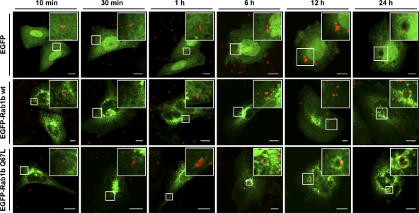
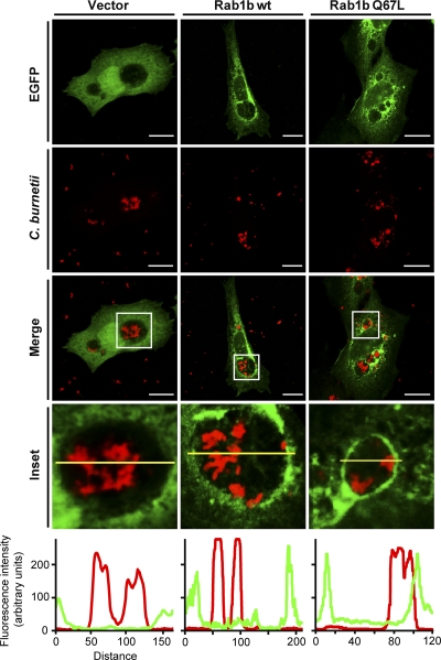
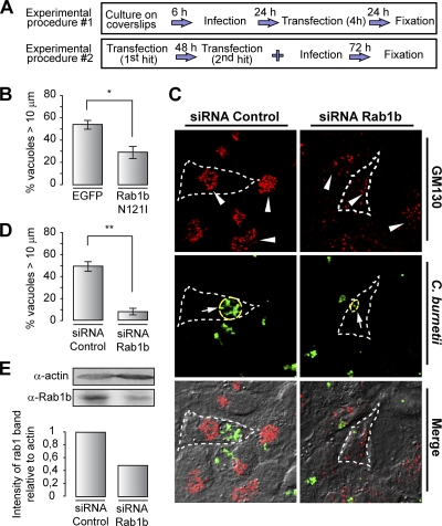
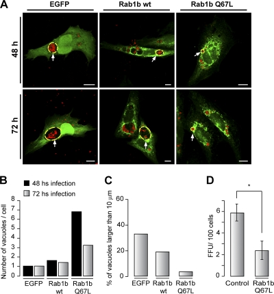
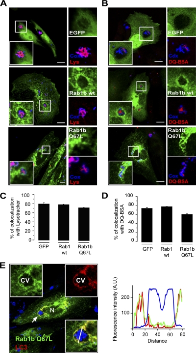
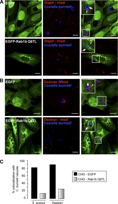
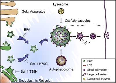
Coxiella burnetii is a Gram-negative obligate intracellular bacterium. After internalization, this bacterium replicates in a large parasitophorous vacuole that has features of both phagolysosomes and autophagosomal compartments. We have previously demonstrated that early after internalization Coxiella phagosomes interact with both the endocytic and the autophagic pathways. In this report, we present evidence that the Coxiella-replicative vacuoles (CRVs) also interact with the secretory pathway. Rab1b is a small GTPase responsible for the anterograde transport between the endoplasmic reticulum and the Golgi apparatus. We present evidence that Rab1b is recruited to the CRV at later infection times (i.e., after 6 h of infection). Interestingly, knockdown of Rab1b altered vacuole growth, indicating that this protein was required for the proper biogenesis of the CRV. In addition, overexpression of the active GTPase-defective mutant (GFP-Rab1b Q67L) affected the development of the Coxiella-replicative compartment inhibiting bacterial growth. On the other hand, disruption of the secretory pathway by brefeldin A treatment or by overexpression of Sar1 T39N, a defective dominant-negative mutant of Sar1, affected the typical spaciousness of the CRVs. Taken together, our results show for the first time that the Coxiella-replicative niche also intercepts the early secretory pathway.


Figures

FIG. 1.
FIG. 1.
Kinetic of infection of C. burnetii under Rab1b overexpression. Stably transfected CHO cells overexpressing EGFP alone, EGFP-Rab1b wt, or the GTPase-defective mutant EGFP-Rab1b Q67L were infected with C. burnetii. The cells were fixed at different infection times (10 min, 30 min, 1 h, 6 h, 12 h, and 24 h), subjected to indirect immunofluorescence using an antibody against C. burnetii (red), and visualized by confocal microscopy. The images show a typical Rab1b distribution in a perinuclear Golgi pattern; however, no colocalization of Rab1b (wt or mutant) with C. burnetii containing compartment was observed at early times (10, 30, or 60 min). Nevertheless, a clear recruitment of EGFP-Rab1b Q67L is observed at later infection times (6, 12, and 24 h). Bars, 10 μm.
FIG. 2.
FIG. 2.
Rab1b decorates the Coxiella-replicative niche at later times of infection. CHO cells stably overexpressing EGFP, EGFP-Rab1b wt, or the active mutant of Rab1 (EGFP-Rab1b Q67L) were infected for 48 h with C. burnetii. After fixation, cells were subjected to indirect immunofluorescence using an antibody against C. burnetii (red) and analyzed by confocal microscopy. As depicted in the insets, the large Coxiella-containing vacuole was strongly labeled by Rab1b Q67L and in a patchy pattern by Rab1b wt. Insets also show a yellow line scan in order to visualize the localization of the Rab1b wt and its defective dominant-active mutant (Q67L) in the vacuole membrane. Images are representative of at least three independent experiments. Bars, 10 μm.
FIG. 3.
FIG. 3.
Overexpression of Rab1b N121I and knockdown of Rab1b affect CRV growth. (A) Outline of the experimental procedures applied in the different assays. (B) CHO cells were infected for 24 h with C. burnetii and subsequently transfected with pEGFP-Rab1 N121I or pEGFP (control). At 48 h postinfection the cells were fixed and subjected to indirect immunofluorescence to detect Coxiella using specific antibodies. Quantification of the vacuole diameter in cells overexpressing EGFP-Rab1b N121I was compared to the control condition (P ≤ 0.05). (C) HeLa cells were transfected with siRNA against Rab1b or an irrelevant siRNA as a negative control. After 48 h, cells were transfected for a second time, infected with C. burnetii, and cultured for an additional 72-h period to allow the development of the large CRV. Subsequently, the cells were fixed and subjected to indirect immunofluorescence for the detection of both Golgi protein GM130 and Coxiella by using specific antibodies. Images were captured by confocal microscopy. The panels show that in contrast to control cells, in cells treated with the siRNA against Rab1b the Golgi apparatus was disassembled (punctate distribution, arrowheads) in the majority of the cells, indicating that Rab1b was effectively depleted. Furthermore, the sizes of the Coxiella-vacuoles were markedly decreased in cells treated with the siRNA against Rab1b (yellow dashed line). (D) Quantification of the vacuole diameter in cells in which Rab1b was depleted compared to the control condition. The data represent the means ± the standard errors of the mean (SEM) of at least three independent experiments in which at least 200 vacuoles were scored in each experiment (P ≤ 0.001). The data represent the means ± the SEM of at least three independent experiments. (E) Western blot of the assay described in panel C and quantification of intensity of the Rab1 bands relative to actin. The data represent one of two independent experiments.
FIG. 4.
FIG. 4.
BFA treatment or overexpression of a dominant-negative mutant of Sar1 alter CRVs growth. CHO cells transiently overexpressing EGFP were infected with C. burnetii. After 48 h of continuous infection, the cells were treated with BFA for 2 or 6 h. After treatment, the cells were fixed and subjected to indirect immunofluorescence with specific antibodies against C. burnetii and GM 130 as a Golgi marker. (A) The images show smaller CRVs (blue) under BFA treatment (6 h) compared to untreated control cells (yellow dashed line). A partial disassembled Golgi (red) is shown in the lower panels (arrowheads). (B) Quantification of the vacuole size (10 μm) of the experiment presented in panel A (P ≤ 0.05). (C) CHO cells infected with C. burnetii for 24 h were transfected with pIRES-DsRed Sar1 T39N. After fixation, the cells were subjected to immunofluorescence using antibodies against C. burnetii (blue) and GM130 as a Golgi marker (green). A Golgi apparatus totally disassembled is shown (arrowheads). The figure shows the reduction in the CRV size (yellow dashed line) under Sar1 T39N overexpression (white dashed line). (D) A bar graph shows a quantification of the >10-μm CRVs in control or Sar1 T39N-overexpressing cells. The data represent the means ± the SEM of at least three independent experiments (P ≤ 0.001). Bars, 15 μm.
FIG. 5.
FIG. 5.
Overexpression of Rab1b Q67L alters the infection profile of C. burnetii. CHO cells transfected with pEGFP plasmids encoding Rab1b wt or the active mutant Rab1b Q67L were infected with C. burnetii for 48 or 72 h (see Materials and Methods), subjected to indirect immunofluorescence to detect the bacteria, and analyzed by confocal microscopy. (A) Confocal images showing the typical formation of one or two large vacuoles containing C. burnetii (yellow dashed line) in CHO cells overexpressing EGFP alone or EGFP-Rab1b wt. In contrast, in cells overexpressing EGFP-Rab1b Q67L, the Coxiella-containing vacuoles are smaller than those of the control condition but are present in large numbers (arrowheads). (B) Quantification of the number of vacuoles per cell at 48 and 72 h of infection from the experiment presented in panel A. (C) Quantification of the size of vacuoles at 48 h postinfection from the experiment presented in panel A. The data represent the means ± the SEM of at least three independent experiments. (D) CHO cells stably overexpressing EGFP (as control) or EGFP-Rab1b Q67L were incubated with C. burnetii. After 48 h of infection, the cells were lysed. Cell lysates with released C. burnetii were used to infect Vero cells. After 72 h of incubation (chase), cells were fixed, and the fluorescent infectious FFU were determined by indirect immunofluorescence, and FFU counts were obtained using confocal microscopy. The data represent the means ± the SEM of at least three independent experiments where at least 200 cells were scored in each experiment (P ≤ 0.05). Bars, 15 μm.
FIG. 6.
FIG. 6.
Coxiella-containing compartments labeled with Rab1b Q67L retain its acidic and degradative characteristics and are also labeled by LC3. CHO cells stably overexpressing EGFP, EGFP-Rab1wt, or EGFP-Rab1b Q67L were infected with C. burnetii. At 48 h of infection, the cells were incubated for 30 min with the acidotropic probe LysoTracker or DQ-BSA for 6 h. After fixation, the cells were examined by indirect immunofluorescence with a specific antibody against C. burnetii and analyzed by confocal microscopy. (A) The insets show the colocalization of LysoTracker (red) and C. burnetii (blue) inside the Coxiella vacuole in all cell types, suggesting that these compartments preserve the acidic properties typical of the Coxiella-replicative niche. (B) Confocal images show the colocalization of C. burnetii (blue) and DQ-BSA (red), indicating the degradative characteristics of the vacuoles. Bars, 20 μm. (C) Quantification of the percentage of colocalization of C. burnetii and LysoTracker. (D) Quantification of the percentage of colocalization of C. burnetii and DQ-BSA. A total of 50 cells were counted in each condition. (E) CHO cells stably overexpressing EGFP-Rab1b Q67L were transiently transfected with pRFP-LC3. At 24 h posttransfection, the cells were infected with C. burnetii for 72 h. Cells were fixed, analyzed by indirect immunofluorescence, and examined by confocal microscopy. Insets show the colocalization of Rab1b Q67L (green) and LC3 (red) at the membrane of the vacuoles containing Coxiella (blue). This result suggests that the interaction with the autophagic pathway is maintained under overexpression of Rab1b Q67L. Bars, 15 μm.
FIG. 7.
FIG. 7.
Overexpression of Rab1b Q67L alters the fusogenic and replicative properties of the C. burnetii compartments. CHO cells stably overexpressing EGFP alone as a control or EGFP-Rab1b Q67L were infected with C. burnetii. At 48 h postinfection, the cells were incubated for 1 h with heat-inactivated Staphylococcus aureus-rhodamine or with 5 μg of dextran-rhodamine/ml to label the phagocytic and endocytic pathways, respectively. (A) The upper panels show colocalization (see inset) of C. burnetii (blue) and S. aureus-rhodamine (red). In contrast, no colocalization of heat-inactivated S. aureus-rhodamine, and the bacterium is observed in the vacuoles of cells overexpressing Rab1b Q67L (lower panels), suggesting that these compartments have its fusogenic properties with the phagocytic pathway altered. (B) The upper panels show that in CHO cells overexpressing EGFP the majority of the Coxiella-vacuoles contain the endocytic marker dextran- rhodamine (red), whereas the absence of colocalization is observed in cells overexpressing Rab1b Q67L (lower panels), suggesting that the fusogenic capacity with the endocytic pathway was altered. Bars, 20 μm. (C) Quantification of the percentage of colocalization of C. burnetii and heat-inactivated Staphylococcus aureus-rhodamine or dextran-rhodamine. A total of 50 cells were counted in each condition.
FIG. 8.
FIG. 8.
Overexpression of a dominant-negative mutant of Sar1 prevents the recruitment of Rab1b Q67L to the C. burnetii vacuole membrane. CHO cells infected with C. burnetii for 24 h were cotransfected with pEGFP-Rab1b Q67L and pIRES-DsRed Sar1 T39N, pEGFP-Rab1b Q67L, and pIRES-DsRed Sar1 H79G or individually transfected with pEGFP, pIRES-DsRed Sar1 T39N, pIRES-DsRed Sar1 H79G, or pEGFP-Rab1b Q67L as controls. At 24 h posttransfection, the cells were fixed and subjected to indirect immunofluorescence with specific antibodies against C. burnetii (blue) and GM130 (red). The images were analyzed by confocal microscopy. (A) The insets in the left panels show clearly the recruitment of Rab1b Q67L to the vacuole membrane. In contrast, no recruitment of Rab1b Q67L to the CRV (see the inset) is observed under overexpression of Sar1 T39N (dashed delineated cell). The middle panels show the different diameters of the vacuoles under Rab1b overexpression, coexpression of Rab1b Q67L and Sar1 T39N (dashed delineated cell), or untransfected cells (arrows). A disassembled Golgi apparatus is showing cells overexpressing Sar1 T39N in contrast with an untransfected cell (arrowheads). (B) Quantification of the size of vacuoles at 48 h of infection from the experiment presented in panel A. The data represent the means ± the SEM of at least three independent experiments (P ≤ 0.001). (C) Quantification of the vacuole size at 48 h postinfection from cells overexpressing EGFP, EGFP-Rab1b Q67L, or DS-Red-Sar1 H69G or coexpressing Rab1b Q67L and DsRed H69G. The data represent the means ± the SEM of at least three independent experiments (P ≤ 0.001). Bars, 15 μm.
FIG. 9.
FIG. 9.
Model showing the interaction among C. burnetii and the endocytic, autophagic, and secretory pathways during the vacuole development process. Upon internalization, bacterium phagosomes interact with the autophagic pathway (the protein LC3 is present on the vacuole membrane). These Coxiella-containing phagosomes also interact with degradative organelles such as the lysosomes. These fusion events contribute to generate a proper environment for the replication process. The secretory pathway through Rab1b-labeled vesicles likely contributes by supplying membrane to generate the spacious Coxiella-vacuole. The highly fusogenic properties of the Coxiella-phagosomes would contribute not only to the formation of the large replicative niche but also to the acquisition of key factors and nutrients to favor the transformation of the bacteria into the replication-competent form (i.e., large cell variant).

References

    1. Allan, B. B., B. D. Moyer, and W. E. Balch. 2000. Rab1 recruitment of p115 into a cis-SNARE complex: programming budding COPII vesicles for fusion. Science 289:444-448. - PubMed
    1. Alvarez, C., R. Garcia-Mata, E. Brandon, and E. Sztul. 2003. COPI recruitment is modulated by a Rab1b-dependent mechanism. Mol. Biol. Cell 14:2116-2127. - PMC - PubMed
    1. Amer, A. O., and M. S. Swanson. 2005. Autophagy is an immediate macrophage response to Legionella pneumophila. Cell Microbiol. 7:765-778. - PMC - PubMed
    1. Beron, W., M. G. Gutierrez, M. Rabinovitch, and M. I. Colombo. 2002. Coxiella burnetii localizes in a Rab7-labeled compartment with autophagic characteristics. Infect. Immun. 70:5816-5821. - PMC - PubMed
    1. Brumell, J. H., and M. A. Scidmore. 2007. Manipulation of Rab GTPase function by intracellular bacterial pathogens. Microbiol. Mol. Biol. Rev. 71:636-652. - PMC - PubMed

LinkOut - more resources