. 2010 Dec;30(24):5608-20.
doi: 10.1128/MCB.01506-09. Epub 2010 Oct 11.

Anaphase-promoting complex/cyclosome participates in the acute response to protein-damaging stress

Affiliations

Anaphase-promoting complex/cyclosome participates in the acute response to protein-damaging stress

Johanna K Ahlskog et al. Mol Cell Biol. 2010 Dec.

Abstract

The ubiquitin E3 ligase anaphase-promoting complex/cyclosome (APC/C) drives degradation of cell cycle regulators in cycling cells by associating with the coactivators Cdc20 and Cdh1. Although a plethora of APC/C substrates have been identified, only a few transcriptional regulators are described as direct targets of APC/C-dependent ubiquitination. Here we show that APC/C, through substrate recognition by both Cdc20 and Cdh1, mediates ubiquitination and degradation of heat shock factor 2 (HSF2), a transcription factor that binds to the Hsp70 promoter. The interaction between HSF2 and the APC/C subunit Cdc27 and coactivator Cdc20 is enhanced by moderate heat stress, and the degradation of HSF2 is induced during the acute phase of the heat shock response, leading to clearance of HSF2 from the Hsp70 promoter. Remarkably, Cdc20 and the proteasome 20S core α2 subunit are recruited to the Hsp70 promoter in a heat shock-inducible manner. Moreover, the heat shock-induced expression of Hsp70 is increased when Cdc20 is silenced by a specific small interfering RNA (siRNA). Our results provide the first evidence for participation of APC/C in the acute response to protein-damaging stress.


Figures
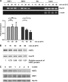
FIG. 1.
FIG. 1.
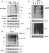
HSF2 DNA-binding activity and protein levels are decreased during heat shock. (A) ChIP was performed on K562 cells heat shocked at 42°C for the indicated times, and the samples were immunoprecipitated with anti-HSF1, anti-HSF2, or nonspecific (NS) antibody. The IP and input samples were subjected to PCR using primers specific for the human Hsp70 and β-actin promoters. The promoter occupancies of HSF1 and HSF2 were statistically analyzed by performing paired two-tailed Student's t test on values obtained by quantitative real-time PCR on ChIP samples. The maximal binding for both HSF1 (light bars) and HSF2 (dark bars) was detected upon 15 min of heat shock and was set to 1. Error bars represent standard errors of the means from four independent experiments. ns, no significance; *, P < 0.05. (B) HEK293 cells were treated with heat shock at 42°C for the indicated times, and total Laemmli lysates were run on SDS-polyacrylamide gels. The protein levels of HSF2 were detected by blotting with anti-HSF2 antibody, and equal loading was confirmed by blotting with anti-Hsc70 antibody. The relative amount of HSF2 protein (indicated below the blot) was determined by densitometry and normalized to that of Hsc70. The highest HSF2 level was detected at the 0-min time point and was set to 1. (C) HEK293 cells were treated with cycloheximide (CHX) (10 μg/ml) at 37°C for the indicated times, and total Laemmli lysates were analyzed by blotting with anti-HSF1 or anti-HSF2; anti-Hsc70 antibody was used to confirm equal loading.
FIG. 2.
FIG. 2.
Ubiquitination of HSF2 is enhanced by heat stress. (A) HEK293 cells were left untreated (C) or treated with 20 μM MG132 (MG) for 5 h. IP was performed with anti-HSF1, anti-HSF2, or no antibody (−), and samples were run on SDS-polyacrylamide gels. Ubiquitinated proteins were subsequently detected with antiubiquitin antibody. The IP membrane was reblotted first with anti-HSF2 antibody and then with anti-HSF1 antibody, and therefore, HSF2 is also seen in the HSF1 blot (**). *, unspecific band. The inputs were blotted with anti-HSF1, anti-HSF2, and anti-Hsc70 antibodies. (B) HEK293 cells were treated with heat shock at 42°C for the indicated times. HSF2 was immunoprecipitated with anti-HSF2 antibody, and the samples were analyzed by SDS-PAGE followed by blotting with antiubiquitin antibody. Input samples were analyzed by immunoblotting (IB) with anti-HSF2 and anti-Hsc70 antibodies. (C) HEK293 cells were transfected with empty vector (mock) or Myc-tagged wild-type (WT) or mutant ubiquitin (Ub) where lysines 11, 48, and 63 were mutated to arginine (K11R, K48R, and K63R, respectively). The transfected cells were heat shocked for 30 min at 42°C. Anti-HSF2 antibody was used for IP, and ubiquitination was detected with anti-Myc antibody. The protein amounts in the input samples were detected with antibodies against HSF2, Myc, and Hsc70. The numerical values outside the blots indicate molecular size markers in kilodaltons.
FIG. 3.
FIG. 3.
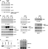
HSF2 interacts with Cdc20, Cdh1, and Cdc27. (A) HEK293 cells were transfected with empty vector (mock) or Cdc20-GFP and left untreated (C) or heat shocked at 42°C for 30 min (HS). The cells were lysed, analyzed by SDS-PAGE, and immunoblotted with antibodies recognizing HSF2, GFP, and Hsc70. The relative amount of HSF2 protein (indicated below the blot) was determined by densitometry and normalized to that of Hsc70. The highest HSF2 level was detected in lane 1 and was set to 1. (B) (Left) HEK293 cells were transfected with empty vector (mock), HSF1-Myc, or Flag-HSF2, followed by no treatment (C) or heat shock at 42°C for 30 min (HS). IP was performed with anti-Cdc20 antibody and analyzed with anti-HSF1 or anti-HSF2 antibody. The input membranes were immunoblotted with antibodies recognizing HSF1, HSF2, Cdc20, and Hsc70. (Middle) Anti-Cdc27 IP was performed on HEK293 cells transfected with Flag-HSF2 and treated as described above. The IP samples were analyzed with anti-HSF2 and anti-Cdc27 antibodies, and the input samples were treated with anti-HSF2 and anti-Hsc70 antibodies. (Right) IP was performed with anti-Cdh1 antibody on cells transfected and treated as described above. Anti-HSF2, anti-Cdh1, and anti-Hsc70 antibodies were used for immunoblotting. (C) An in vitro pulldown assay was performed on reaction mixtures containing 35S-labeled HSF2 and cold in vitro-translated Myc-tagged empty vector (mock), Cdc20, or Cdh1. Myc-tagged proteins were immunoprecipitated using Myc-agarose, and HSF2 was detected by autoradiography. The input sample contains 20% of the HSF2 volume used in the pulldown samples. (D) An in vitro ubiquitination assay was performed on in vitro-translated HSF2 as a substrate and immunopurified APC/C as an E3 ligase from HeLa cells that were untreated, heat shocked at 42°C for 30 min (HS), or arrested with nocodazole (Noc) (100 ng/ml) for 6 h. The reaction samples were incubated at 37°C for 45 min and run on SDS-polyacrylamide gels, followed by immunoblotting with anti-HSF2 antibody. Numbers on the left indicate molecular size markers in kilodaltons.
FIG. 4.
FIG. 4.
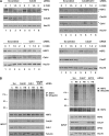
Downregulation of APC/C subunits increases HSF2 stability. (A) Scrambled siRNA or siRNA oligonucleotides downregulating Cdc20 or Cdh1 (left), Cdc20 and Cdh1 (top right), or Cdc27 (bottom right) were transfected into HEK293 cells, followed by treatment with cycloheximide (CHX) (10 μg/ml). Laemmli lysates were harvested at the indicated time points and analyzed by SDS-PAGE. Protein levels of HSF2, Cdc20, Cdh1, Cdc27, and Hsc70 were detected by blotting with specific antibodies. *, unspecific band. (B) HEK293 cells were transfected with scrambled (Scr), Cdc20, and/or Cdh1 siRNA (left) or scrambled (Scr), Cdc27, or APC2 siRNA (right) and left untreated (C) or heat shocked for 30 min at 42°C (HS). The assay detecting HSF2 ubiquitination was performed as described in the legend for Fig. 2B. The input samples were blotted with anti-HSF2, anti-Cdc20, anti-Cdh1, anti-Cdc27, and anti-APC2 antibodies. Equal loading was controlled with antitubulin or anti-Hsc70 antibody. Numbers on the left indicate molecular size markers in kilodaltons.
FIG. 5.
FIG. 5.
HSF2 becomes more resistant to heat shock-induced degradation when APC/C coactivators are silenced. Laemmli lysates from HEK293 cells that were transfected with scrambled, Cdc20, or Cdc20 and Cdh1 siRNA and subjected to heat shock at 42°C for the indicated times were run on SDS-polyacrylamide gels. Immunoblotting was performed with anti-HSF2, antitubulin, anti-Cdc20, and anti-Cdh1 antibodies. The relative amount of HSF2 protein (indicated below the blots) was determined by densitometry and normalized to that of tubulin. The highest HSF2 level for each transfection was detected at the 0-min time point and was set to 1.
FIG. 6.
FIG. 6.
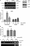
Cdc20 and proteasome subunit α2 are recruited to the Hsp70 promoter in response to heat stress. (A) ChIP for HSF2 binding to the Hsp70 promoter was performed on HeLa cells transfected with scrambled (Scr) or Cdc20 siRNA, followed by heat shock treatment at 42°C for the indicated times. The top left panel shows a representative ChIP experiment, where NS denotes a nonspecific antibody. The samples were analyzed as described in the legend for Fig. 1A except that the value of binding at the 0-min time point in the scrambled siRNA-transfected cells was set to 1 (bottom left). Error bars represent standard errors of the means from four independent experiments. For the representative blot of protein levels shown in the right panel, Laemmli samples of the transfected cells were collected prior to heat shock treatment and run on SDS-polyacrylamide gels, followed by immunoblotting with antibodies for HSF2, Cdc20, and Hsc70. *, unspecific band. (B) K562 cells were heat shocked at 42°C for the indicated times, and four independent experiments were analyzed by ChIP with anti-Cdc20 antibody and quantitative real-time PCR as described above. (C) ChIP with antibody against the α2 subunit of the proteasome was performed on K562 cells that were left untreated or treated with heat shock for 30 min at 42°C. NS denotes a nonspecific antibody control. The IP and input samples were subjected to PCR using primers specific for the human Hsp70 and β-actin promoters.
FIG. 7.
FIG. 7.
Altered transcription of Hsp70 and Sat III upon Cdc20 knockdown. HeLa cells were transfected with scrambled (Scr) and Cdc20 siRNA oligonucleotides and heat shocked for 1 h at 42°C. The mRNAs for Hsp70 (A) and Satellite III (B) were determined by quantitative real-time RT-PCR. The relative expression levels were calculated, and the value for the scrambled control was set to 1. Error bars represent standard deviations from five (A) or four (B) independent experiments. *, P < 0.05; **, P < 0.01. A representative blot for Cdc20 knockdown is shown in Fig. 6A.

References

    1. Abravaya, K., B. Phillips, and R. I. Morimoto. 1991. Attenuation of the heat shock response in HeLa cells is mediated by the release of bound heat shock transcription factor and is modulated by changes in growth and in heat shock temperatures. Genes Dev. 5:2117-2127. - PubMed
    1. Adhikary, S., F. Marinoni, A. Hock, E. Hulleman, N. Popov, R. Beier, S. Bernard, M. Quarto, M. Capra, S. Goettig, U. Kogel, M. Scheffner, K. Helin, and M. Eilers. 2005. The ubiquitin ligase HectH9 regulates transcriptional activation by Myc and is essential for tumor cell proliferation. Cell 123:409-421. - PubMed
    1. Ahn, S. G., P. C. Liu, K. Klyachko, R. I. Morimoto, and D. J. Thiele. 2001. The loop domain of heat shock transcription factor 1 dictates DNA-binding specificity and responses to heat stress. Genes Dev. 15:2134-2145. - PMC - PubMed
    1. Ahn, S. G., and D. J. Thiele. 2003. Redox regulation of mammalian heat shock factor 1 is essential for Hsp gene activation and protection from stress. Genes Dev. 17:516-528. - PMC - PubMed
    1. Åkerfelt, M., E. Henriksson, A. Laiho, A. Vihervaara, K. Rautoma, N. Kotaja, and L. Sistonen. 2008. Promoter ChIP-chip analysis in mouse testis reveals Y chromosome occupancy by HSF2. Proc. Natl. Acad. Sci. U. S. A. 105:11224-11229. - PMC - PubMed

Publication types

MeSH terms

LinkOut - more resources