. 2010 Dec;30(24):5698-709.
doi: 10.1128/MCB.00165-10. Epub 2010 Oct 11.

Oligomerization of the mitochondrial protein voltage-dependent anion channel is coupled to the induction of apoptosis

Affiliations

Oligomerization of the mitochondrial protein voltage-dependent anion channel is coupled to the induction of apoptosis

Nurit Keinan et al. Mol Cell Biol. 2010 Dec.

Abstract

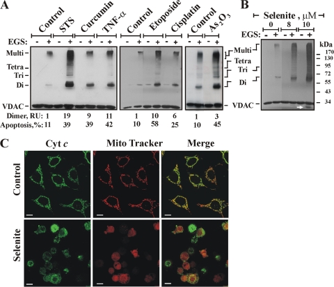
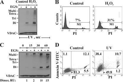
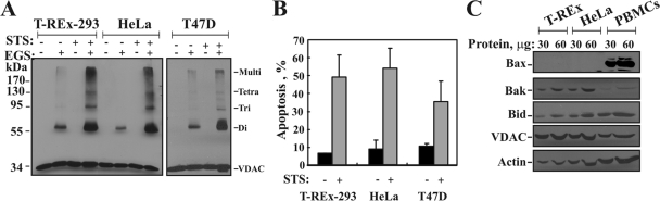
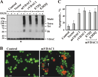
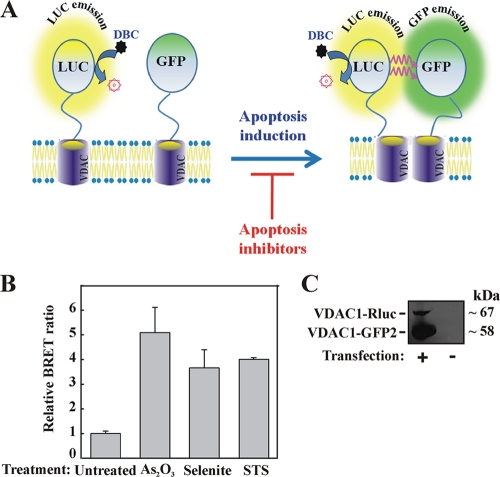
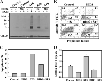
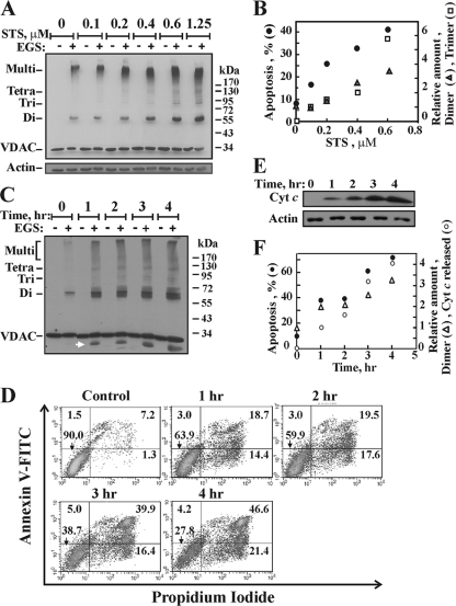
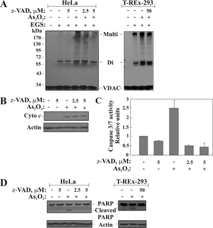
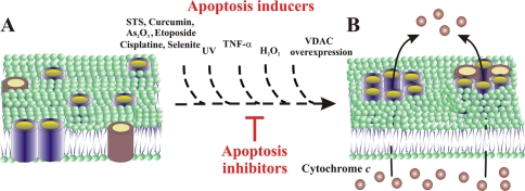
Accumulating evidence implicates that the voltage-dependent anion channel (VDAC) functions in mitochondrion-mediated apoptosis and as a critical player in the release of apoptogenic proteins, such as cytochrome c, triggering caspase activation and apoptosis. The mechanisms regulating cytochrome c release and the molecular architecture of the cytochrome c-conducting channel remain unknown. Here the relationship between VDAC oligomerization and the induction of apoptosis was examined. We demonstrated that apoptosis induction by various stimuli was accompanied by highly increased VDAC oligomerization, as revealed by cross-linking and directly monitored in living cells using bioluminescence resonance energy transfer technology. VDAC oligomerization was induced in all cell types and with all apoptosis inducers used, including staurosporine, curcumin, As(2)O(3), etoposide, cisplatin, selenite, tumor necrosis factor alpha (TNF-α), H(2)O(2), and UV irradiation, all acting through different mechanisms yet all involving mitochondria. Moreover, correlation between the levels of VDAC oligomerization and apoptosis was observed. Furthermore, the apoptosis inhibitor 4,4'-diisothiocyanostilbene-2,2'-disulfonic acid (DIDS) inhibited VDAC oligomerization. Finally, a caspase inhibitor had no effect on VDAC oligomerization and cytochrome c release. We propose that VDAC oligomerization is involved in mitochondrion-mediated apoptosis and may represent a general mechanism common to numerous apoptogens acting via different initiating cascades. Thus, targeting the oligomeric status of VDAC, and hence apoptosis, offers a therapeutic strategy for combating cancers and neurodegenerative diseases.


Figures

FIG. 1.
FIG. 1.
Apoptosis induction is associated with VDAC oligomerization. (A) Cells were exposed either to STS (1.25 μM) for 2.5 h or to curcumin (40 μM), TNF-α (12 ng/ml), etoposide (2 μM), cisplatin (40 μM), or As2O3 (30 μM) for 16 h. Cells washed with PBS at 2.5 to 3 mg/ml were incubated with EGS (250 to 300 μM) at 30°C for 15 min and were then subjected to SDS-PAGE and immunoblotting using anti-VDAC antibodies. The positions of VDAC monomers to multimers are indicated. Percentages of apoptotic cells (assayed via AcrOr-EthBro staining; n = 3) and relative amounts of dimers are given at the bottom. RU, relative units. (B and C) HeLa cells were exposed to selenite at the indicated concentrations for 17 h and were then analyzed for VDAC oligomerization by assessment of EGS-based cross-linking (B) or immunocytochemical assessment of Cyto c release (C). Control and selenite-treated HeLa cells were stained with the mitochondrial marker MitoTracker red dye, followed by immunostaining with anti-cytochrome c antibodies and Alexa Fluor 488-conjugated secondary antibodies (green), and visualized by confocal microscopy. The arrow in panel B indicates an anti-VDAC1 antibody-labeled protein band migrating below the position of monomeric VDAC.
FIG. 2.
FIG. 2.
H2O2- and UV-induced VDAC oligomerization. (A and B) T-REx-293 cells were incubated for 5 h with 1 mM H2O2, harvested, and subjected either to cross-linking with EGS (250 μM) as for Fig. 1 (A) or to apoptosis analysis via PI uptake and FACS analysis (B). The results of one experiment representative of three similar experiments are shown. (C) Cells were exposed to UV irradiation for the indicated times and were analyzed after 24 h for VDAC oligomerization by using EGS-based cross-linking and immunoblotting, as described for panel A. The relative amounts of dimers are given (RU, relative units), and the positions of VDAC monomers to tetramers and multimers are indicated. (D) Apoptosis, induced by UV irradiation, was assayed using annexin V-FITC-PI staining and FACS analysis. Results from one experiment representative of two similar experiments are shown.
FIG. 3.
FIG. 3.
STS induces VDAC oligomerization in all cell types used. (A) T-REx-293, HeLa, and T47D cells (2.5 mg/ml) were incubated in the absence or presence of STS (1.25 μM; 5 h) and were subjected to cross-linking with EGS (250 μM) and to immunoblotting using anti-VDAC antibodies. The positions of molecular size protein standards are provided. (B) Quantitative analysis of apoptosis (assayed via AcrOr-EthBro staining) (n = 3). (C) T-REx-293, HeLa, and human peripheral blood mononuclear cells (PBMCs; isolated using Ficoll-Paque density gradient centrifugation) (30 and 60 μg) were subjected to immunoblotting using anti-Bax, anti-Bak, anti-Bid, and anti-VDAC antibodies. As a loading control, actin levels in the samples were determined using antiactin antibodies. Note that the increase in the amount of protein loaded was seen for all immunoblotted proteins, except for VDAC, due to the high affinity of the antibodies used, which yielded a saturated signal over 30 μg.
FIG. 4.
FIG. 4.
VDAC1 overexpression induces VDAC oligomerization and apoptotic cell death. (A) T-REx-293 cells were transfected to overexpress murine VDAC1 (mVDAC1), rat VDAC1 (rVDAC1), E72Q-mVDAC1, or E202Q-mVDAC1. At 72 h following transfection, cells were incubated with EGS (75 μM) for 15 min and were then subjected to SDS-PAGE and immunoblotting. At the relatively low EGS concentration used, VDAC oligomers were obtained in cells overexpressing VDAC1 but not in nontransfected control cells. An additional band, above the dimer, appeared in cells overexpressing VDAC1 (indicated by a hyphen). (B) Apoptosis in cells overexpressing native or mutated VDAC1 was analyzed by use of AcrOr-EthBro staining 105 h following transfection. The arrow indicates cells in an early apoptotic state, reflected by degraded nuclei (stained green with acridine orange). The arrowhead indicates cells in the late apoptotic state (stained orange with acridine orange and ethidium bromide). Bars, 15 μm. (C) Quantitative analysis of apoptosis showed that at 105 h posttransfection, about 55% of the cells presented apoptotic characteristics.
FIG. 5.
FIG. 5.
VDAC1 oligomerization and BRET2-based assay. (A) Schematic representation showing energy transfer between VDAC1-luciferase (RLuc, a light-producing enzyme) as the donor and VDAC1-GFP2 (fluorophore) as the acceptor, which occurs only when the donor and the acceptor are spatially close. The BRET2 signal is obtained when the two VDAC1 molecules interact physically. Compounds enhancing apoptosis lead to VDAC1 oligomerization and thus enhance the BRET2 signal, while apoptosis inhibitors inhibit VDAC1 oligomerization and therefore decrease the BRET2 signal. The luciferase substrate DBC emits light upon cleavage and thus causes excitation of the proximal GFP2 protein, thereby generating the BRET2 signal. (B) STS, selenite, and As2O3 enhance the BRET2 signal. T-REx cells expressing hVDAC1 shRNA were cotransfected with plasmids encoding rVDAC1-Rluc (0.1 μg) and rVDAC1-GFP2 (0.8 μg). Luciferase and GFP signals were measured 72 h posttransfection. The BRET2 signals obtained in cells treated with STS (0.6 μM; 3 h), selenite (8 μM; 16 h), or As2O3 (20 μM; 16 h) are shown. “Untreated” refers to cells transfected with the rVDAC1-Rluc plasmid and treated with the appropriate amount of dimethyl sulfoxide. BRET2 signals were measured, and BRET2 ratios were calculated as described in Materials and Methods. The results were collected from three 96-well plates (STS) or one 96-well plate (selenite and As2O3). (C) Cellular expression levels of VDAC1-Rluc and VDAC1-GFP2, analyzed by immunoblotting using anti-VDAC1antibodies.
FIG. 6.
FIG. 6.
DIDS inhibits VDAC oligomerization and apoptosis. (A) HeLa cells were incubated with DIDS (100 or 200 μM) for 1 h, after which they were incubated with or without STS (1.25 μM; 2 h), harvested, cross-linked with EGS (250 μM; 15 min), and then analyzed by immunoblotting using anti-VDAC antibodies. The dramatic inhibition of STS-induced VDAC oligomerization by DIDS is shown. (B) Representative FACS analysis of apoptotic cell death, assayed using annexin V-FITC-propidium iodide staining. (C) Quantitative analysis of apoptosis, as measured by FACS analysis (from panel B). Results of one experiment representative of three similar experiments are shown. (D) Ratios of the BRET2 signals obtained for cells treated with STS (0.8 μM; 3.0 h) with or without pretreatment with DIDS (100 μM; 1 h). T-REx-293 cells expressing VDAC1 shRNA were cotransfected with plasmids encoding rVDAC1-Rluc (0.1 μg) and rVDAC1-GFP2 (0.8 μg). Data are represented as means ± standard errors of the means.
FIG. 7.
FIG. 7.
Correlation between the extent of apoptosis and the level of VDAC oligomerization. (A) Immunoblot analysis shows the levels of VDAC oligomerization (cross-linking with 300 μM EGS for 15 min) induced by STS at various concentrations (0.1 to 0.6 μM for 16 h or 1.25 μM for 2.5 h) in T-REx-293 cells. (B) Quantitative analysis of the immunostained VDAC dimers and trimers and the extent of apoptosis, presented as a function of the STS concentration. (C to F) HeLa cells were exposed to H2O2 (12 mM) for the indicated times, after which VDAC oligomerization (C), apoptotic cell death (D), and Cyto c release (E) were analyzed and presented as a function of time (F).
FIG. 8.
FIG. 8.
Caspase activity is not required for VDAC oligomerization. Cells were preincubated with or without zVAD-fmk (2.5 or 5 μM with HeLa cells and 50 μM with T-REx cells, for 1.5 h), followed by As2O3 treatment (30 μM for 17 h). (A and B) Cells were analyzed for VDAC oligomerization (A) and Cyto c release (B). (C) The enzymatic activities of the caspases were followed by measuring the bioluminescent signals using a microplate reader, as described in Materials and Methods. (D) Caspase activity, as reflected by PARP cleavage, was analyzed by immunoblotting. The immunoblot was probed with a rabbit anti-PARP polyclonal antibody. Immunoblotting of actin, as a loading control, is also presented.
FIG. 9.
FIG. 9.
Model for apoptotic signals inducing VDAC1 oligomerization-mediated Cyto c release. (A) Side view of membranal VDAC1 (in blue) and a proapoptotic protein (in red), both predominantly in the monomeric state. (B) When an apoptotic signal (e.g., STS, curcumin, As2O3, etoposide, cisplatin, selenite, TNF-α, H2O2, UV irradiation, or VDAC1 overexpression) is encountered, oligomerization of VDAC as homo- or hetero-oligomers is enhanced, leading to pore formation between VDAC β-barrel monomers. While apoptosis inducers facilitate homo- and/or hetero-oligomer formation, apoptosis inhibitors (e.g., DIDS) inhibit such oligomerization and thereby inhibit Cyto c release and apoptotic cell death.

References

    1. Abu-Hamad, S., N. Arbel, D. Calo, L. Arzoine, A. Israelson, N. Keinan, R. Ben-Romano, O. Friedman, and V. Shoshan-Barmatz. 2009. The VDAC1 N-terminus is essential both for apoptosis and the protective effect of anti-apoptotic proteins. J. Cell Sci. 122:1906-1916. - PubMed
    1. Abu-Hamad, S., S. Sivan, and V. Shoshan-Barmatz. 2006. The expression level of the voltage-dependent anion channel controls life and death of the cell. Proc. Natl. Acad. Sci. U. S. A. 103:5787-5792. - PMC - PubMed
    1. Abu-Hamad, S., H. Zaid, A. Israelson, E. Nahon, and V. Shoshan-Barmatz. 2008. Hexokinase-I protection against apoptotic cell death is mediated via interaction with the voltage-dependent anion channel-1: mapping the site of binding. J. Biol. Chem. 19:13482-13490. - PubMed
    1. Angers, S., A. Salahpour, E. Joly, S. Hilairet, D. Chelsky, M. Dennis, and M. Bouvier. 2000. Detection of β2-adrenergic receptor dimerization in living cells using bioluminescence resonance energy transfer (BRET). Proc. Natl. Acad. Sci. U. S. A. 97:3684-3689. - PMC - PubMed
    1. Annis, M. G., E. L. Soucie, P. J. Dlugosz, J. A. Cruz-Aguado, L. Z. Penn, B. Leber, and D. W. Andrews. 2005. Bax forms multispanning monomers that oligomerize to permeabilize membranes during apoptosis. EMBO J. 24:2096-2103. - PMC - PubMed

Publication types

MeSH terms