. 2010 Dec 17;285(51):39623-36.
doi: 10.1074/jbc.M110.140467. Epub 2010 Oct 11.

Drosophila Myc interacts with host cell factor (dHCF) to activate transcription and control growth

Affiliations

Drosophila Myc interacts with host cell factor (dHCF) to activate transcription and control growth

Michael Furrer et al. J Biol Chem. .

Abstract

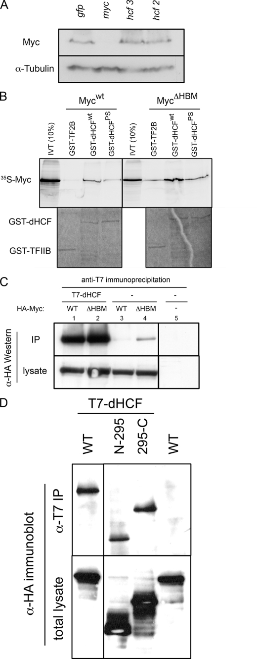
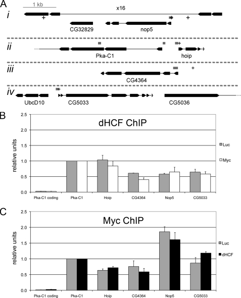
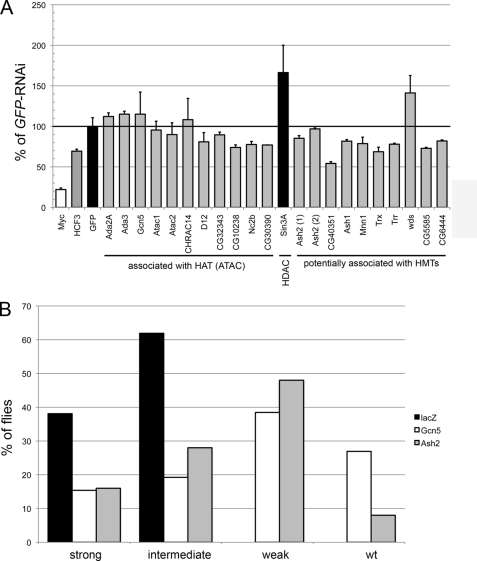
The Myc proto-oncoproteins are transcription factors that recognize numerous target genes through hexameric DNA sequences called E-boxes. The mechanism by which they then activate the expression of these targets is still under debate. Here, we use an RNAi screen in Drosophila S2 cells to identify Drosophila host cell factor (dHCF) as a novel co-factor for Myc that is functionally required for the activation of a Myc-dependent reporter construct. dHCF is also essential for the full activation of endogenous Myc target genes in S2 cells, and for the ability of Myc to promote growth in vivo. Myc and dHCF physically interact, and they colocalize on common target genes. Furthermore, down-regulation of dHCF-associated histone acetyltransferase and histone methyltransferase complexes in vivo interferes with the Myc biological activities. We therefore propose that dHCF recruits such chromatin-modifying complexes and thereby contributes to the expression of Myc targets and hence to the execution of Myc biological activities.


Figures

FIGURE 1.
FIGURE 1.
dHCF is required for the full expression of a Myc reporter. A, schematic of the Myc-responsive luciferase system. The dark line represents genomic sequences from −322 to +61 (start of the open-reading frame) of the Myc target CG5033, and the small gray rectangle shows the position of the single Myc binding site (E-box) in this region. B, schematic of dHCF and the constructs used in this work. The different domains of dHCF have been described before (P175: proline mutated in a temperature-sensitive mutant of vertebrate HCF-1; SAS1N: self-association domain 1N; FnIII/SAS1C: fibronectin-type III domain/self-association domain 1C; NLS: bipartite nuclear localization signal; Refs. 19, 20, 44). Numbers correspond to amino acid positions. In vitro shows the part of dHCF that was used for in vitro translation and in a fusion with GST. The short black lines indicate the extents of dsRNAs used for RNAi (see “Experimental Methods”): 1, from the original screen; 2 & 3, used for tissue culture experiments and in transgenic flies; V, for transgenic experiments. C, efficiency of dHCF knockdown. S2 cells were transfected with pTub-GAL4, UAST-T7-dHCF-FLAG, UAS-GFP either alone (lane −) or together with the indicated hcf dsRNAs; ctr lane lacks ectopically expressed proteins. 48 h later, cell lysates were analyzed by immunoblotting with anti-T7-epitope and anti-GFP antiserum. D, changes of dHCF levels affect the Myc-dependent reporter. S2 were co-transfected with the Myc-dependent luciferase reporters shown in A, together with the indicated dsRNAs or with pACXT-T7-dHCF, and assayed 48 h later for relative luciferase activity (wt-RLuc/ΔE-FLuc, with the gfp-dsRNA sample being set to 100%). Error bars show standard deviations from duplicate transfections.
FIGURE 2.
FIGURE 2.
dHCF synergizes with Myc in the induction of growth. A, top panel, scanning electron micrographs (S.E.) of representative eyes overexpressing Myc and/or dHCF under the control of GMR-GAL4; all eyes are at the same magnification. Bottom panel, ommatidial areas of eyes corresponding to the genotypes shown above. Shown are averages and standard deviations from 4 to 6 independent eyes per genotype. B, average areas of 45-h-old wing imaginal disc clones overexpressing Myc and/or dHCF (n = 25, except for the co-expression of Myc and dHCF: n = 17).
FIGURE 3.
FIGURE 3.
Knockdown of dHCF in vivo impairs (Myc-dependent) growth. A, expression of dHCF-dsRNA in bristle precursor cells with sca-GAL4 reduces the average area of posterior scutellar bristles (n = 6, 7, 4, for control, hcf2, hcf3, respectively); the size reduction is borderline significant (p = 0.06 for both hcf-samples as compared with control). B, knockdown of dHCF alleviates wing defects caused by Myc overexpression under the control of ap-GAL4. Adult Myc-overexpressing wings were assigned to the phenotypic classes shown in the top panel, according to their appearance under a dissecting microscope (n = 21 for control and 25 for each of the dHCF samples). C, average area of salivary gland nuclei that have been overexpressing Myc and/or dHCF-dsRNA for 45 h (n = 34–48). D, expression of dHCF-dsRNA during eye development significantly reduces the area of dmP0 (hypomorphic Myc-mutant) ommatidia, but not of wild-type ommatidia (n = 4–7). * and ** indicate that comparisons with the corresponding control genotype (without dHCF-RNAi) are significant with p < 0.05 and p < 0.01, respectively.
FIGURE 3.
FIGURE 3.
Knockdown of dHCF in vivo impairs (Myc-dependent) growth. A, expression of dHCF-dsRNA in bristle precursor cells with sca-GAL4 reduces the average area of posterior scutellar bristles (n = 6, 7, 4, for control, hcf2, hcf3, respectively); the size reduction is borderline significant (p = 0.06 for both hcf-samples as compared with control). B, knockdown of dHCF alleviates wing defects caused by Myc overexpression under the control of ap-GAL4. Adult Myc-overexpressing wings were assigned to the phenotypic classes shown in the top panel, according to their appearance under a dissecting microscope (n = 21 for control and 25 for each of the dHCF samples). C, average area of salivary gland nuclei that have been overexpressing Myc and/or dHCF-dsRNA for 45 h (n = 34–48). D, expression of dHCF-dsRNA during eye development significantly reduces the area of dmP0 (hypomorphic Myc-mutant) ommatidia, but not of wild-type ommatidia (n = 4–7). * and ** indicate that comparisons with the corresponding control genotype (without dHCF-RNAi) are significant with p < 0.05 and p < 0.01, respectively.
FIGURE 4.
FIGURE 4.
dHCF physically interacts with Myc. A, dHCF knockdown does not affect Myc levels. S2 cells were transfected with the indicated dsRNAs and 48 h later processed for Western blotting against endogenous Myc and α-tubulin as a loading control. B, in vitro translated Mycwt binds to GST-dHCFwt and GST-dHCFPS, but not to the negative control protein GST-TFIIB; MycΔHBM binds preferentially GST-dHCF, but shows significant background binding to GST-TFIIB. The upper panel is a phosphorimager picture of input and bound 35S-labeled Myc proteins; each binding reaction contained 20% of in vitro synthesized Myc. The lower panel shows the same gel after Coomassie Blue staining to reveal the relative amounts of GST-fusion proteins. C, anti-T7-epitope antibodies precipitate HA-MycWT and HA-MycΔBHM when they are co-expressed with T7-dHCF, but not when they are expressed alone. Labels above the lanes indicate the transfected proteins (T7-dHCF and/or HA-MycWT/ΔHBM). As in B, HA-MycΔHBM shows some non-HBM-specific binding. Immunoprecipitates correspond to 50% of a 3-cm well, lysates to 20%. D, two non-overlapping fragments of Myc associate with dHCF. HA-tagged versions of Myc containing amino acids 1–295 or 295–717 (as indicated) both were co-precipitated with T7-dHCF, indicating that at least two regions of Myc mediate the interaction with dHCF.
FIGURE 5.
FIGURE 5.
dHCF and Myc co-localize at the promoters of Myc-regulated genes. A, schematic representation of the fragments probed in chromatin immunoprecipitations (ChIP assays). Exons are depicted as boxes and introns as thin lines. Gray boxes show positions of the PCR fragments and + signs consensus E-boxes. The fragment located in the coding region of Pka-C1 (3rd exon) serves as a negative control. All 4 chromosomal regions (i-iv) are depicted at the same scale. B and C, relative binding of dHCF (B) and Myc (C) to the indicated regions. SL2 cells were incubated with the indicated dsRNAs (Myc, dHCF, or Luciferase, which served as a negative control), and chromatin extracts prepared 3 days later for ChIP with polyclonal antibodies against dHCF or Myc. The diagrams represent the average of three biological replicates, and standard errors are shown as error bars.
FIGURE 6.
FIGURE 6.
Histone methyltransferases and acetyltransferases affect Myc-dependent processes. A, down-regulation of potentially dHCF-associated HATs (ATAC complex) and HMTs moderately reduces the activity of the Myc-dependent luciferase reporter. Luciferase assays were carried out in duplicate 48 h after co-transfection of the indicated dsRNAs. B, suppression of Myc-induced wing defects by co-expression of ash2- or GCN5-dsRNAs. Neither of these RNAi-transgenes had a strong effect on wing morphology when expressed on their own (although a second independent UAS-GCN5-IR insertion, expressing the same dsRNA, produced strongly deformed wings on its own upon overexpression by ap-GAL4, and it also did not rescue the Myc overexpression defect, presumably because it down-regulated GCN5 too strongly). The experiment was carried out and evaluated as indicated in Fig. 3B (n = 21, 26, 31 for lacZ, GCN5, ash2, respectively). Note that the full composition of the Drosophila dHCF-HMT complex has not been published, and some of the listed proteins were included because of their similarity to vertebrate or yeast HMT-components (Ash2/Hs Ash2L; CG40531/Sc Set1; Ash1, Trx, Trr/SET-domain containing HMTs; Mnn1/Hs Men1; wds/Hs WDR5; CG5585/Hs RBBP5; CG6444/Sc Sdc1); the ATAC components are taken from Refs. , .

Similar articles

Cited by

References

    1. Oster S. K., Ho C. S., Soucie E. L., Penn L. Z. (2002) Adv. Cancer Res. 84, 81–154 - PubMed
    1. Pirity M., Blanck J. K., Schreiber-Agus N. (2006) Curr. Top Microbiol. Immunol. 302, 205–234 - PubMed
    1. Trumpp A., Refaeli Y., Oskarsson T., Gasser S., Murphy M., Martin G. R., Bishop J. M. (2001) Nature 414, 768–773 - PubMed
    1. Benassayag C., Montero L., Colombié N., Gallant P., Cribbs D., Morello D. (2005) Mol. Cell Biol. 25, 9897–9909 - PMC - PubMed
    1. Gallant P. (2009) Adv. Cancer Res. 103, 111–144 - PubMed

Publication types

MeSH terms

LinkOut - more resources