. 2010 Dec 10;285(50):39051-60.
doi: 10.1074/jbc.M110.163782. Epub 2010 Oct 11.

Angiotensin-converting enzyme overexpression in mouse myelomonocytic cells augments resistance to Listeria and methicillin-resistant Staphylococcus aureus

Affiliations

Angiotensin-converting enzyme overexpression in mouse myelomonocytic cells augments resistance to Listeria and methicillin-resistant Staphylococcus aureus

Derick Okwan-Duodu et al. J Biol Chem. .

Abstract

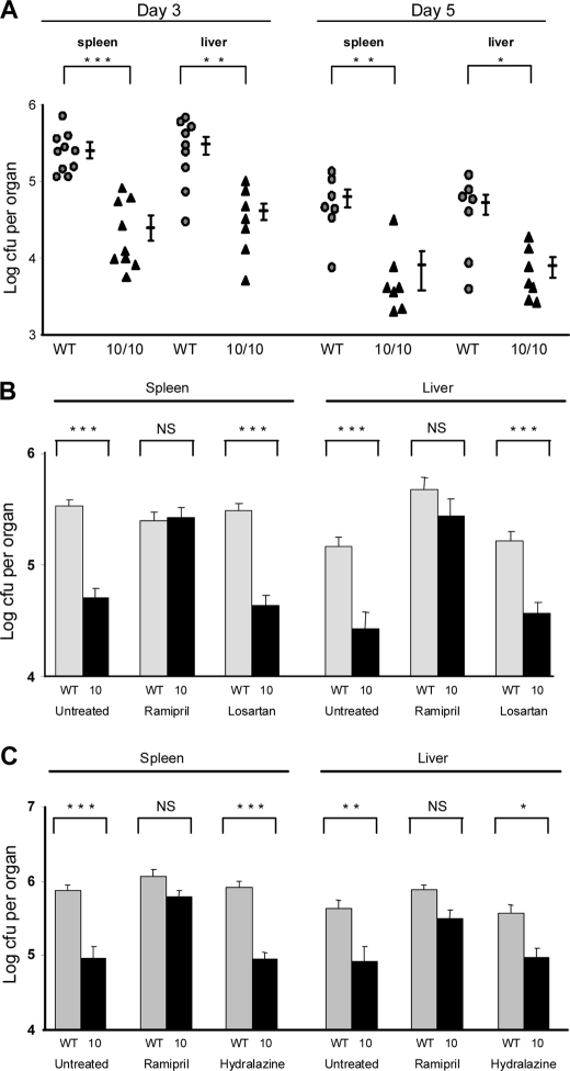
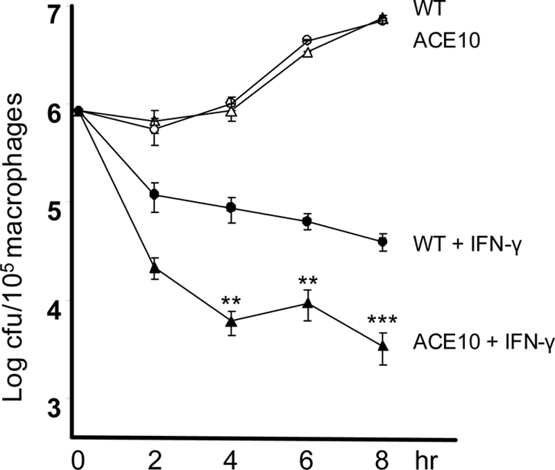
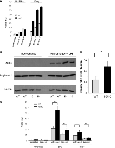
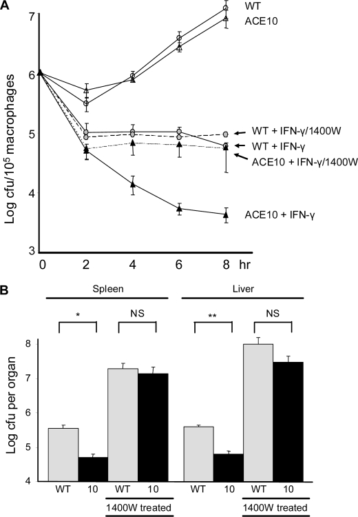
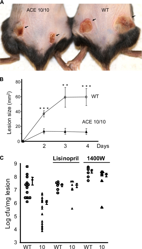
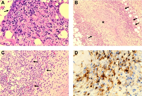
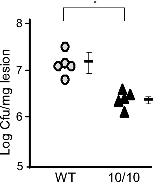
Gene targeting in ES cells was used to substitute control of angiotensin converting enzyme (ACE) expression from the endogenous promoter to the mouse c-fms promoter. The result is an animal model called ACE 10/10 in which ACE is overexpressed by monocytes, macrophages, and other myelomonocytic lineage cells. To study the immune response of these mice to bacterial infection, we challenged them with Listeria monocytogenes or methicillin-resistant Staphylococcus aureus (MRSA). ACE 10/10 mice have a significantly enhanced immune response to both bacteria in vivo and in vitro. For example, 5 days after Listeria infection, the spleen and liver of ACE 10/10 mice had 8.0- and 5.2-fold less bacteria than wild type mice (WT). In a model of MRSA skin infection, ACE 10/10 mice had 50-fold less bacteria than WT mice. Histologic examination showed a prominent infiltrate of ACE-positive mononuclear cells in the skin lesions from ACE 10/10. Increased bacterial resistance in ACE 10/10 is directly due to overexpression of ACE, as it is eliminated by an ACE inhibitor. Critical to increased immunity in ACE 10/10 is the overexpression of iNOS and reactive nitrogen intermediates, as inhibition of iNOS by the inhibitor 1400W eliminated all in vitro and in vivo differences in innate bacterial resistance between ACE 10/10 and WT mice. Increased resistance to MRSA was transferable by bone marrow transplantation. The overexpression of ACE and iNOS by myelomonocytic cells substantially boosts innate immunity and may represent a new means to address serious bacterial infections.


Figures

FIGURE 1.
FIGURE 1.
In vivo challenge with L. monocytogenes. A, WT and ACE 10/10 (10/10) mice were inoculated i.v. with 4 × 10³ L. monocytogenes, strain EGD. Groups of mice were sacrificed 3 or 5 days after inoculation, and the number of cfu in the spleen and liver was determined. Values for individual WT (circles) and ACE 10/10 (triangles) mice are shown, as well as the group means and S.E. *, p < 0.05; **, p < 0.005; ***, p < 0.0005. ACE 10/10 have a significantly lower bacterial burden than WT. B, WT (gray bars) and ACE 10/10 (10, black bars) were implanted with an osmotic mini-pump and treated with either saline, the ACE inhibitor ramipril or the AT1 receptor antagonist losartan. After 7 days, the mice were challenged with 4 × 10³ Listeria i.v. Three days later, the number of bacteria in the spleen and in the liver was determined. Ramipril eliminated the difference between WT and ACE 10/10, whereas losartan had no significant effect (n = 6). C, WT and ACE 10/10 (10) were implanted with osmotic mini-pumps and treated with either ramipril or hydralazine. Mice then received 3 × 10⁴ Listeria i.v. Five days after inoculation, the bacterial counts in the spleen and liver were determined. Ramipril eliminated differences between ACE 10/10 and WT mice, but no such effect was found with hydralazine (n = 6). NS is not significant.
FIGURE 2.
FIGURE 2.
In vitro killing of Listeria by macrophages. TPMs were cultured overnight with either IFN-γ (filled triangle, ACE 10/10; filled circle, WT) or without IFN-γ priming (open triangle, ACE 10/10; open circle, WT). At time 0, cells were infected with Listeria at a multiplicity of 10 for 1 h. Then, after washing, medium containing gentamicin was added to kill the remaining extracellular bacteria. After further incubation, such that the total time since first exposure to bacteria was 2 to 8 h, the antibiotic was removed, the cells were lysed, and cfus were determined. In the presence of IFN-γ priming, there is substantially better killing of Listeria by ACE 10/10 macrophages than by WT cells (n = 8 mice). **, p < 0.005; ***, p < 0.001.
FIGURE 3.
FIGURE 3.
Macrophage production of nitrites and iNOS. A, TPMs from WT (light gray) or ACE 10/10 (black) mice were cultured overnight with or without IFN-γ. After 18 h, groups of cells were treated with Listeria at a multiplicity of either 10 or 50. Separate groups of cells were left untreated. Four hours later, the culture supernatant was collected, and nitrite levels were measured. In the absence of IFN-γ priming, there was no significant difference in nitrite levels between cells from WT and ACE 10/10. However, with IFN-γ treatment, ACE 10/10 macrophages made from 2.2- to 2.9-fold more nitrites than identically treated cells from WT mice (n = 6, p < 0.005 for all groups of IFN-γ-treated cells comparing ACE 10/10 with WT). B, TPMs from WT and ACE 10/10 (10) were cultured for 18 h with or without 1 μg/ml of LPS. Cell lysates were then probed for iNOS, arginase I, and β-actin by Western blot. C, densitometry was used to quantitate the average density ratio of iNOS relative to the expression of β-actin. There is increased expression of iNOS by ACE 10/10 macrophages (n = 5). *, p < 0.05. D, WT (gray bars) and ACE 10/10 (black bars) mice were treated with lisinopril for 7 days. TPMs from these mice and from untreated mice were then stimulated with either LPS or IFN-γ for 18 h. Nitrite accumulation in the supernatant was then determined by Griess assay. ACE inhibition abrogates the elevated nitrite production by ACE 10/10 macrophages (n = 5 mice per group with or without inhibitor). *, p = 0.05; **, p < 0.01. ns is not significant.
FIGURE 4.
FIGURE 4.
In vitro and in vivo killing of Listeria is inhibited by the iNOS inhibitor 1400W. A, TPMs from ACE 10/10 (triangles) or WT (circles) mice were cultured overnight. Some cells were cultured with IFN-γ or the combination of IFN-γ plus 1400W, as indicated in the figure. After 18 h, cells were mixed with Listeria at a multiplicity of 10 for 1 h. After washing, media containing gentamicin was added to kill the remaining extracellular bacteria. At the indicated times after the first addition of Listeria, gentamicin was removed, the cells were lysed, and cfus were determined. Although inhibition of iNOS has no significant effect on the WT cells, treatment of ACE 10/10 cells renders these cells equivalent to WT in the killing of Listeria (n = 5 mice). ACE 10/10:IFN-γ + 1400W versus WT:IFN-γ + 1400W, p > 0.4 at all time points). B, WT (gray bars) and ACE 10/10 mice (10, black bars) were inoculated i.v. with 3 × 10⁴ Listeria. On the day of infection, some mice were treated with the iNOS inhibitor 1400W in the drinking water. The mice were sacrificed on day 5, and the bacterial burden in the spleen and liver was determined. Inhibition of iNOS with 1400W increased the bacterial burden in all groups. In the presence of 1400W, the difference between WT and ACE 10/10 mice was eliminated (n = 6). *, p < 0.05; **, p < 0.005. NS is not significant.
FIGURE 5.
FIGURE 5.
Skin infection with MRSA. Mice were infected subcutaneously with 1 × 10⁹ MRSA, clone USA 300, in the hind flanks. A, a representative comparison of skin lesions present in ACE 10/10 and WT mice 4 days after MRSA infection. B, lesion size of WT (circles) and ACE 10/10 (10, triangles) were measured on days 2, 3, and 4 after infection (n ≥ 14 mice per group). **, p < 0.005; ***, p < 0.0005. C, four days after skin infection, WT (circles) and ACE10/10 (triangles) were sacrificed, and bacterial counts in the lesion were determined. ACE 10/10 mice averaged >50-fold less bacteria within lesions (n ≥ 14, p < 0.001). These differences were eliminated when mice were treated with either the ACE inhibitor lisinopril or the iNOS inhibitor 1400W.
FIGURE 6.
FIGURE 6.
Histology of skin after MRSA infection. Four days after subcutaneous infection with MRSA, mice were sacrificed, and histologic sections of the lesions were prepared. A, WT mice show extensive acute inflammation within the superficial and deep dermis. There is abundant polymorphonuclear leukocytes with karyorrhexis, tissue necrosis, and pus formation. Occasional masses of bacteria are present (arrow). B, ACE 10/10 mice also had areas of necrosis and pus formation. However, at the periphery of the necrosis, there was a mononuclear cell infiltrate. Pictured in B is a marked example where a central region of necrosis (indicated by an asterisk) is surrounded by a cuff of mononuclear cells (indicated by arrows). C, a higher power of the mononuclear infiltrate (arrows) is shown. D, immunohistochemical staining of the tissue sections with an anti-ACE antibody showed that these cells were positive for ACE expression.
FIGURE 7.
FIGURE 7.
Transplantation of ACE10/10 bone marrow into WT mice confers increased resistance to MRSA. Recipient C57BL/6 mice were lethally irradiated and then immediately transplanted with bone marrow from either ACE 10/10 (10/10) or littermate WT mice. After engraftment, the mice were challenged subcutaneously with 1 × 10⁹ MRSA. Four days later, mice were sacrificed, and bacterial burden in the skin lesion was determined. WT mice receiving bone marrow from ACE 10/10 mice (triangles) showed a significantly reduced bacteria burden in the skin lesion, compared with WT mice receiving WT bone marrow (circles) (n = 5). *, p < 0.05.

References

    1. Corvol P., Williams T. A., Soubrier F. (1995) Methods Enzymol. 248, 283–305 - PubMed
    1. Bernstein K. E., Xiao H. D., Frenzel K., Li P., Shen X. Z., Adams J. W., Fuchs S. (2005) Circ. Res. 96, 1135–1144 - PubMed
    1. Leehey D. J., Singh A. K., Alavi N., Singh R. (2000) Kidney Int. Suppl. 77, S93–S98 - PubMed
    1. Metzger R., Bohle R. M., Chumachenko P., Danilov S. M., Franke F. E. (2000) Atherosclerosis 150, 21–31 - PubMed
    1. Stegbauer J., Lee D. H., Seubert S., Ellrichmann G., Manzel A., Kvakan H., Muller D. N., Gaupp S., Rump L. C., Gold R., Linker R. A. (2009) Proc. Natl. Acad. Sci. U.S.A. 106, 14942–14947 - PMC - PubMed

Publication types