. 2010 Dec 17;285(51):39888-97.
doi: 10.1074/jbc.M110.128348. Epub 2010 Oct 11.

High mobility group box 1 release from hepatocytes during ischemia and reperfusion injury is mediated by decreased histone deacetylase activity

Affiliations

High mobility group box 1 release from hepatocytes during ischemia and reperfusion injury is mediated by decreased histone deacetylase activity

John Evankovich et al. J Biol Chem. .

Abstract

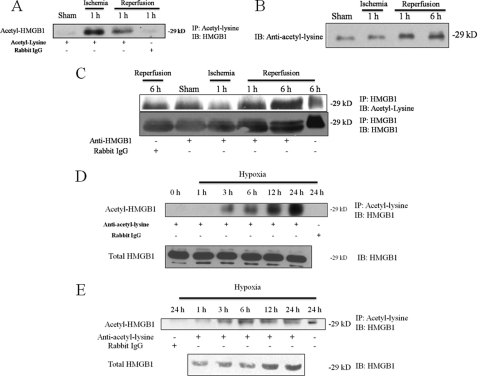
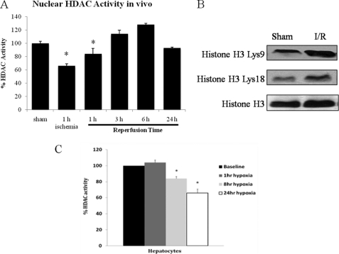
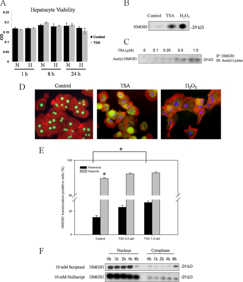
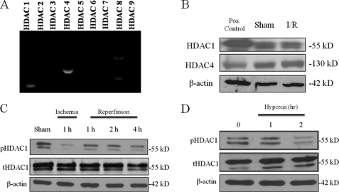
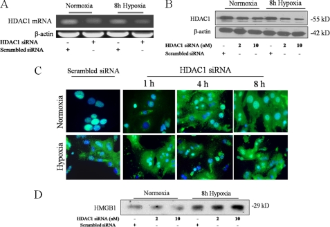
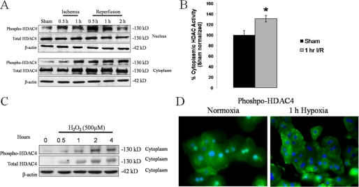
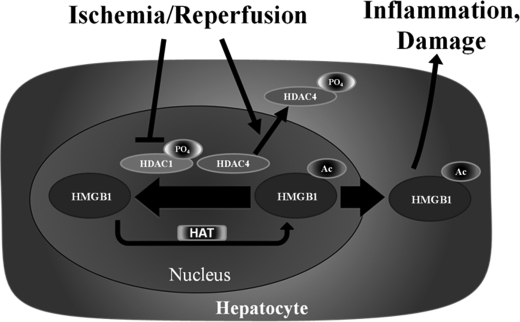
The mobilization and extracellular release of nuclear high mobility group box-1 (HMGB1) by ischemic cells activates inflammatory pathways following liver ischemia/reperfusion (I/R) injury. In immune cells such as macrophages, post-translational modification by acetylation appears to be critical for active HMGB1 release. Hyperacetylation shifts its equilibrium from a predominant nuclear location toward cytosolic accumulation and subsequent release. However, mechanisms governing its release by parenchymal cells such as hepatocytes are unknown. In this study, we found that serum HMGB1 released following liver I/R in vivo is acetylated, and that hepatocytes exposed to oxidative stress in vitro also released acetylated HMGB1. Histone deacetylases (HDACs) are a family of enzymes that remove acetyl groups and control the acetylation status of histones and various intracellular proteins. Levels of acetylated HMGB1 increased with a concomitant decrease in total nuclear HDAC activity, suggesting that suppression in HDAC activity contributes to the increase in acetylated HMGB1 release after oxidative stress in hepatocytes. We identified the isoforms HDAC1 and HDAC4 as critical in regulating acetylated HMGB1 release. Activation of HDAC1 was decreased in the nucleus of hepatocytes undergoing oxidative stress. In addition, HDAC1 knockdown with siRNA promoted HMGB1 translocation and release. Furthermore, we demonstrate that HDAC4 is shuttled from the nucleus to cytoplasm in response to oxidative stress, resulting in decreased HDAC activity in the nucleus. Together, these findings suggest that decreased nuclear HDAC1 and HDAC4 activities in hepatocytes following liver I/R is a mechanism that promotes the hyperacetylation and subsequent release of HMGB1.


Figures

FIGURE 1.
FIGURE 1.
HMGB1 is acetylated and released in warm liver I/R injury and hypoxia. Co-immunoprecipitation analysis from mouse liver tissue lysates and serum following I/R. Mice were divided into sham or I/R groups. I/R groups underwent 60 min of warm ischemia followed by reperfusion. Blot shown is representative of three experiments with similar results. A, ischemic liver tissue lysates were immunoprecipitated with an anti-acetyl antibody and immunoblotted for HMGB1. Anti-rabbit IgG was used as a negative control. The blot shown is representative of three experiments with similar results. B, Western blot of mouse serum samples following I/R for anti-acetyl-lysine. C, co-immunoprecipitation of mouse serum following I/R. Samples were pulled down with anti-HMGB1 and immunoblotted with anti-acetyl-lysine. The blot was then stripped and re-probed for HMGB1. D, whole cell lysates of hepatocytes exposed to 1% hypoxia were immunoprecipitated with an anti-acetyl-lysine antibody and immunoblotted for HMGB1. The blot shown is representative of three experiments with similar results. E, co-immunoprecipitation of cell culture supernatants following stimulus with hypoxia (1% O2) for acetylated HMGB1.
FIGURE 2.
FIGURE 2.
Nuclear HDAC activity is decreased after liver I/R and hypoxia. A, nuclear protein was extracted from mice livers subjected to a time course of ischemia/reperfusion. HDAC activity was determined by colorimetric assay. *, p < 0.05. Assay shown is representative of three experiments with similar results. B, Western blot of acetylated (Lys-9 and Lys-18) and total H3 in sham and 1 h I/R mice. C, hepatocytes were exposed to hypoxia (1% O2) for 1, 4, 8, and 24 h and nuclear protein was analyzed for HDAC activity. *, p < 0.05. Assay shown is representative of three experiments with similar results.
FIGURE 3.
FIGURE 3.
Inhibition of HDAC results in nuclear-cytosolic translocation of HMGB1 and HMGB1 release. A, hepatocyte cell viability was examined by Crystal Violet staining in normoxia and hypoxia following treatment with 1 μm TSA. B, Western blot analysis of rat hepatocyte cell supernatants for HMGB1 after TSA treatment or 500 μm H202 treatment. Blot shown is representative of three experiments with similar results. C, co-immunoprecipitation of cell supernatants following treatment with TSA. Cells were immunoprecipitated with anti-HMGB1 and immunoblotted for anti-acetyl-lysine. Blot shown is representative of three experiments with similar results. D, rat hepatocytes were treated with 1 μm TSA or 500 μm H202 and stained for HMGB1. Green, HMGB1; blue, nuclei; red, F-actin. Imaging shown is representative of three experiments with similar results. E, high content analysis for HMGB1 translocation in hepatocytes cell cultures. Cells were immunostained for HMGB1 and staining intensities were quantified in both nuclear and cytosplasmic compartments using a CellomicsTM Arrayscan® platform. “HMGB1 translocation-positive” and “HMGB1 translocation-negative” cell populations were identified using Spotfire Decisionsite Software. *, p < 0.05. F, Western blot analysis for HMGB1 of nuclear and cytoplasmic protein from primary rat hepatocytes treated with 10 mm of the HDAC inhibitor Scriptaid, or its inactive analog, Nullscript and subjected to hypoxia (1% O2) for various time points.
FIGURE 4.
FIGURE 4.
HDAC1 and HDAC4 are expressed in mouse hepatocytes. A, RT-PCR for HDAC 1–9 in rat hepatocytes. B, Western blot analysis of mouse liver nuclear protein extracts for HDAC 1 and HDAC4. Jurkat nuclear extracts were used as positive controls for HDAC1 and -4. The blot shown is representative of three experiments with similar results. C, Western blot analysis of nuclear extracts from ischemic liver tissue following I/R. Immunoblots were performed for phospho-HDAC1 and total HDAC1. The blot shown is representative of three experiments with similar results. D, mouse hepatocytes were subjected to hypoxia for and analyzed for pHDAC1 and tHDAC1 by Western blot analysis. The blot shown is representative of three experiments with similar results.
FIGURE 5.
FIGURE 5.
Specific inhibition of HDAC1 promotes translocation of nuclear HMGB1 to the cytoplasm and increases HMGB1 release. Mouse hepatocytes were transfected with scrambled siRNA or HDAC1 siRNA and subjected to RT-PCR 24 h post-transfection (A) or Western blot (B) analysis 40 h post-transfection. Blots shown are representative of three experiments with similar results. C, hepatocytes were transfected with 10 μm HDAC1 siRNA analyzed by immunofluorescent staining 40–48 h later. Cells were analyzed in normoxia or after 1, 4, and 8 h of hypoxia (1% O2) by immunostaining. Green, HMGB1; blue, nuclei. Imaging shown is representative of three experiments with similar results. D, Western blot for HMGB1 in cell supernatants of hepatocytes transfected with HDAC1 siRNA following 8 h of hypoxia. The blot shown is representative of three experiments with similar results.
FIGURE 6.
FIGURE 6.
HDAC4 is shuttled from the nucleus to the cytoplasm during I/R. A, Western blot analysis of liver nuclear and cytoplasmic fractions for phospho-HDAC4 during I/R. B, total HDAC activity as measured by colorimetric assay at 1 h reperfusion. C, in vitro, hepatocytes were stimulated with 500 μm H202 and Western blot analysis was performed on cytoplasmic fractions for phospho-HDAC4 and total HDAC4. D, hepatocytes were subjected to hypoxia for 1 h and stained for phospho-HDAC4.
FIGURE 7.
FIGURE 7.
I/R induces release of acetylated-HMGB1 from hepatocytes through HDAC1 deactivation and HDAC4 shuttling. The proposed model showing release of acetylated HMGB1 following I/R. Both de-activation of nuclear HDAC1 and cytosplasmic shuttling of HDAC4 contribute to a reduction in overall nuclear HDAC activity, tilting the acetylation/deacetylation balance of HMGB1 toward net acetylation and release.

References

    1. Bustin M., Hopkins R. B., Isenberg I. (1978) J. Biol. Chem. 253, 1694–1699 - PubMed
    1. Wang H., Bloom O., Zhang M., Vishnubhakat J. M., Ombrellino M., Che J., Frazier A., Yang H., Ivanova S., Borovikova L., Manogue K. R., Faist E., Abraham E., Andersson J., Andersson U., Molina P. E., Abumrad N. N., Sama A., Tracey K. J. (1999) Science 285, 248–251 - PubMed
    1. Tsung A., Sahai R., Tanaka H., Nakao A., Fink M. P., Lotze M. T., Yang H., Li J., Tracey K. J., Geller D. A., Billiar T. R. (2005) J. Exp. Med. 201, 1135–1143 - PMC - PubMed
    1. Levy R. M., Mollen K. P., Prince J. M., Kaczorowski D. J., Vallabhaneni R., Liu S., Tracey K. J., Lotze M. T., Hackam D. J., Fink M. P., Vodovotz Y., Billiar T. R. (2007) Am. J. Physiol. Regul. Integr. Comp. Physiol. 293, R1538–R1544 - PubMed
    1. Hori O., Brett J., Slattery T., Cao R., Zhang J., Chen J. X., Nagashima M., Lundh E. R., Vijay S., Nitecki D. (1995) J. Biol. Chem. 270, 25752–25761 - PubMed

Publication types