. 2010 Dec 10;285(50):38876-88.
doi: 10.1074/jbc.M110.179150. Epub 2010 Oct 11.

Temperature and Mg2+ sensing by a novel PhoP-PhoQ two-component system for regulation of virulence in Edwardsiella tarda

Affiliations

Temperature and Mg2+ sensing by a novel PhoP-PhoQ two-component system for regulation of virulence in Edwardsiella tarda

Smarajit Chakraborty et al. J Biol Chem. .

Abstract

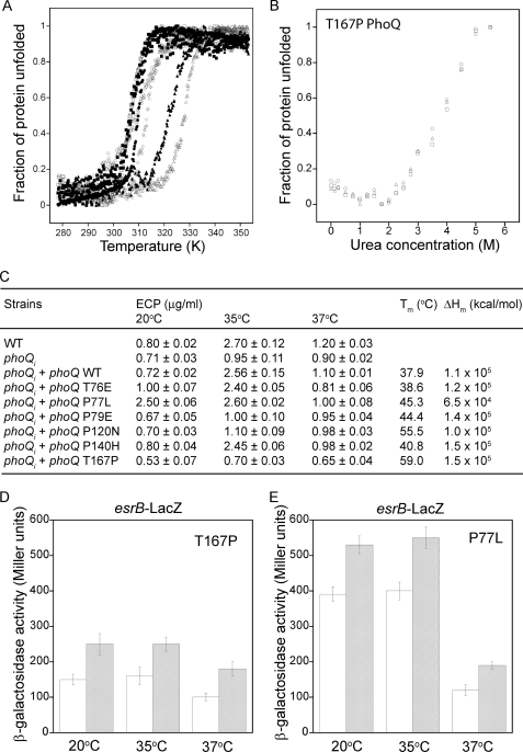
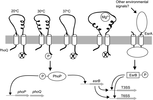
The PhoP-PhoQ two-component system is commonly used by bacteria to sense environmental factors. Here we show that the PhoP-PhoQ system of Edwardsiella tarda detects changes in environmental temperature and Mg(2+) concentration as well as regulates the type III and VI secretion systems through direct activation of esrB. Protein secretion is activated from 23 to 35 °C or at low Mg(2+) concentrations, but it is suppressed at or below 20 °C, at or above 37 °C, or at high Mg(2+) concentrations. The effects of temperature and Mg(2+) concentration are additive. The PhoQ sensor domain has a low T(m) of 37.9 °C, and it detects temperatures through a conformational change of its secondary structure. Mutation of specific Pro or Thr residues increased the stability of the PhoQ sensor drastically, altering its temperature-sensing ability. The PhoQ sensor detects Mg(2+) concentration through the direct binding of Mg(2+) to a cluster of acidic residues (DDDSAD) and through changes that likely affect its tertiary structure. Here, we describe for the first time the use of PhoP-PhoQ as a temperature sensor for bacterial virulence control.


Figures

FIGURE 1.
FIGURE 1.
Sequence alignment of the PhoQ sensor domains. The sequence of the PhoQ periplasmic sensor region from E. tarda (PhoQ_ET; GU324976) was compared with those from Y. pestis (PhoQ_YP; NP_669110), S. typhimurium (PhoQ_ST; NP_460200), and E. coli (PhoQ_EC; NP_753417). The secondary structural elements from the crystal structure of the E. coli PhoQ sensor are shown underneath the sequences, whereas the predicted secondary structural elements of the E. tarda PhoQ sensor are shown on top of the sequences. Asterisks below the sequences indicate identical residues. Negatively charged residues in the acidic patches are in bold. Pro and Thr residues in the E. tarda PhoQ sensor that are not conserved with those in E. coli and S. typhimurium and were selected for mutation studies are boxed.
FIGURE 2.
FIGURE 2.
PhoP-PhoQ regulates T3SS and T6SS through direct binding of PhoP to the promoter region of ersB. A, silver-stained SDS-PAGE shows the ECP (T3SS: EseB, EseC and EseD; T6SS: EvpC and EvpP) secretion profiles of E. tarda PPD130/91 (WT), E. tarda carrying an insertion mutation in phoP (phoPi) or phoQ (phoQi), and the mutant E. tarda with complementation of the corresponding wild type gene (phoPi + phoP or phoQi + phoQ). The incubation temperature was 35 °C. Lane BL contains a blank control with only DMEM and no bacterial cells. B, shown is the promoter region of the E. tarda 130/91 esrB gene, a putative PhoP box. The sequences corresponding to the (G/T)GTTTA direct repeats are shaded. The classic PhoP box from the mgtA promoter and the imperfect but orthodox PhoP box from the orgB promoter of S. typhimurium are shown for comparison. T and A residues that are conserved among PhoP boxes are in bold. C, shown is an electrophoretic mobility shift assay of PhoP protein on a 5′ 6-carboxyfluorescein-labeled DNA fragment (470 bp, from nt −467 to +3) from the promoter region of esrB (upper panel) and another DNA fragment with the same boundaries but with the PhoP box (nt −311 to −295) removed (lower panel). D, shown is an electrophoretic mobility shift assay of PhoP protein on a 5′ 6-carboxyfluorescein-labeled DNA fragment (454 bp, from nt −451 to +3) from the promoter region of phoP (upper panel) and another DNA fragment with the same boundaries but with the PhoP box (nt −192 to −176) removed (lower panel). E, shown are transcription levels of the esrB-LacZ reporter gene fusion measured by β-galactosidase activity from bacterial cells cultured at 30 °C in TSB medium. WT, E. tarda PPD130/91; phoPi, E. tarda with insertion mutation in the phoP gene; phoPi + phoP, E. tarda phoPi mutant complemented with wild type phoP.
FIGURE 3.
FIGURE 3.
The secretion of T3SS and T6SS ECPs by E. tarda is temperature- and Mg2+ concentration-dependent through transcriptional activation of esrB, phoP, and phoQ. A, silver-stained SDS-PAGE shows T3SS and T6SS ECP secretion by E. tarda PPD130/91 at different incubation temperatures as indicted above the lanes. The lane BL contains a blank control with only DMEM and no bacterial cells. B, Western blot analysis shows the secretion of EseB from T3SS (upper panel) and EvpC from T6SS (lower panel) by E. tarda at the temperatures of 20, 30, and 37 °C in the presence of 1 mm (L) or 10 mm (H) Mg2+ using N-minimal medium. C and D, the β-galactosidase activities of the reporter genes esrB-LacZ and phoP-LacZ were determined in N-minimal medium under different incubation temperatures in the presence of 1 mm (striped bars) or 10 mm (empty bars) Mg2+. Both genes showed reduced activity at 20 and 37 °C compared with 23, 25, and 35 °C. The activities of both genes are also repressed at higher concentrations of Mg2+, and this effect is additive with that of the temperature.
FIGURE 4.
FIGURE 4.
Thermal and urea denaturation of the E. tarda PhoQ sensor domain. A, shown is a far-UV CD spectra of the PhoQ sensor domain at 20 °C (open circle), 30 °C (closed squares), 35 °C (open squares), and 37 °C (closed circles). B, shown is thermal denaturation of the E. tarda PhoQ sensor domain monitored by CD at 206.5 nm in the presence (closed circles) or absence (open circles) of 10 mm Mg2+. C, shown is urea denaturation of the E. tarda PhoQ sensor domain monitored by CD at 210 nm at 20 °C (circles), 30 °C (squares), and 37 °C (triangles) in the presence (open symbols) or absence (closed symbols) of 10 mm Mg2+. D, shown is urea denaturation of the E. tarda PhoQ sensor domain monitored by fluorescence at 350 nm at 20 °C (circles), 30 °C (squares), and 37 °C (triangles) in the presence (open symbols) or absence (closed symbols) of 10 mm Mg2+.
FIGURE 5.
FIGURE 5.
Thermal denaturation of Thr or Pro residue mutants of the E. tarda PhoQ sensor domain. A, shown is thermal denaturation of the wild type and mutant E. tarda PhoQ sensor domains monitored by CD at 202 nm for P79E (open rhombuses) and P120N (filled triangles), at 206.5 nm for T167P (open triangles), and at 218 nm for wild type (open squares), T76E (closed squares), P77L (open circles), and P140H (closed circles). All samples had a concentration of 10 mm Mg2+. B, shown is urea denaturation of the E. tarda PhoQ T167P mutant sensor domain monitored by CD at 210 nm at 20 °C (open circles), 30 °C (open squares), and 37 °C (open triangles) in the presence of 10 mm Mg2+. C, shown is the amount of ECP obtained at the incubation temperatures of 20, 35, and 37 °C from wild type E. tarda, the E. tarda phoQi mutant, and the E. tarda phoQi mutant complemented with various phoQ gene mutations. The Tm and ΔHm values obtained from the curve-fitting of the thermal denaturation data from the wild type and mutant PhoQs are also listed. D and E, the β-galactosidase activity of the reporter gene esrB-LacZ was determined in N-minimal medium under different incubation temperatures in the presence of 1 mm (striped bars) or 10 mm (empty bars) Mg2+ for E. tarda phoQi + phoQ T167P or phoQi + phoQ P77L.
FIGURE 6.
FIGURE 6.
The cluster of acidic residues in PhoQ is responsible for sensing Mg2+ concentration and antimicrobial peptide but not acidic pH. A, binding of Mg2+ to the PhoQ sensor domain was monitored by the absolute change in fluorescence at 350 nm at 20 °C (filled rhombuses), 30 °C (closed squares), and 37 °C (closed circles). Mg2+ binding by the acidic cluster mutant, PhoQ NNNSANA, was monitored at 30 °C (filled triangles). B, Western blot analysis shows the secretion of EseB from T3SS (upper panel) and EvpC from T6SS (lower panel) by the E. tarda phoQi mutant strain complemented with either phoQ NNNSANA or wild type phoQ in the absence (L) or presence (H) of 10 mm Mg2+ at 30 °C. C, the activity of the reporter gene esrB-LacZ at different temperatures in the presence of 1 mm (striped bars) or 10 mm (empty bars) Mg2+ using the E. tarda phoQi mutant complemented with phoQ NNNSANA is shown. D, Western blot analysis shows the secretion of proteins from T3SS (EseB) and T6SS (EvpC) by E. tarda phoQi mutant complemented with either phoQ NNNSANA or wild type phoQ grown in minimal medium at pH 5.5 (100 mm MES) or pH 7.5 (100 mm Tris-HCl) and in the absence or presence of 5 μg/ml antimicrobial peptide KR-20 (AMP). E, the activity of the reporter gene esrB-LacZ using E. tarda phoQi mutant complemented with either phoQ NNNSANA (striped bars) or wild type phoQ (empty bars) grown at pH 5.5 or 7.5 and in the absence or presence of 5 μg/ml antimicrobial peptide KR-20 (AMP).
FIGURE 7.
FIGURE 7.
Model illustrating the temperature and Mg2+ regulation of T3SS and T6SS by the PhoP-PhoQ system. The PhoQ sensor senses changes in temperature through conformational changes in its thermally unstable secondary structures (coils in the diagram). There should be less secondary structure at 37 °C as compared with 30 °C, and the conformational change at 20 °C should be different from that at 37 °C to distinguish the different temperatures. The overall tertiary structure, or shape, of the PhoQ sensor remains unchanged at different temperatures. In addition to temperature, the PhoQ sensor can also detect changes in Mg2+ concentration through direct binding of Mg2+ to a cluster of acidic residues that likely change the tertiary structure of the protein. The signals from environmental temperature and Mg2+ concentration are additive with each other. Activation of the PhoQ histidine kinase over the temperature range from 23 to 35 °C at low Mg2+ concentrations leads to autophosphorylation and the transfer of a phosphate group from PhoQ to PhoP. The phosphorylated PhoP binds directly to the PhoP box within the promoter region of esrB to activate its transcription. PhoP also self-regulates by binding to another PhoP box within its own promoter region to up-regulate the expression of both PhoP and PhoQ. The EsrB protein then integrates the signal from another two-component system, EsrA-EsrB, to activate the transcription of genes from both T3SS and T6SS.

References

    1. Janda J. M., Abbott S. L. (1993) Clin. Infect. Dis. 17, 742–748 - PubMed
    1. Thune R. L., Stanley L. A., Cooper R. K. (1993) Annu. Rev. Fish Dis. 3, 37–68
    1. Srinivasa Rao P. S., Lim T. M., Leung K. Y. (2003) Infect. Immun. 71, 1343–1351 - PMC - PubMed
    1. Srinivasa Rao P. S., Yamada Y., Tan Y. P., Leung K. Y. (2004) Mol. Microbiol. 53, 573–586 - PubMed
    1. Tan Y. P., Zheng J., Tung S. L., Rosenshine I., Leung K. Y. (2005) Microbiology 151, 2301–2313 - PubMed

Publication types

LinkOut - more resources