. 2010 Dec 10;285(50):38969-77.
doi: 10.1074/jbc.M110.160986. Epub 2010 Oct 11.

Long range interactions regulate Igf2 gene transcription during skeletal muscle differentiation

Affiliations

Long range interactions regulate Igf2 gene transcription during skeletal muscle differentiation

Damir T Alzhanov et al. J Biol Chem. .

Abstract

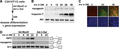
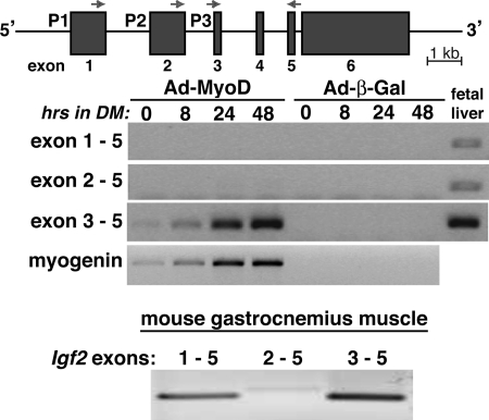
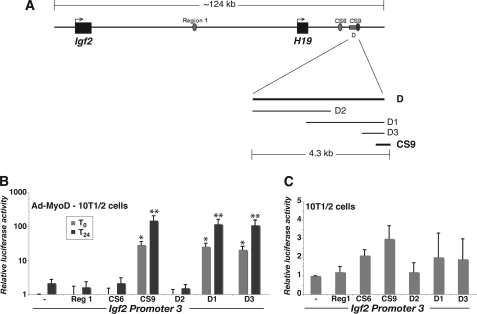
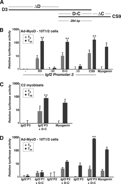
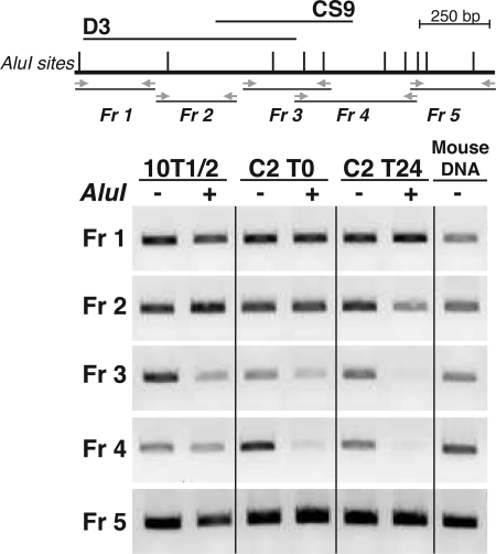
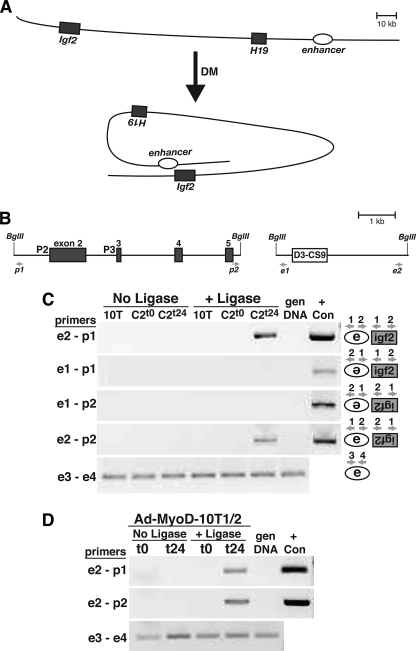
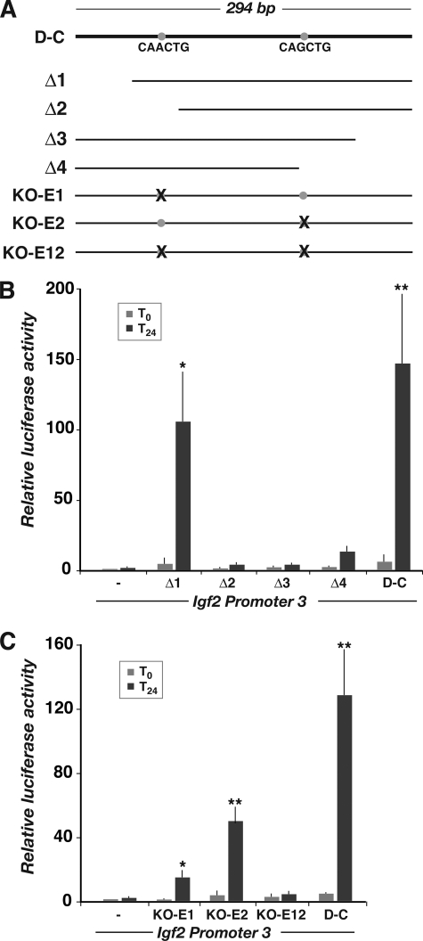
The differentiation, maintenance, and repair of skeletal muscle is controlled by interactions between genetically determined transcriptional programs regulated by myogenic transcription factors and environmental cues activated by growth factors and hormones. Signaling through the insulin-like growth factor 1 (IGF1) receptor by locally produced IGF2 defines one such pathway that is critical for normal muscle growth and for regeneration after injury. IGF2 gene and protein expression are induced as early events in muscle differentiation, but the responsible molecular mechanisms are unknown. Here we characterize a distal DNA element within the imprinted mouse Igf2-H19 locus with properties of a muscle transcriptional enhancer. We find that this region undergoes a transition to open chromatin during differentiation, whereas adjacent chromatin remains closed, and that it interacts in differentiating muscle nuclei but not in mesenchymal precursor cells with the Igf2 gene found more than 100 kb away, suggesting that chromatin looping or sliding to bring the enhancer in proximity to Igf2 promoters is also an early event in muscle differentiation. Because this element directly stimulates the transcriptional activity of an Igf2 promoter-reporter gene in differentiating myoblasts, our results indicate that we have identified a bona fide distal transcriptional enhancer that supports Igf2 gene activation in skeletal muscle cells. Because this DNA element is conserved in the human IGF2-H19 locus, our results further suggest that its muscle enhancer function also is conserved among different mammalian species.


Figures

FIGURE 1.
FIGURE 1.
Activation of Igf2 gene transcription during muscle differentiation. Shown are the results of time course experiments with 10T1/2 mesenchymal stem cells infected with Ad-MyoD or Ad-β-Gal and incubated in DM for up to 2 days. A, experimental scheme. B, induction of muscle proteins myogenin and troponin-T and constant expression of Akt during differentiation, as assessed by immunoblotting. C, myotube formation measured by immunocytochemistry for myogenin (green) and troponin-T (red), and nuclear staining by Hoechst dye (blue). D, time course of transcription for Igf2, myogenin, and S17 genes, measured by semi-quantitative RT-PCR. The locations of relevant exons and PCR primers are shown.
FIGURE 2.
FIGURE 2.
Promoter 3-specific induction of Igf2 gene expression during muscle differentiation. Top panel, schematic of the mouse Igf2 gene. The three promoters and six exons are indicated, as are the locations of exon-specific PCR primers. Middle panel, results of time course gene expression experiments by RT-PCR for Igf2 and myogenin using 10T1/2 mesenchymal stem cells infected with Ad-MyoD or Ad-β-Gal and incubated in DM for up to 48 h. For Igf2, only transcripts containing exons 3 and 5 gave a positive result in differentiating myoblasts, demonstrating that only Igf2 promoter 3 is activated in this muscle differentiation model. Mouse day 18 fetal liver RNA serves as a positive control for active transcription from each Igf2 promoter (14). Bottom panel, results of RT-PCR experiments measuring promoter-specific Igf2 transcripts using gastrocnemius muscle from 8-week-old male mice. Transcripts directed by promoter 3 and containing exons 3 and 5 are more abundant than mRNAs derived from promoters 1 or 2.
FIGURE 3.
FIGURE 3.
Identification of DNA elements that mediate muscle-specific induction of Igf2 gene transcription. A, schematic of Igf2-H19 locus on mouse chromosome 7. Region 1, CS6, CS9, and D sites are indicated, as is dissection of the D segment into D1–D3. B, results of luciferase reporter gene experiments with Ad-MyoD-infected 10T1/2 cells incubated in DM for 0 or 24 h, using mouse Igf2 promoter 3 ± Region 1 (Reg1), CS6, CS9, or D DNA segments (means ± S.D., n = 5–8 experiments; *, p < 0.001; **, p < 0.0002 versus IGF2 promoter 3). Note the log scale on the ordinate. C, results of luciferase reporter gene experiments with 10T1/2 cells, using the same recombinant plasmids as in B. Note the linear scale on the ordinate.
FIGURE 4.
FIGURE 4.
Functional dissection of the putative Igf2 muscle enhancer. A, diagram of D3 and CS9 regions (see Fig. 3A for genomic context). B, results of luciferase reporter gene experiments with Ad-MyoD-infected 10T1/2 cells incubated in DM for 0 or 24 h, using mouse Igf2 promoter 3 (P3) ± D3, ΔD, D-C, ΔC, CS9, or the mouse myogenin promoter (mean ± S.D., n = 5 experiments; *, p < 0.01; **, p < 0.001 versus Igf2 P3). C, results of luciferase reporter gene experiments with C2 myoblasts incubated in DM for 0 or 48 h, using mouse Igf2 P3, Igf2 P3 + D-C, or the mouse myogenin promoter (mean ± S.D., n = 5 experiments; *, p < 0.01; **, p < 0.005 versus IGF2 P3). D, results of luciferase reporter gene experiments with Ad-MyoD-infected 10T1/2 cells incubated in DM for 0 or 24 h, using mouse Igf2 P1, P2, or P3 ± D-C, or the mouse myogenin promoter (mean ± S.D., n = 3 experiments; *, p < 0.01; **, p < 0.005 versus Igf2 P3). Note the log scales on the ordinates for B and C.
FIGURE 5.
FIGURE 5.
Mapping chromatin changes during muscle differentiation in the putative Igf2 muscle enhancer. Top panel, schematic of the D3 and CS9 region 3′ to H19 showing location of AluI sites (gray vertical lines) and PCR primers (horizontal arrows) defining fragments (Fr) 1–5. Bottom panel, results of restriction endonuclease accessibility assays in 10T1/2 cells, and in C2 myoblasts before (T0) and 24 h after onset of differentiation (T24). Mouse genomic DNA serves as a positive control for PCR. The results shown are representative of three independent experiments.
FIGURE 6.
FIGURE 6.
The distal enhancer physically interacts with Igf2 promoter 3 during muscle differentiation. A, schematic of Igf2-H19 locus showing putative chromosomal looping during muscle differentiation and potential inducible association of the D-C enhancer region with Igf2 promoter 3. B, higher resolution view of mouse Igf2 exons 2–5 (left panel), and the putative distal enhancer (D3-CS9, right panel), showing locations of BglII sites and PCR primers used in the chromatin conformation and capture assays. C, results of chromatin conformation capture experiments using 10T1/2 mesenchymal stem cells (10T) or C2 myoblasts incubated in DM for 0 or 24 h. D, results of chromatin conformation and capture experiments using Ad-MyoD-infected 10T1/2 cells incubated in DM for 0 or 24 h. For C and D, the results are presented ± incubation of chromatin with DNA ligase. Also, PCR primer pairs for each group of experiments are indicated to the left of each panel, and in C the orientation of association between the enhancer and promoter being tested in each panel is diagrammed to the right. gen DNA, mouse genomic DNA (negative control); + Con, positive control for each primer pair, generated by overlap extension PCR (29). Primer pairs e3-e4 serve as positive controls for DNA quality and quantity. The results depicted are representative of three independent experiments for both C and D.
FIGURE 7.
FIGURE 7.
Functional dissection of the putative Igf2 muscle enhancer. A, diagram of the 294-base pair region of overlap between the D3 and CS9 segments. The locations of putative E-boxes are indicated by gray circles, and the DNA sequences are listed below. The serial deletions and mutations used in promoter-reporter experiments are diagramed below. B and C, results of luciferase reporter gene experiments using Ad-MyoD-infected 10T1/2 cells incubated in DM for 0 or 24 h. B, results with deletions Δ1–Δ4 (mean ± S.D., n = 5 experiments; *, p < 0.01; **, p < 0.005 versus Igf2 P3). C, results with the E-box mutations listed above (mean ± S.D., n = 3 experiments; *, p < 0.05; **, p < 0.02 versus Igf2 P3).

References

    1. Lassar A., Münsterberg A. (1994) Curr. Opin. Cell Biol. 6, 432–442 - PubMed
    1. Naya F. J., Olson E. (1999) Curr. Opin Cell Biol. 11, 683–688 - PubMed
    1. McKinsey T. A., Zhang C. L., Olson E. N. (2002) Trends Biochem. Sci 27, 40–47 - PubMed
    1. Palacios D., Puri P. L. (2006) J. Cell. Physiol. 207, 1–11 - PubMed
    1. Glass D. J. (2003) Nat. Cell Biol. 5, 87–90 - PubMed

Publication types

LinkOut - more resources