. 2010 Dec 17;285(51):39655-62.
doi: 10.1074/jbc.M110.170290. Epub 2010 Oct 11.

Peptidylarginine deiminase 2 suppresses inhibitory {kappa}B kinase activity in lipopolysaccharide-stimulated RAW 264.7 macrophages

Affiliations

Peptidylarginine deiminase 2 suppresses inhibitory {kappa}B kinase activity in lipopolysaccharide-stimulated RAW 264.7 macrophages

Hye Jeong Lee et al. J Biol Chem. .

Abstract

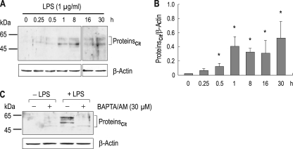
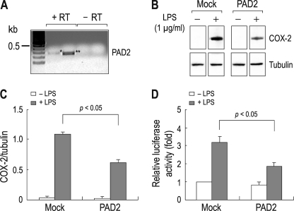
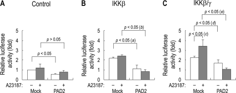
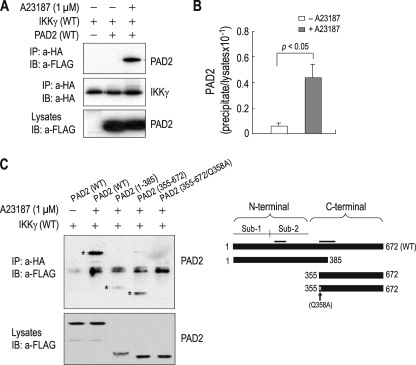
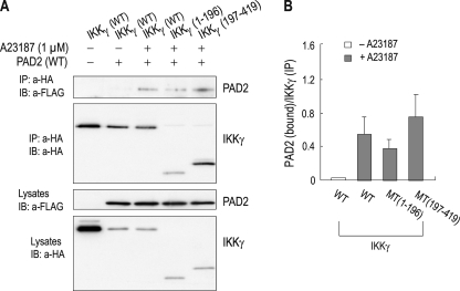
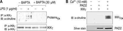
Peptidylarginine deiminases (PADs) are enzymes that convert arginine to citrulline in proteins. In this study, we examined PAD-mediated citrullination and its effect on pro-inflammatory activity in the macrophage cell line RAW 264.7. Citrullination of 45-65-kDa proteins was induced when cells were treated with lipopolysaccharide (LPS; 1 μg/ml). Protein citrullination was suppressed by the intracellular calcium chelator BAPTA/AM (30 μM). LPS treatment up-regulated COX-2 levels in cells. Interestingly, overexpressing PAD2 reduced LPS-mediated COX-2 up-regulation by 50%. PAD2 overexpression also reduced NF-κB activity, determined by NF-κB-driven luciferase activity. The effect of PAD2 on NF-κB activity was further examined by using HEK 293 cells transfected with NF-κB luciferase, IκB β/γ kinase (IKKβ/γ) subunits, and PAD2. IKKβ increased NF-κB activity, but this increase was markedly suppressed when PAD2 was present in cells. IKKβ-mediated NF-κB activation was further enhanced by IKKγ in the presence of calcium ionophore A23187. However, this stimulatory effect of IKKβ/γ was abolished by PAD2. Coimmunoprecipitation of cell lysates showed that IKKγ and PAD2 can coimmunoprecipitate in the presence of the Ca(2+) ionophore. IKKγ coimmunoprecipitated truncation mutants, PAD2(1-385) and PAD2(355-672). The substitution of Gln-358 (a putative ligand for Ca(2+) binding) with an Ala abolished coimmunoprecipitation. Conversely, PAD2 coimmunoprecipitated truncation mutants IKKγ(1-196) and IKKγ(197-419). In other experiments, treating RAW 264.7 cells with LPS induced citrullination in the immunoprecipitates of IKKγ. In vitro citrullination assay showed that incubation of purified PAD2 and IKKγ proteins in the presence of Ca(2+) citrullinated IKKγ. These results demonstrate that PAD2 interacts with IKKγ and suppresses NF-κB activity.


Figures

FIGURE 1.
FIGURE 1.
Lipopolysaccharide-induced citrullination in RAW 264.7 cells. A, time course of citrullination after LPS treatment. Cells were treated with LPS (1 μg/ml) for 0.25–30 h and lysed for immunoblot analysis with the anti-citrulline antibody (n = 4). β-Actin served as an internal control. Vertical stripes or dots at 0.25 (lane 2), 0.5 (lane 3), 16 (lane 6) and 30 h (lane 7) are artifacts. Proteincit, citrullinated proteins. B, densitometric measurements of citrullinated proteins. The intensity of immunoreactive bands was normalized to the intensity of β-actin (n = 4). Artifacts were not included for quantitation. C, effect of the Ca2+ chelator BAPTA/AM on citrullination. Cells were incubated with 30 μm BAPTA/AM or none for 1 h before LPS treatment (1 μg/ml; 4 h). *, p < 0.05.
FIGURE 2.
FIGURE 2.
Suppression of NF-κB activity by PAD2 overexpressed in RAW 264.7 cells. A, RT-PCR for PAD2. Expression of PAD2 mRNA was determined by PCR with two sets of primers amplifying 277 bp (*; lane 2) and 246 bp (**; lane 3) PAD2 cDNA fragments. Total RNA extracted from untreated cells was used. PCR without RT reaction (−RT) served as negative controls. PCR products were sequenced to confirm the specificity of the products. B, effect of PAD2 overexpression on COX-2 production. Cells transfected with the full-length mouse PAD2 were treated with LPS (1 μg/ml) for 4 h. COX-2 production was determined by immunoblot. Cells transfected with vector only (Mock) served as controls. Tubulin served as an internal control for immunoblot. C, comparison of COX-2 expression between control cells and PAD2-expressing cells. Densitometric measurements of immunoreactive bands were done (n = 3). The intensity of COX-2 was normalized to the intensity of tubulin. D, NF-κB activity in control cells and PAD2-expressing cells. Cells transfected with the NF-κB reporter plasmid and PAD2 or none were incubated for 24 h and then treated with LPS for 4 h. NF-κB activity was measured by luciferase assay (n = 4) as described previously (22). The activity was presented relative to the value in mock cells without PAD2 and LPS (leftmost bar).
FIGURE 3.
FIGURE 3.
Suppression of IKK activity by PAD2. A, NF-κB activity in HEK 293 cells transfected with the NF-κB reporter and either PAD2 or vector only (Mock). B and C, NF-κB activity in HEK 293 cells transfected with IKKβ (B) or IKKβ/γ (C) in addition to the plasmids described in A. The values were presented relative to the value in mock cells that have no PAD2 and the calcium ionophore A23187 (leftmost bar in panel A). Twenty-four hours after transfection, cells were treated with the calcium ionophore A23187 (1 μm) for 1 h to induce PAD2 activation. All data were obtained from three independent experiments. (a), p < 0.05; (b), p < 0.05; (c), p < 0.05; (d) and (e), p < 0.05.
FIGURE 4.
FIGURE 4.
Interaction of PAD2 and IKKγ. A, coimmunoprecipitation of PAD2 and IKKγ. HEK 293 cells were transfected with the plasmid containing HA-tagged IKKγ and either FLAG-tagged PAD2 or vector only. Twenty-four hours later, cells were treated with 1 μm A23187 for 1 h, lysed, and immunoprecipitated with the anti-HA antibody (a-HA). The immunoprecipitates (IP) were then immunoblotted (IB) with the anti-FLAG antibody (a-FLAG). One-tenth of lysates was loaded as input controls (bottom panel). B, densitometric measurements of PAD2 bound to IKKγ. The intensity of PAD2 in the immunoprecipitates was normalized to the corresponding intensity in the lysates (n = 4). C, coimmunoprecipitation of truncation mutants of PAD2 with IKKγ. PAD2(1–385) contains the N-terminal 385 amino acids, whereas PAD2(355–672) contains the C-terminal 323 amino acids. PAD2(355–672)/Q358A has an Ala instead of Gln-358 (a putative ligand for Ca2+ binding). The molecular masses of PAD2(1–385) and PAD2(355–672) are 42 and 35 kDa, respectively. The 50-kDa bands in all lanes are IgG heavy chains. *, PAD2 coimmunoprecipitated with IKKγ. The schematic diagram for truncation mutants is shown in the right panel. Putative ligand sites for Ca2+ binding are clustered in two areas (bars). Sub-1, Subdomain-1; Sub-2, Subdomain-2.
FIGURE 5.
FIGURE 5.
Binding assay of PAD2 and truncation mutants of IKKγ. A, coimmunoprecipitation of PAD2 and truncation mutants of IKKγ. IKKγ(1–196) contains the N-terminal 196 amino acids of the protein, whereas IKKγ(197–419) contains the remaining 223 amino acids. The molecular masses of IKKγ(1–196) and IKKγ(197–419) are 22 and 25 kDa, respectively. Coimmunoprecipitation was performed with the protocol described in the legend for Fig. 4. For control cells (lane 1), a 5-fold higher concentration of IKKγ was used for transfection to ensure that the lack of PAD2 in the immunoprecipitates (IP) is not due to insufficient amounts of IKKγ. IB, immunoblot; a-HA, anti-HA antibody; a-FLAG, anti-FLAG antibody. B, relative ratio of PAD2 to IKKγ in the immunoprecipitates. The pixel intensity of PAD2 was normalized to the intensity of IKKγ in the same immunoprecipitates (n = 4). MT, truncation mutant of IKKγ.
FIGURE 6.
FIGURE 6.
Citrullination of IKKγ by PAD2. A, detection of citrullination in IKKγ immunoprecipitates (IP) from RAW 264.7 cells. Cells were incubated with 1 μg/ml LPS for 4 h after pretreatment with BAPTA/AM (30 μm) for 1 h, and immunoprecipitation was performed with an anti-IKKγ antibody (a-IKKγ). The immunoprecipitates were then immunoblotted (IB) with anti-citrulline antibody (a-citrulline). Proteincit, citrullinated proteins. B, in vitro citrullination assay. Purified IKKγ and PAD2 proteins were incubated in reaction buffer containing 10 mm Ca2+ for 2 h. The reaction mixture was then immunoblotted with the anti-citrulline antibody. Silver staining was done with sister mixture loaded on the gel (bottom panel). IKKγcit, citrullinated IKKγ.

References

    1. Rogers G. E. (1962) Nature 194, 1149–1151 - PubMed
    1. Vossenaar E. R., Zendman A. J., van Venrooij W. J., Pruijn G. J. (2003) Bioessays 25, 1106–1118 - PubMed
    1. Tarcsa E., Marekov L. N., Mei G., Melino G., Lee S. C., Steinert P. M. (1996) J. Biol. Chem. 271, 30709–30716 - PubMed
    1. Arita K., Hashimoto H., Shimizu T., Nakashima K., Yamada M., Sato M. (2004) Nat. Struct. Mol. Biol. 11, 777–783 - PubMed
    1. Arita K., Shimizu T., Hashimoto H., Hidaka Y., Yamada M., Sato M. (2006) Proc. Natl. Acad. Sci. U.S.A. 103, 5291–5296 - PMC - PubMed

Publication types

MeSH terms