. 2010 Nov 15;185(10):5723-31.
doi: 10.4049/jimmunol.1001612. Epub 2010 Oct 11.

CCR10 is important for the development of skin-specific gammadeltaT cells by regulating their migration and location

Affiliations

CCR10 is important for the development of skin-specific gammadeltaT cells by regulating their migration and location

Yan Jin et al. J Immunol. .

Abstract

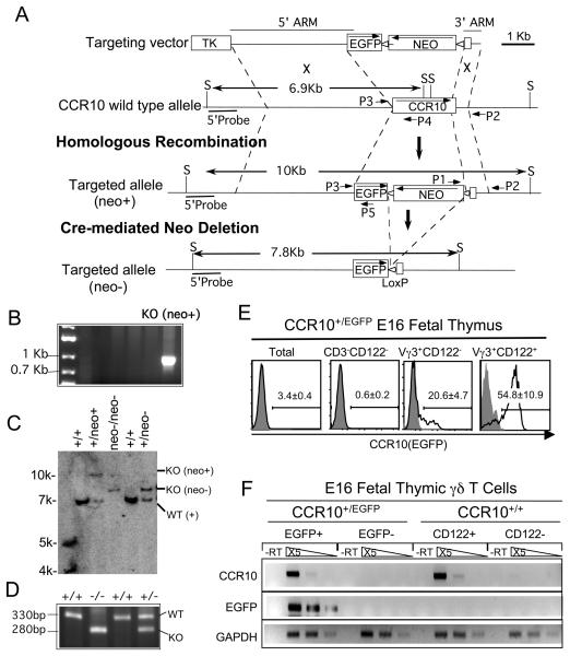
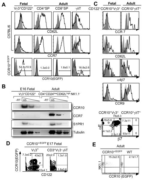
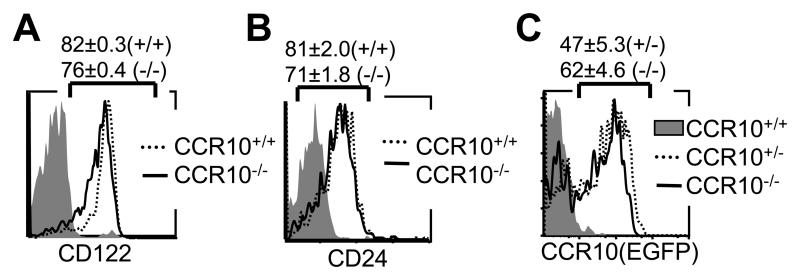
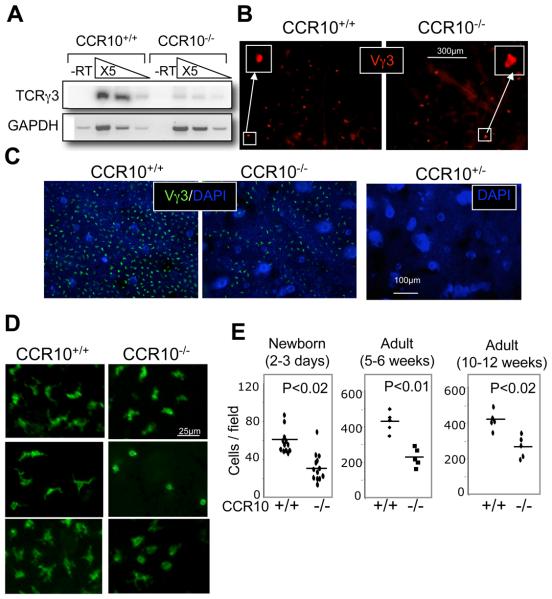
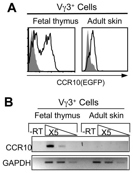
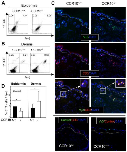
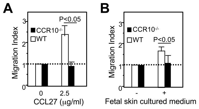
Unlike conventional αβ T cells, which preferentially reside in secondary lymphoid organs for adaptive immune responses, various subsets of unconventional T cells, such as the γδ T cells with innate properties, preferentially reside in epithelial tissues as the first line of defense. However, mechanisms underlying their tissue-specific development are not well understood. We report in this paper that among different thymic T cell subsets fetal thymic precursors of the prototypic skin intraepithelial Vγ3(+) T lymphocytes (sIELs) were selected to display a unique pattern of homing molecules, including a high level of CCR10 expression that was important for their development into sIELs. In fetal CCR10-knockout mice, the Vγ3(+) sIEL precursors developed normally in the thymus but were defective in migrating into the skin. Although the earlier defect in skin-seeding by sIEL precursors was partially compensated for by their normal expansion in the skin of adult CCR10-knockout mice, the Vγ3(+) sIELs displayed abnormal morphology and increasingly accumulated in the dermal region of the skin. These findings provide definite evidence that CCR10 is important in sIEL development by regulating the migration of sIEL precursors and their maintenance in proper regions of the skin and support the notion that unique homing properties of different thymic T cell subsets play an important role in their peripheral location.


Figures

Figure 1
Figure 1
Generation of CCR10-knockout mice with a knocked-in EGFP reporter. A. Targeting and screening strategies for generation of CCR10-knockout/EGFP-knockin mice. B. The screening for CCR10 knockout ES cell clones by a genomic PCR with primers P1 and P2 (the Panel A) that would amplify a 0.8Kb band from the CCR10 knockout allele but not wild type CCR10 allele or CCR10 knockout construct inserted into the genome. C. Southern blot analysis of CCR10 knockout ES cells or mice. Genomic DNAs of the cells and mice were digested with restriction enzyme Sac I (S) and probed with a 0.8Kb DNA fragment located 5′ upstream of the 5′ arm in the CCR10 allele (5′Probe, panel A). The Southern blot gave bands of 6.9, 7.9 and 10 Kb sizes of the wild-type, neo+ CCR10 targeted allele (neo+) and neo-deleted targeted CCR10 allele (neo−) respectively. +/+: wild type ES cells and mice; neo+: recombinant CCR10 knockout ES cells with the neo cassette; neo−/neo−: homozygous CCR10-knockout/EGFP-knockin mice with the neo cassette deleted; +/neo−: heterozygous CCR10-knockout/EGFP-knockin mice with the neo cassette deleted. D. Identification of heterozygous and homozygous CCR10 knockout mice by a genomic PCR with primers P3, P4 and P5 (panel A), which amplifies a 330bp wild-type and a 280bp knockout band. E. Flow cytometric (FACS) analysis of EGFP expression in fetal thymic Vγ3+ γδ T cells before and after the positive selection. Thirty-one CCR10+/EGFP fetuses were analyzed. F. Correlated expression of the knocked-in EGFP and endogenous CCR10 genes. Different fetal thymic Vγ3+ cell populations were purified from CCR10+/EGFP and CCR10+/+ mice based on their EGFP or CD122 expression (as indicated) by cell-sorters. Levels of CCR10 and EGFP transcripts in the sorted populations were analyzed by semi-quantitative RT-PCR. -RT, no reverse transcription. GAPDH was used as loading controls. N=2.
Figure 2
Figure 2
Differential expression of CCR10 and other homing molecules among positively selected fetal thymic Vγ3+ and other thymic T cell subsets. A. FACS analysis of CCR10 (EGFP), CD62L, and CCR7 expression on positively selected E16 fetal thymic Vγ3+ (N=8) vs. adult (N=3) thymic CD4+, CD8+ or γδ T cells. The CCR10 (EGFP) analysis was on CCR10+/EGFP mice (N≥4). B. Semi-quantitative RT-PCR determination of CCR7, CCR10 and S1PR1 expression in mature E16 fetal thymic CD122+ Vγ3+ and adult thymic CD24lowCD62LhighNK1.1 CD4+ cells of C57BL/6 mice. The experiments were repeated twice. C. FACS analysis of α4β7, CCR9, CCR7, CD62L, αE and β7 expression on CCR10(EGFP)+ E16 fetal (N=10) thymic Vγ3+ vs. adult (N=2) thymic γδT cells. D. Comparison of CCR10 (EGFP) and CD122 expression on E16 fetal thymic Vγ3+ vs. Vγ3 γδT cells of CCR10+/EGFP mice. More than five mice were analyzed. E. Expression of CCR10 (EGFP) on the gated adult NK1.1+ CD3+ thymocytes. The experiments were repeated twice.
Figure 3
Figure 3
Normal development of Vγ3+ sIEL precursors in fetal thymi of CCR10 knockout mice. A and B. Comparison of the CD24 and CD122 expression on E16 fetal Vγ3+ thymocytes of CCR10EGFP/EGFP (CCR10−/−) and wild type (CCR10+/+) littermates by flow cytometry. More than five mice of each genotype were analyzed. C. FACS analysis of EGFP expression on gated Vγ3+ γδ T cells of CCR10EGFP/EGFP mice vs. CCR10+/EGFP (CCR10+/−) mice. The staining of wild type cells was used as a negative control for the EGFP expression.
Figure 4
Figure 4
Impaired sIEL development in CCR10EGFP/EGFP mice. A. Comparison of TCRγ3 transcripts in fetal skin of CCR10EGFP/EGFP and wild type mice by semi-quantitative RT-PCR. Skin RNAs of E18 fetal CCR10EGFP/EGFP and wild-type mice were subjected to semi-quantitative RT-PCR analysis for rearranged TCRγ3. -RT, no reverse transcription. GAPDH was used as a loading control. The experiments were repeated twice. B. Immunofluorescent microscopic analysis of epidermal sheets of the 2-3 day old newborn CCR10EGFP/EGFP and wild type mice for Vγ3+ sIELs. Epidermal sheets of the back skin were stained with biotin conjugated anti-Vγ3 antibodies followed with Alexa 647 conjugated streptavidin. C. Immunofluorescent microscopic analysis of epidermal sheets of 5-6 week old adult CCR10EGFP/EGFP and wild type mice for Vγ3+ sIELs. Ear epidermal sheets were co-stained with FITC-conjugated anti-Vγ3 antibody and DAPI. The picture on the right was stained with DAPI and used as a control for endogenous EGFP signals. D. High-magnification immunofluorescent microscopy of ear epidermal sheets of adult CCR10EGFP/EGFP and wild type mice for morphology of Vγ3+ sIELs. Images of three pairs of littermates were shown. E. Quantitative comparison of numbers of Vγ3+ sIELs in the CCR10EGFP/EGFP and wild type mice of different ages. The number of Vγ3+ sIELs was calculated from the immunofluorescent microscopy of epidermal sheets with at least five fields counted for each mouse. One dot represents average number of Vγ3+ sIELs per field from one mouse.
Figure 5
Figure 5
Regulated CCR10 expression in sIELs and their thymic precursors. A. FACS analysis of CCR10 (EGFP) expression on the adult Vγ3+ sIELs and fetal thymic CD122+ Vγ3+ T cells of CCR10+/EGFP mice. B. Comparison of levels of CCR10 transcripts in adult sIELs and positively selected fetal thymic sIEL precursors. RNAs of purified adult Vγ3+ sIELs and E16 fetal thymic CD122+Vγ3+ cells of wild type mice were analyzed by semi-quantitative RT-PCR for CCR10 transcripts. The experiments were repeated twice.
Figure 6
Figure 6
Abnormal accumulation of Vγ3+ γδ T cells in dermal regions of CCR10 knockout mice. A. FACS analysis for Vγ3+ sIELs in cells isolated from epidermis of the CCR10EGFP/EGFP and wild type mice. B. FACS analysis for Vγ3+ T cells in cells isolated from the dermal regions of the CCR10EGFP/EGFP and wild type mice. A set of representative plots of at least three experiments is shown. C. Immunofluorescent microscopy of skin sections of the CCR10EGFP/EGFP and wild type mice co-stained with FITC-conjugated anti-Vγ3 and Alexa-647 conjugated anti-CD3 antibodies. The dashed lines run along borders of the epidermis and dermis. Isotype controls are shown at the bottom. D. Quantification of epidermal and dermal Vγ3+ cells of the CCR10EGFP/EGFP and wild type mice based on the immunofluorescent microscopic analyses in the panel C. Five mice of each genotype were analyzed with more than 10 fields counted for each mouse.
Figure 7
Figure 7
Defective migration of CCR10EGFP/EGFP fetal thymic Vγ3+ cells towards CCL27 and skin in the in vitro chemotaxis assay. The migration index is calculated as a ratio of numbers of Vγ3+ cells migrating into the bottom chamber in presence of CCL27 (A), conditioned medium of fetal skin culture (B) vs. medium only. The experiments were repeated twice.

References

    1. Hayday AC. [gamma][delta] cells: a right time and a right place for a conserved third way of protection. Annual Review of Immunology. 2000;18:975–1026. - PubMed
    1. Raulet D, Spencer D, Hsiang Y-H, Goldman J, Bix M, Liao N-S, Zijlstra M, Jaenisch R, Correa I. Control of γδ T cell development. Immunol. Rev. 1991;120:185–204. - PubMed
    1. Kim CH, Witherden DA, Havran WL. Characterization and TCR variable region gene use of mouse resident nasal gammadelta T lymphocytes. J Leukoc Biol. 2008;84:1259–1263. - PMC - PubMed
    1. Xiong N, Raulet DH. Development and selection of gammadelta T cells. Immunol Rev. 2007;215:15–31. - PubMed
    1. Girardi M, Oppenheim DE, Steele CR, Lewis JM, Glusac E, Filler R, Hobby P, Sutton B, Tigelaar RE, Hayday AC. Regulation of cutaneous malignancy by gammadelta T cells. Science. 2001;294:605–609. - PubMed

Publication types

MeSH terms

Substances