. 2010 Oct 26;107(43):18475-80.
doi: 10.1073/pnas.1012946107. Epub 2010 Oct 11.

A chromatin localization screen reveals poly (ADP ribose)-regulated recruitment of the repressive polycomb and NuRD complexes to sites of DNA damage

Affiliations

A chromatin localization screen reveals poly (ADP ribose)-regulated recruitment of the repressive polycomb and NuRD complexes to sites of DNA damage

Danny M Chou et al. Proc Natl Acad Sci U S A. .

Abstract

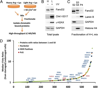
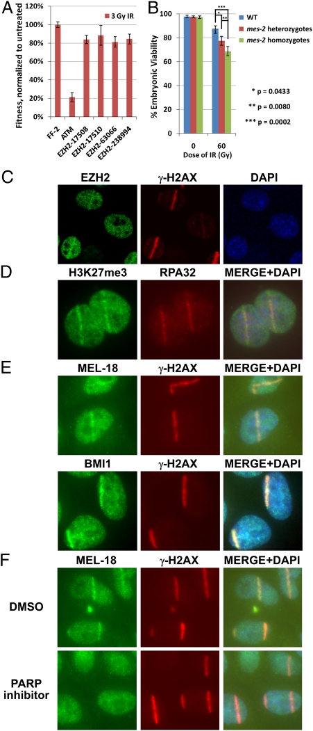
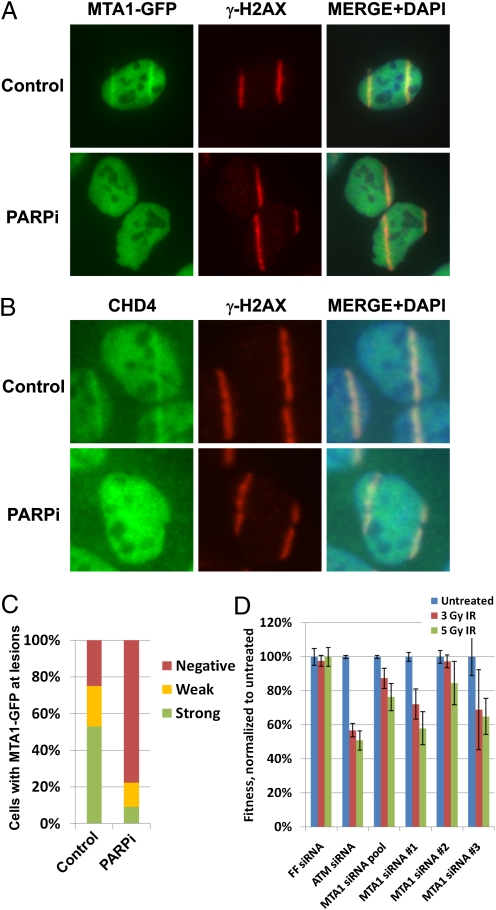
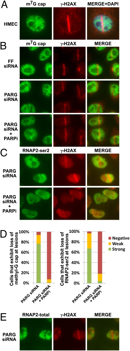
Many proteins that respond to DNA damage are recruited to DNA lesions. We used a proteomics approach that coupled isotopic labeling with chromatin fractionation and mass spectrometry to uncover proteins that associate with damaged DNA, many of which are involved in DNA repair or nucleolar function. We show that polycomb group members are recruited by poly(ADP ribose) polymerase (PARP) to DNA lesions following UV laser microirradiation. Loss of polycomb components results in IR sensitivity of mammalian cells and Caenorhabditis elegans. PARP also recruits two components of the repressive nucleosome remodeling and deacetylase (NuRD) complex, chromodomain helicase DNA-binding protein 4 (CHD4) and metastasis associated 1 (MTA1), to DNA lesions. PARP plays a role in removing nascent RNA and elongating RNA polymerase II from sites of DNA damage. We propose that PARP sets up a transient repressive chromatin structure at sites of DNA damage to block transcription and facilitate DNA repair.


Conflict of interest statement

The authors declare no conflict of interest.

Figures

Fig. 1.
Fig. 1.
A proteomics screen to identify proteins that are recruited to sites of DNA damage. (A) Overview of the proteomics strategy used to identify proteins that are preferentially recruited to sites of DNA damage. Arg, arginine; Lys, lysine. (B) UV induced monoubiquitination of FANCD2 as well as phosphorylation of Chk1 and H2AX. (C) Untreated HeLa cells were mixed with cells exposed to UV light and fractionated. The effectiveness of separation of soluble, chromatin-bound, and nuclear matrix-associated proteins was determined by Western blotting. (D) Proteins in the chromatin fraction with a median vista ratio >1 are plotted in the order of increasing ratio. A ratio >1 suggests enrichment in the chromatin fraction after DNA damage.
Fig. 2.
Fig. 2.
PcG proteins confer protection against ionizing radiation and accumulate at DNA lesions in a PARP1/2-dependent manner. (A) U2OS cells expressing dsRed and the indicated shRNA were mixed with U2OS cells expressing control FF shRNA, were mock-treated or exposed to IR, and were analyzed by FACS after 10 d in a competition assay. Relative fitness reflects the percentage of IR-treated red cells remaining in the mixture relative to mock-treated cells. (B) mes-2 mutant animals show increased sensitivity to ionizing radiation. Embryonic viability of the offspring of untreated and irradiated animals is quantified 24 h postirradiation by assessing the number of live offspring relative to the number of eggs laid by each animal. Error bars indicate SEM for 15–30 animals in three independent experiments. (C) MEFs were microirradiated with a UV laser, fixed after 10 min, and immunostained with anti-EZH2 and anti-γH2AX antibodies. (D) MEFs were microirradiated with a UV laser, fixed after 5 min, and immunostained with anti-H3K27me3 and anti-RPA32 antibodies. (E) Endogenous MEL-18 and BMI1 were recruited within 10–12 min to UV laser-induced sites of damage in HeLa cells. (F) HeLa cells treated with either DMSO or 1 μM KU-0058948 (PARP1/2 inhibitor) were microirradiated with a UV laser and immunostained with anti-MEL-18 and anti-γH2AX antibodies.
Fig. 3.
Fig. 3.
NuRD components CHD4 and MTA1 accumulate at DNA lesions following UV laser microirradiation. (A) U2OS cells expressing MTA1-GFP pretreated for 1 h with either DMSO (control) or 1 μM PARP inhibitor KU-0058948 were microirradiated with a UV laser, fixed after 10 min, and immunostained with anti-GFP and anti-γH2AX antibodies. (B) U2OS cells pretreated for 1 h with DMSO or 2 μM PARP inhibitor KU-0058948 were microirradiated with a UV laser, fixed after 10 min, and immunostained with anti-CHD4 and anti-γH2AX antibodies. (C) Quantification of the data shown in A. (D) U2OS cells expressing GFP were transfected with the indicated siRNAs, mixed with cells expressing dsRed, mock-treated or exposed to various doses of IR, and analyzed by FACS after 9 d in a competition assay.
Fig. 4.
Fig. 4.
PARP activity is required for inhibition of transcription at sites of DNA damage. (A) Human mammary epithelial cells were microirradiated with a UV laser, fixed after 10–15 min, and immunostained with an antibody that detects the mRNA m7G cap and an anti-γH2AX antibody. (B) HeLa cells were transfected with the indicated siRNAs and were mock-treated or incubated with the PARP inhibitor (PARPi) 1 h before microirradiation, fixed 20 min after irradiation, and immunostained with the anti-m7G cap and anti-γH2AX antibodies. (C) HeLa cells transfected with PARG siRNAs were either mock treated or incubated with the PARP inhibitor (PARPi) 1 hour prior to microirradiation, fixed after 20 min, and immunostained with anti-RNA polymerase II (RNAP2)-ser-2 and anti-γH2AX antibodies. (D) Quantification of the data shown in B and C. (E) HeLa cells transfected with PARG siRNAs were microirradiated, fixed after 20 min, and immunostained with anti-RNAP2 and anti-γH2AX antibodies.

References

    1. Harper JW, Elledge SJ. The DNA damage response: Ten years after. Mol Cell. 2007;28:739–745. - PubMed
    1. van Attikum H, Gasser SM. Crosstalk between histone modifications during the DNA damage response. Trends Cell Biol. 2009;19:207–217. - PubMed
    1. Wang B, Elledge SJ. Ubc13/Rnf8 ubiquitin ligases control foci formation of the Rap80/Abraxas/Brca1/Brcc36 complex in response to DNA damage. Proc Natl Acad Sci USA. 2007;104:20759–20763. - PMC - PubMed
    1. Huen MS, et al. RNF8 transduces the DNA-damage signal via histone ubiquitylation and checkpoint protein assembly. Cell. 2007;131:901–914. - PMC - PubMed
    1. Kolas NK, et al. Orchestration of the DNA-damage response by the RNF8 ubiquitin ligase. Science. 2007;318:1637–1640. - PMC - PubMed

Publication types

MeSH terms

Substances