. 2010 Oct 26;107(43):18694-9.
doi: 10.1073/pnas.1009500107. Epub 2010 Oct 11.

Contribution of calcium-dependent facilitation to synaptic plasticity revealed by migraine mutations in the P/Q-type calcium channel

Affiliations

Contribution of calcium-dependent facilitation to synaptic plasticity revealed by migraine mutations in the P/Q-type calcium channel

Paul J Adams et al. Proc Natl Acad Sci U S A. .

Abstract

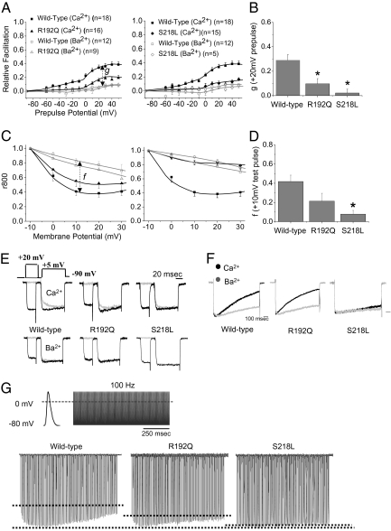
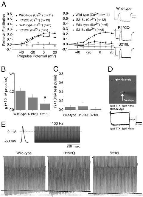
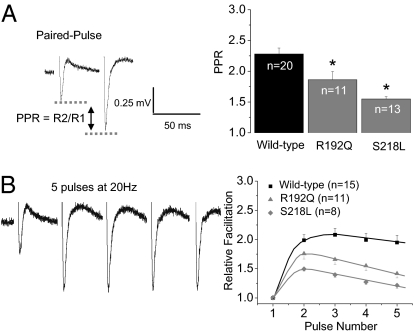
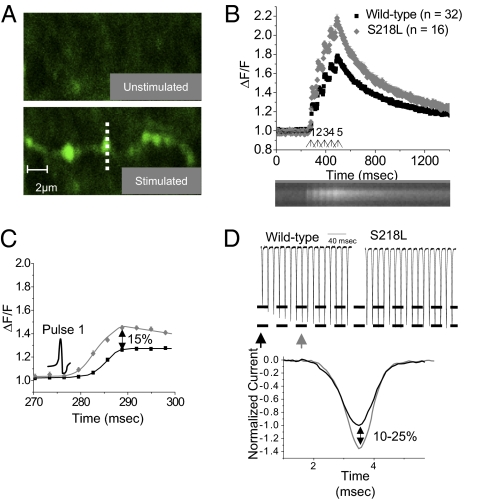
The dynamics, computational power, and strength of neural circuits are essential for encoding and processing information in the CNS and rely on short and long forms of synaptic plasticity. In a model system, residual calcium (Ca(2+)) in presynaptic terminals can act through neuronal Ca(2+) sensor proteins to cause Ca(2+)-dependent facilitation (CDF) of P/Q-type channels and induce short-term synaptic facilitation. However, whether this is a general mechanism of plasticity at intact central synapses and whether mutations associated with human disease affect this process have not been described to our knowledge. In this report, we find that, in both exogenous and native preparations, gain-of-function missense mutations underlying Familial Hemiplegic Migraine type 1 (FHM-1) occlude CDF of P/Q-type Ca(2+) channels. In FHM-1 mutant mice, the alteration of P/Q-type channel CDF correlates with reduced short-term synaptic facilitation at cerebellar parallel fiber-to-Purkinje cell synapses. Two-photon imaging suggests that P/Q-type channels at parallel fiber terminals in FHM-1 mice are in a basally facilitated state. Overall, the results provide evidence that FHM-1 mutations directly affect both P/Q-type channel CDF and synaptic plasticity and that together likely contribute toward the pathophysiology underlying FHM-1. The findings also suggest that P/Q-type channel CDF is an important mechanism required for normal synaptic plasticity at a fast synapse in the mammalian CNS.


Conflict of interest statement

The authors declare no conflict of interest.

Figures

Fig. 1.
Fig. 1.
R192Q and S218L FHM-1 mutations inhibit Ca2+-dependent modulation of human recombinant Cav2.1 channels. (A) Using the paired-pulse protocol previously described (9), both the R192Q and S218L FHM-1 mutations are found to occlude CDF across several prepulse potentials shown as relative facilitation versus prepulse potential; results are means ± SEM. (B) Pure CDF (g) following a +20 mV prepulse is significantly reduced by the R192Q (g = 0.097 ± 0.042; *P < 0.05) and S218L (g = 0.023 ± 0.031; *P < 0.05) mutations compared with WT (g = 0.290 ± 0.046). (C) Using 1-s square test-pulses between −10 mV and +30 mV, the current remaining at 800 ms (r800) across several prepulse potentials is increased by both FHM-1 mutations relative to WT; data are means ± SEM. (D) Pure CDI (f) relative to WT (f = 0.418 ± 0.067) is modestly reduced by the R192Q mutation (f = 0.214 ± 0.082) but significantly reduced by the S218L mutation (f = 0.083 ± 0.038; *P < 0.05). (E) Paired-pulse protocol used and representative traces for peak CDF obtained following a +20 mV prepulse for WT, R192Q, and S218L Cav2.1 channels using Ca2+ (Top) and Ba2+ (Bottom) as charge carrier; traces normalized to the end of the test pulse. (F) Representative traces for CDI in which traces are normalized to the peak of a +10 mV test pulse. (G) APW used and representative traces for WT, R192Q, and S218L Cav2.1 channels. First hashed line represents the level of Ca2+ response during the first AP, and the second line represents the peak (maximum facilitation). The 100-Hz APW was derived from APs recorded in the calyx of Held (38, 54). n refers to the number of cells recorded. All statistics were obtained with use of one-way ANOVA.
Fig. 2.
Fig. 2.
P/Q-type current CDF is altered in acutely dissociated PCs from FHM-1 R192Q and S218L knock-in mice. (A) Both the R192Q and S218L mutations occlude CDF across several prepulse potentials; results are means ± SEM (representative traces, far right). (B) Pure CDF (g) relative to WT (g = 0.210 ± 0.028) is not significantly reduced by R192Q (g = 0.136 ± 0.03), whereas the S218L mutation results in a significant reduction (g = 0.0686 ± 0.035; *P < 0.05). (C) There was no appreciable CDI of P/Q-type currents in PCs from WT (f = 0.058 ± 0.045) or R192Q (f = 0.074 ± 0.091) and S218L (f = 0.023 ± 0.028) mice. (D) P/Q-type currents were isolated from freshly dissociated cerebellar PCs identified by their characteristic large size and tear-shaped morphology. Exemplar trace below shows that the pharmacologically isolated P/Q-type currents were completely blocked with 0.2 μM ω-Aga-IVA. (E) APW used and representative traces for WT, R192Q, and S218L Cav2.1 channels. First hashed line represents the level of Ca2+ response during the first AP, and the second line represents the peak (maximum facilitation). n refers to the number of cells recorded. All statistics were obtained with use of a one-way ANOVA.
Fig. 3.
Fig. 3.
FHM-1 mutant mice exhibit attenuated PPF at the PF–PC synapse. (A, Left) Exemplar field recording of synaptic responses from the PCs evoked by two extracellular stimuli of approximately 15 V, 180 μs delivered at a 50-ms interval to PFs in the molecular layer (WT mouse). The PPR is a quantification of facilitation obtained by dividing response 2 (R2) by response 1 (R1). Right: PPR is significantly reduced relative to WT (2.36 ± 0.096) by both the R192Q (1.86 ± 0.13; *P < 0.05) and S218L mutations (1.60 ± 0.075; *P < 0.05); results are means ± SEM. (B) Left: Exemplar field recording of synaptic responses from the PCs evoked by five extracellular stimuli of approximately 15 V, 180 μs delivered at 20 Hz (WT mouse). (Right) RF was measured by dividing the peak response from each stimulus by the response obtained from the first stimulus, plotted versus pulse number; results are means ± SEM. Both mutations reduce RF during five pulses at 20 Hz. n refers to the number of slices recorded (from eight WT, four R192Q, and five S218L mice). All statistics were obtained with use of a one-way ANOVA.
Fig. 4.
Fig. 4.
The kinetics of Ca2+ influx at PF terminals from S218L mice suggests a basal facilitation of P/Q-type channels. (A) PFs were stimulated with a silver wire electrode and two-photon imaging was used to measure Ca2+ transients in presynaptic terminals of the PFs; postsynaptic activity was blocked with 100 μM MCPG, 20 μM CNQX, and 50 μM APV. Line scans were used to measure individual terminals (representative terminal indicated by hashed line). Four sets of five 50-μs stimuli at 20 Hz were given and the Ca2+ transients were averaged for each terminal. (B) The average Ca2+ influx at PF terminals (normalized to the background fluorescence) is enhanced in S218L mice relative to WT littermates; results are means ± SEM (arrows represent the five stimuli). A representative average Ca2+ transient response measured for one terminal in line-scan mode is shown (Lower). (C) An expanded scale of B to show Ca2+ transient elicited during the first AP. Ca2+ transients in mutant terminals are larger in magnitude than WT terminals (peak enhanced approximately 15% relative to WT), as expected if mutant channels are in a basally facilitated gating mode. Stimulus is shown by AP illustration. n refers to the number of presynaptic terminals recorded (from five WT and five S218L mice). (D) Expanded view of CDF of recombinant channels during APW from Fig. 1G. Ca2+ currents through WT recombinant Cav2.1 channels increases as Ca2+ influx causes CDF (Left), whereas recombinant Cav2.1 channels containing the S218L mutation have a maximal Ca2+ response regardless of APs (Right). Bottom: Ca2+ currents through facilitated P/Q-type channels (gray trace) are larger than through unfacilitated channels (black trace) during evoked APs. A 10–25% increase in Ca2+ current amplitude was the range obtained from recombinant Cav2.1 channels in HEK cells (Fig. 1G) and endogenous P/Q-type currents in PCs (Fig. 2E). The unfacilitated trace is the Ca2+ response of a WT channel obtained during the first AP, and the facilitated trace is the Ca2+ response obtained during the 10th AP (indicated by arrows, Top, Left).

References

    1. Zucker RS, Regehr WG. Short-term synaptic plasticity. Annu Rev Physiol. 2002;64:355–405. - PubMed
    1. Cuttle MF, Tsujimoto T, Forsythe ID, Takahashi T. Facilitation of the presynaptic calcium current at an auditory synapse in rat brainstem. J Physiol. 1998;512:723–729. - PMC - PubMed
    1. Xu J, Wu LG. The decrease in the presynaptic calcium current is a major cause of short-term depression at a calyx-type synapse. Neuron. 2005;46:633–645. - PubMed
    1. Tsujimoto T, Jeromin A, Saitoh N, Roder JC, Takahashi T. Neuronal calcium sensor 1 and activity-dependent facilitation of P/Q-type calcium currents at presynaptic nerve terminals. Science. 2002;295:2276–2279. - PubMed
    1. Mochida S, Few AP, Scheuer T, Catterall WA. Regulation of presynaptic Ca(V)2.1 channels by Ca2+ sensor proteins mediates short-term synaptic plasticity. Neuron. 2008;57:210–216. - PubMed

Publication types

MeSH terms