. 2010 Dec 10;285(50):39536-43.
doi: 10.1074/jbc.M110.167619. Epub 2010 Oct 11.

A novel heme-responsive element mediates transcriptional regulation in Caenorhabditis elegans

Affiliations

A novel heme-responsive element mediates transcriptional regulation in Caenorhabditis elegans

Jason Sinclair et al. J Biol Chem. .

Abstract

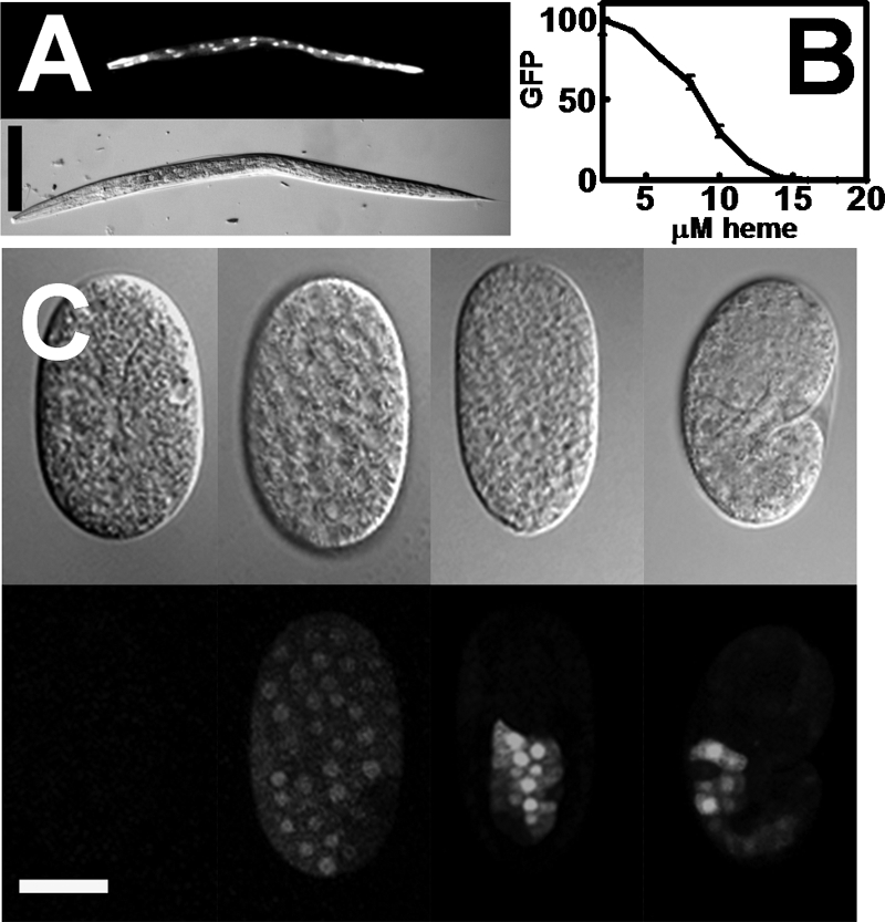
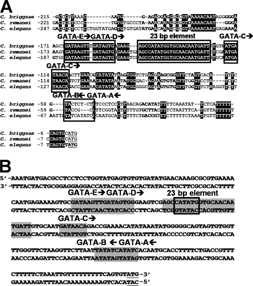
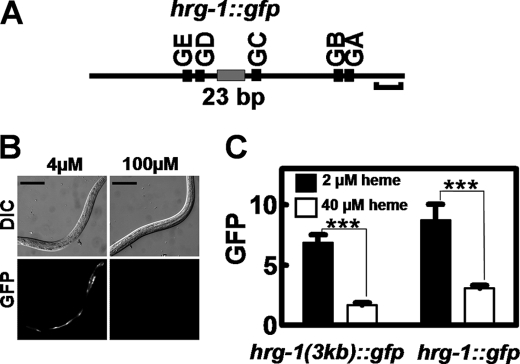
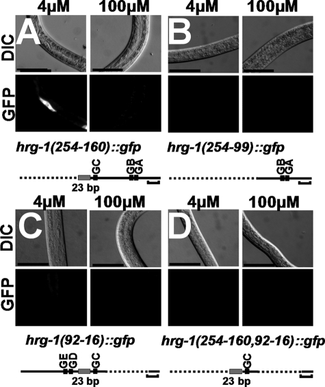
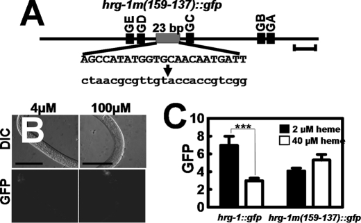
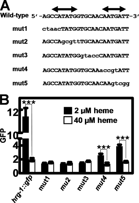
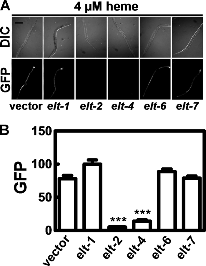
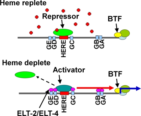
Hemes are prosthetic groups that participate in diverse biochemical pathways across phylogeny. Although heme can also regulate broad physiological processes by directly modulating gene expression in Metazoa, the regulatory pathways for sensing and responding to heme are not well defined. Caenorhabditis elegans is a heme auxotroph and relies solely on environmental heme for sustenance. Worms respond to heme availability by regulating heme-responsive genes such as hrg-1, an intestinal heme transporter that is up-regulated by >60-fold during heme depletion. To identify the mechanism for the heme-dependent regulation of hrg-1, we interrogated the hrg-1 promoter. Deletion and mutagenesis studies of the hrg-1 promoter revealed a 23-bp heme-responsive element that is both necessary and sufficient for heme-dependent regulation of hrg-1. Furthermore, our studies show that the heme regulation of hrg-1 is mediated by both activation and repression in conjunction with ELT-2 and ELT-4, transcription factors that specify intestinal expression.


Figures

FIGURE 1.
FIGURE 1.
hrg-1 is transcriptionally up-regulated in low heme. A, IQ6011 worms were grown in mCeHR-2 medium supplemented with 4 μm heme for 48 h and then analyzed for GFP by fluorescence microscopy. Upper panel, GFP; lower panel, DIC. Scale bar = 100 μm. B, synchronized IQ6011 L1 larvae were grown in varying heme concentrations for 72 h in mCeHR-2 medium. GFP (arbitrary units) was quantified using the COPAS BIOSORT instrument. C, IQ6011 worms were grown at 4 μm heme for 48 h and then bleached to obtain embryos. Embryos at varying developmental stages were analyzed for fluorescence (GFP; lower panels) and DIC (upper panels). Scale bar = 20 μm.
FIGURE 2.
FIGURE 2.
The hrg-1 promoter contains conserved putative cis-acting DNA elements. A, ClustalW/Boxshade 3.2.1 alignment of the hrg-1 5′-flanking sequences of C. briggsae, C. remanei, and C. elegans. Reversed letters indicate conserved nucleotides. Boxes indicate putative regulatory elements. Arrows show the orientation of the GATA sites. The ATG start codon is underlined. B, diagram depicting the five putative GATA sites and the 23-bp element in the 5′-flanking region of hrg-1. Shaded letters indicate putative regulatory elements. The box indicates an E-box. Arrows show the orientation of the GATA sites. The ATG start codon is underlined.
FIGURE 3.
FIGURE 3.
A 254-bp region within the hrg-1 promoter is sufficient for heme responsiveness. A, schematic diagram showing 254 bp of the hrg-1 5′-flanking sequence. GA–GE, GATA sites. Horizontal bracket = 20 bp. B, transgenic C. elegans strains carrying hrg-1::gfp were made axenic and simultaneously synchronized. L1 larvae were placed in either 4 or 100 μm heme for 72 h. Scale bars = 100 μm. GFP in the worms was analyzed by microscopy. Upper panels, DIC; lower panels, GFP. C, GFP was quantified in C. elegans transformed with hrg-1::gfp and supplemented 2 or 40 μm heme. The y axis is the GFP level in arbitrary units. ***, p < 0.001.
FIGURE 4.
FIGURE 4.
The HERE is within a 67-bp region. Deletion constructs (A–D) were synthesized to determine the specific region that contained the HERE. GATA sites (GA–GE) are shown by black boxes, and the 23-bp element is shown by a gray box. Horizontal brackets = 20 bp. For microscopy, GFP (lower panels) and DIC (upper panels) were analyzed in worms grown at 4 or 100 μm heme. Longer exposures of worms in C are shown in supplemental Fig. S3.
FIGURE 5.
FIGURE 5.
Mutation of the 23-bp conserved element abolishes expression of hrg-1. A, schematic diagram of the wild-type 23-bp element and the corresponding mutation indicated in lowercase letters. GA–GE, GATA sites. B, transgenic C. elegans strains transformed with hrg-1m(159–137)::gfp were made axenic, and synchronized L1 larvae were placed in medium supplemented with either 4 or 100 μm heme for 72 h. GFP expression in the worms was analyzed by microscopy. Scale bars = 100 μm. C, quantification of GFP in C. elegans transformed with hrg-1m(159–137)::gfp and supplemented with 2 or 40 μm heme. The y axis is the GFP level in arbitrary units. ***, p < 0.001.
FIGURE 6.
FIGURE 6.
The 23-bp element is sufficient for heme response. A, schematic diagram of a concatamer of six direct repeats of the 23-bp element fused to the egl-18 basal promoter. This chimeric promoter was placed upstream of gfp and the unc-54 3′ UTR. B, transgenic C. elegans strains expressing either egl-18::gfp or egl-18(+23)::gfp were placed in medium supplemented with either 4 or 100 μm heme for 120 h, and GFP expression in the worms was analyzed by microscopy. Scale bars = 100 μm. C, GFP was quantified in C. elegans transformed with egl-18::gfp or egl-18(+23)::gfp and supplemented with 2 or 40 μm heme. The y axis is the GFP level in arbitrary units. ***, p < 0.001.
FIGURE 7.
FIGURE 7.
Heme responsiveness in the hrg-1 promoter is mediated by a 15-bp region containing an E-box. A, schematics for the synthesis of mut1 to mut5 by mutating five consecutive nucleotides at a time within the 23-bp HERE. Double-headed arrows indicate the location of inverse repeats. B, GFP was quantified in C. elegans transformed with mut1, mut2, mut3, mut4, or mut5 and supplemented with 2 or 40 μm heme. The y axis is the GFP level in arbitrary units. ***, p < 0.001.
FIGURE 8.
FIGURE 8.
ELT-2 and ELT-4 are essential for the heme regulation of hrg-1. A, synchronized IQ6011 L3 larvae were fed HT115(DE3) bacteria grown in 4 μm heme and induced to produce double-stranded RNA against elt-1, elt-2, elt-4, elt-6, or elt-7, and GFP was analyzed by microscopy. B, quantification of the GFP expression in A. The y axis is the GFP level in arbitrary units. ***, p < 0.001.
FIGURE 9.
FIGURE 9.
Proposed model of hrg-1 regulation by heme. Under heme-replete conditions, a repressor binds the 23-bp HERE, and hrg-1 expression is suppressed. In heme-depleted conditions, an enhancer may compete with the repressor for binding the HERE, resulting in the recruitment of ELT-2 and/or ELT-4 to activate transcription of hrg-1. BTF, basal transcription factors; GA–GE, GATA elements; red arrow, enhancement and possible interaction with GATA-binding factors; blue arrow, transcription; red plus signs, heme.

References

    1. Ajioka R. S., Phillips J. D., Kushner J. P. (2006) Biochim. Biophys. Acta 1763, 723–736 - PubMed
    1. Nakajima O., Takahashi S., Harigae H., Furuyama K., Hayashi N., Sassa S., Yamamoto M. (1999) EMBO J. 18, 6282–6289 - PMC - PubMed
    1. Rodgers K. R. (1999) Curr. Opin. Chem. Biol. 3, 158–167 - PubMed
    1. Wenger R. H. (2000) J. Exp. Biol. 203, 1253–1263 - PubMed
    1. Gilles-Gonzalez M. A., Gonzalez G. (2004) J. Appl. Physiol. 96, 774–783 - PubMed

Publication types

MeSH terms

LinkOut - more resources