Review
. 2010 Nov;7(11):629-41.
doi: 10.1038/nrgastro.2010.154. Epub 2010 Oct 12.

Mechanisms of disease: Helicobacter pylori virulence factors

Affiliations
Review

Mechanisms of disease: Helicobacter pylori virulence factors

Yoshio Yamaoka. Nat Rev Gastroenterol Hepatol. 2010 Nov.

Abstract

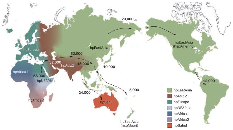
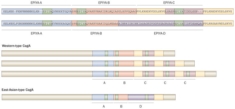
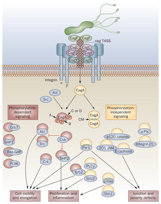
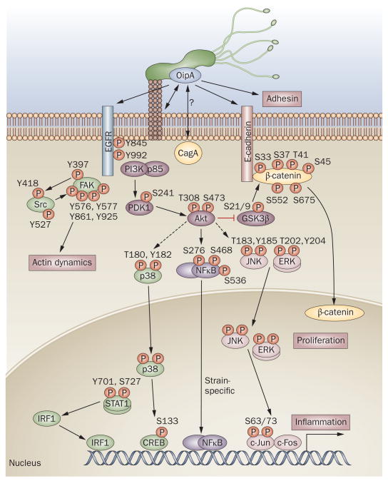
Helicobacter pylori plays an essential role in the development of various gastroduodenal diseases; however, only a small proportion of people infected with H. pylori develop these diseases. Some populations that have a high prevalence of H. pylori infection also have a high incidence of gastric cancer (for example, in East Asia), whereas others do not (for example, in Africa and South Asia). Even within East Asia, the incidence of gastric cancer varies (decreasing in the south). H. pylori is a highly heterogeneous bacterium and its virulence varies geographically. Geographic differences in the incidence of gastric cancer can be explained, at least in part, by the presence of different types of H. pylori virulence factor, especially CagA, VacA and OipA. However, it is still unclear why the pathogenicity of H. pylori increased as it migrated from Africa to East Asia during the course of evolution. H. pylori infection is also thought to be involved in the development of duodenal ulcer, which is at the opposite end of the disease spectrum to gastric cancer. This discrepancy can be explained in part by the presence of H. pylori virulence factor DupA. Despite advances in our understanding of the development of H. pylori-related diseases, further work is required to clarify the roles of H. pylori virulence factors.


Conflict of interest statement

Competing interests

The author declares no competing interests.

Figures

Figure 1
Figure 1
Distribution of Helicobater pylori genotypes before Columbus found the New World and human migration to America and Oceania began. There are seven modern H. pylori population types—hpEurope, hpEastAsia, hpAfrica1, hpAfrica2, hpAsia2, hpNEAfrica and hpSahul. hpEurope includes almost all H. pylori strains isolated from ethnic Europeans, including people from countries colonized by Europeans. Most H. pylori isolates from East Asia belong to hpEastAsia, which includes hspMaori (Polynesians, Melanesians, and native Taiwanese), hspAmerind (American Indians) and hspEAsia (East Asia) subpopulations. hpAsia2 strains are isolated in South, Southeast and Central Asia; hpAfrica1 in West Africa, South Africa and African Americans. hpNEAfrica is predominantly made up of isolates from Northeast Africa. hpAfrica2 is very distinct from any other type and has currently only been isolated in South Africa. hpSahul is a novel group specific to H. pylori strains isolated from Australian Aborigines and Highlanders of New Guinea. H. pylori is predicted to have spread from East Africa over the same time period as anatomically modern humans (~58,000 years ago), and has remained intimately associated with their human hosts ever since. Estimated global patterns of H. pylori migration are indicated by arrows and the numbers show the estimated time since they migrated (years ago). The broken arrow indicates an unconfirmed migration pattern. Detailed analyses of migration patterns have been explained previously.,
Figure 2
Figure 2
Structural polymorphism in CagA. Western-type CagA contain EPIYA-A, EPIYA-B, and EPIYA-C segments. By contrast, East-Asian-type CagA contain the EPIYA-A, EPIYA-B and EPIYA-D segments, but not the EPIYA-C segment. The EPIYA motif in each segment (shown in green) represents the tyrosine phosphorylation sites of CagA. The sequence flanking the tyrosine phosphorylation site of the EPIYA-D segment (EPIYATIDF), but not the EPIYA-C segment (EPIYATIDD), matches perfectly the consensus high-affinity binding sequence for the SH2 domains of SHP2. In Western countries, the incidence of gastric cancer is significantly higher in patients infected with strains containing multiple EPIYA-C segments than in patients infected with strains containing a single EPIYA-C segment (that is, ABCCC versus ABC). By contrast, almost all East Asian strains contain a single EPIYA-D segment. CagA forms dimers in cells in a phosphorylation-independent manner, and the CagA multimerization (CM) sequence (also named the conserved repeat responsible for phosphorylation-independent activity [CRPIA] or MARK2/PAR1b kinase inhibitor [MKI]) in yellow was identified as the site responsible for dimerization, for inhibition of MARK2/PAR1b kinase and for the interaction of CagA with activated c-Met.
Figure 3
Figure 3
The pathogenesis of CagA-related signaling. In the early steps of CagA recognition, CagF binds CagA., CagL, CagY and probably CagI utilize host integrin β1 as a cell-surface receptor, which triggers delivery of CagA into target cells., Injected CagA can then undergo tyrosine phosphorylation by Src and Abl family kinases at the EPIYA motifs located near the C-terminal end (in EPIYA-A [A], EPIYA-B [B], EPIYA-C [C] or EPIYA-D [D] segments). CagA binds to and activates or inactivates multiple signaling proteins in both a phosphorylation-dependent and phosphorylation-independent manner. There are currently 20 known cellular binding partners of CagA, 10 of which form phosphorylation-dependent interactions with CagA. CagA itself forms dimers in a phosphorylation-independent manner, via the CagA multimerization (CM) sequence (also known as MKI or CRPIA). CM sequence is also essential for formation of the CagA–Par1 and CagA–c-Met complexes. The proposed functions of complexes formed by CagA with Grb7, SHP1, Ras-GAP, PI3K, α-Pix and integrin β1 are unknown. Experimental data obtained via natural infection versus transfection of CagA are conflicting—PI3K, Grb2 and Crk did not interact with CagA in transfection experiments.
Figure 4
Figure 4
The pathogenesis of OipA-related signaling. Phosphorylation by OipA reportedly regulates various epithelial cell signaling pathways, most of which are also important for cag PAI-related signaling. p38/STAT1 pathways are regulated independently of the cag PAI. OipA and the cag PAI are both necessary for full activation of the IL-8 promoter, but act via different pathways that diverge upstream of IRF1: only OipA is involved in the STAT1/IRF1/ISRE pathway. OipA is believed to function as an adhesin; but its target receptors have not been identified. OipA phosphorylation sites are shown for all molecules. EGFR contains more than 10 phosphorylation sites but only the two investigated as part of OipA-related signaling pathways, are shown. OipA is reportedly involved in phosphorylation of FAK at tyrosine (Y) 397, Y576, Y577, Y861 and Y925, but not Y407; whereas the cag PAI is involved in phosphorylation of FAK at Y407 only. However, another study reported that FAK phosphorylation at Y397 was cag PAI dependent. Broken arrows indicate unconfirmed interactions. Abbreviations: S, serine; T, threonine.
Figure 5
Figure 5
Type IV secretion system in the Helicobacter pylori plasticity zone. Gene arrangement in the plasticity region of H. pylori strain Shi470, which has a complete dupA cluster (named tfs3a [type IV secretion 3a]) and functional dupA gene sequence, in comparison with corresponding regions of genome sequences of other H. pylori strains (G27, J99, P12 and 26695). Genes encoding type IV secretion system (T4SS) components are represented by arrows, with frameshift mutations indicated by asterisks. Genes encoding proteins that have 90–95% sequence similarity to the Shi470 proteins are shown in green and genes encoding proteins that have 50–75% sequence similarity are shown in pink. It is possible that only those H. pylori strains that have both a functional dupA gene sequence and a complete tfs3a are pathogenic and have the same action as the cag PAI and ComB T4SS. The numbers shown represent the gene number of each strain deposited in GenBank (GenBank accession numbers for Shi470, G27, J99, P12 and 26695 are CP001072, CP001173, AE001439, CP001217 and AE000511, respectively).

References

    1. International Agency for Research on Cancer. The Globocan Project. 2010 [online], http://globocan.iarc.fr/
    1. Jemal A, Siegel R, Xu J, Ward E. Cancer Statistics. CA Cancer J Clin. 2010 doi: 10.3322/caac.20073. - DOI - PubMed
    1. Malaty HM. Epidemiology of Helicobacter pylori infection. Best Pract Res Clin Gastroenterol. 2007;21:205–214. - PubMed
    1. El-Omar EM. Role of host genes in sporadic gastric cancer. Best Pract Res Clin Gastroenterol. 2006;20:675–686. - PubMed
    1. Graham DY. Helicobacter pylori infection in the pathogenesis of duodenal ulcer and gastric cancer: a model. Gastroenterology. 1997;113:1983–1991. - PubMed

Publication types

MeSH terms