. 2010 Oct 12:7:265.
doi: 10.1186/1743-422X-7-265.

Intracellular expression of IRF9 Stat fusion protein overcomes the defective Jak-Stat signaling and inhibits HCV RNA replication

Affiliations

Intracellular expression of IRF9 Stat fusion protein overcomes the defective Jak-Stat signaling and inhibits HCV RNA replication

Bret Poat et al. Virol J. .

Abstract

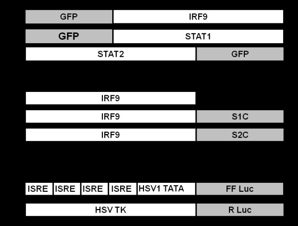
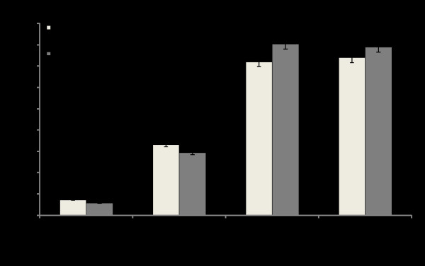
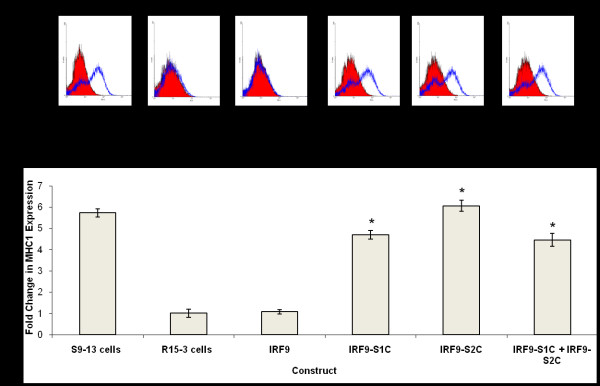
Interferon alpha (IFN-α) binds to a cell surface receptor that activates the Jak-Stat signaling pathway. A critical component of this pathway is the translocation of interferon stimulated gene factor 3 (a complex of three proteins Stat1, Stat2 and IRF9) to the nucleus to activate antiviral genes. A stable sub-genomic replicon cell line resistant to IFN-α was developed in which the nuclear translocation of Stat1 and Stat2 proteins was prevented due to the lack of phosphorylation; whereas the nuclear translocation of IRF9 protein was not affected. In this study, we sought to overcome defective Jak-Stat signaling and to induce an antiviral state in the IFN-α resistant replicon cell line by developing a chimera IRF9 protein fused with the trans activating domain (TAD) of either a Stat1 (IRF9-S1C) or Stat2 (IRF9-S2C) protein. We show here that intracellular expression of fusion proteins using the plasmid constructs of either IRF9-S1C or IRF9-S2C, in the IFN-α resistant cells, resulted in an increase in Interferon Stimulated Response Element (ISRE) luciferase promoter activity and significantly induced HLA-1 surface expression. Moreover, we show that transient transfection of IRF9-S1C or IRF9-S2C plasmid constructs into IFN-α resistant replicon cells containing sub-genomic HCV1b and HCV2a viruses resulted in an inhibition of viral replication and viral protein expression independent of IFN-α treatment. The results of this study indicate that the recombinant fusion proteins of IRF9-S1C, IRF9-S2C alone, or in combination, have potent antiviral properties against the HCV in an IFN-α resistant cell line with a defective Jak-Stat signaling.


Figures

Figure 1
Figure 1
Structure of plasmid constructs. (A) The plasmid constructs containing the full-length clones of IRF9, Stat1 and Stat2 fused with GFP either at the N-terminal or C-terminal ends. (B) Full length IRF9 clone fused with the TAD of either Stat1 or Stat2. IRF9-S1C contains the full length IRF9 molecule fused in frame with the 38 C-terminal amino acids of Stat1. IRF9-S2C also contains the full length IRF9 molecule fused in frame with the 104 C-terminal amino acids of Stat2. (C) The ISRE reporter plasmid containing four copies of ISRE sequences positioned upstream of the HSV thymidine kinase (TK) promoter TATA box that drives the expression of FL. The bottom construct, used as a transfection control contains RL downstream of a HSV TK promoter.
Figure 2
Figure 2
Nuclear translocation of STAT-GFP constructs. The S9-13 and R15-3 cell lines were transfected with IRF9, Stat1, or Stat2 GFP fusion constructs (-) and (+) IFN-α (1,000 IU/ml) and their nuclear translocation was observed under a confocal microscope. The images are represented as the superimposition of Green Fluorescent Protein (green), To-Pro3 633 (far red), and the differential interference contrast images (DIC) (gray scale). Fluorescence green and red microscopic picture of the same area were taken and superimposed using Abode Photoshop. Left panel shows high resolution picture showing that the IRF9-GFP fusion protein translocates to the nucleus of both S9-13 and R15-3 cells in an IFN-α independent manner. Middle panel shows stat1-GFP fusion protein efficiently localized to the nucleus of S9-13 cells within 30 minutes after IFN-α treatment. Stat1-GFP was unable to localize to the nucleus in R15-3 cells. Right panel shows stat2-GFP efficiently localized to the nucleus of S9-13 cells within 30 minutes after IFN-α treatment. Stat2-GFP was unable to localize to the nucleus in R15-3 cells.
Figure 3
Figure 3
Phosphorylation of Stat1-GFP and Stat2-GFP fusion proteins in S9-13 and R15-3 cells determined by co-immunoprecipitation. S9-13 and R15-3 cells were transfected with either Stat1-GFP or Stat2-GFP. After 48 hours IFN-α (1,000 IU/ml) was added. Protein lysates were prepared, subjected to immunoprecipitation with a GFP antibody, and western blot analysis was performed using a p-STAT1 antibody.
Figure 4
Figure 4
Analysis of ISRE-luciferase promoter activation due to intracellular expression of IRF9-Stat fusion proteins. R15-3 cells were transfected with IRF9, IRF9-S1C or IRF9-S2C plasmid and cultured (-) and (+) IFN-α (1,000 IU/ml). At 48 hours FL and RL activity were measured. Values are expressed in RL normalized units and error bars represent the SEM from three experiments. The Student's t-test was used to compare IRF9 with and without IFN to IRF9-S1C, IRF9-S2C and IRF9-S1C plus IRF9-S2C with and without IFN. Values <.05 were considered significant.
Figure 5
Figure 5
Intracellular expression of IRF9-S1 or IRF9-S2C induced HLA-1 surface expression in the R15-3 cells. S9-13 and R15-3 cells were transfected and cultured (-) and (+) IFN-α (1,000 IU/ml). After 48 hours, HLA-1 surface expression was quantified by flow cytometry. (A) Shows the HLA-1 surface expression in the IRF9-Stat fusion transfected R15-3 cells. The red cell population represents IFN-α naïve cells or untransfected cells and the blue cell population represents IFN-α treated or transfected cells. IRF9-S1C, IRF9-S2C, and IRF9-S1C plus IRF9-S2C transfected R15-3 cells demonstrated significant increases in HLA-1 surface expression. (B) Each value represents the mean fluorescence intensity from six experiments. The student's t-test was used to compare the fold increase of transfected R15-3 cells to untransfected R15-3 cells plus IFN-α. Asterisk (*) indicates transfected R15-3 cells with significantly different HLA-1 surface expression relative to untransfected R15-3 cells. Significance was considered at p-values < 0.05.
Figure 6
Figure 6
Ribonuclease protection assay and Real-time RT-PCR of sub-genomic HCV 1b RNA. (A) Intracellular expression of IRF9-S1C and IRF9-S2C or both in R15-3 cells inhibits HCV negative strand RNA detection by RPA. R15-3 cells were transfected treated (-) and (+) IFN-α (1,000 IU/ml), and total RNA was isolated at 72 hours post-transfection. The upper panel shows the RPA results of HCV negative strand detection. The bottom panel shows GAPDH mRNA levels by RPA. (B) Real-time RT-PCR was performed to quantify HCV RNA in R15-3 transfected cells. Cellular RNA was isolated at 72 hours, retrotranscribed, and assayed by real time PCR. S9-13 and R15-3 cells (+) and (-) IFN-α were used as (+) and (-) controls, respectively. Error bars represent SD. Asterisk (*) indicates transfected R15-3 cells with significantly lower HCV RNA relative to untransfected R15-3 cells treated with IFN. Significance was considered at p-values < 0.05.
Figure 7
Figure 7
Immunostaining of HCV NS3 and Western blot of HCV NS5B proteins. (A) The antiviral activity of the IRF9-Stat fusion constructs in R15-3 cells containing HCV 1b sub-genomic RNA HCV. At 48 hours post-transfection the cells were mounted onto a glass slide, stained for HCV NS3 protein and counterstained with hematoxylin. Panel i and ii: Huh-7 cells (-) and (+) IFN-α (1,000 IU/ml). Panel iii and iv: R15-3 cells (-) and (+) IFN-α. Panel v: IRF9 transfected R15-3 cells Panel vi: IRF9-S1C transfected R15-3 cells Panel vii: IRF9-S2C transfected R15-3 cells Panel viii: IRF9-S1C plus IRF9-S2C transfected R15-3 cells. (B) Upper panel: HCV NS5B expression levels in R15-3 transfected cells at 72 hours post-transfection. Lower panel: Shows β-actin protein expression levels using equal amounts of protein lysate.
Figure 8
Figure 8
The antiviral activity of the IRF9-Stat fusion constructs in an IFN resistant cell line containing HCV 2a sub-genomic RNA (R4-GFP). (A) GFP expression levels in R4-GFP cells transfected with IRF9, IRF9-S1C and IRF9-S2C at 72 hrs post-transfection. (B) Flow cytometric analysis of mean fluorescence intensity of R4-GFP transfected cells at 72 hrs. S3-GFP and R4-GFP cells treated with and without IFN-α were used as controls. The error bars represent the SEM from three experiments. The Student's t-test was used to compare R4-GFP transfected cells to R4-GFP cells plus IFN-α. P-values < 0.05 were considered significant.
Figure 9
Figure 9
MTT Assay of IRF9-Stat fusion constructs in resistant cell line. R15-3 cells were transfected with IRF9 constructs and at 72 hours post-transfection cell viability was determined by the MTT assay. Each value represents experiments performed in triplicate. Error bars represent SEM.
Figure 10
Figure 10
Intracellular expression of IRF9-Stat fusion constructs induced eIF2α phosphorylation in the R15-3 cell line at 72 hours post-transfection. Upper panel: Western blot of p-eIF2α in R15-3 cells transfected with different IRF9 plasmids in the absence and presence of IFN-α (1,000 IU/ml). Bottom Panel: β-actin protein expression levels.

Similar articles

Cited by

References

    1. Alter MJ. Epidemiology of hepatitis C virus infection. World J Gastroenterol. 2007;13:2436–2441. - PMC - PubMed
    1. Sy T, Jamal M. Epidemiology of Hepatitis C virus infection. Int J Med Sci. 2006;3:41–46. - PMC - PubMed
    1. Post J, Ratnarajah S, Lloyd AR. Immunological determinants of the outcomes from primary hepatitis C infection. Cell Mol Life Sci. 2009;66:733–756. doi: 10.1007/s00018-008-8270-4. - DOI - PMC - PubMed
    1. Te HS, Jensen DM. Epidemiology of hepatitis B and C viruses: a global overview. Clin Liver Dis. 2010;14:1–21. doi: 10.1016/j.cld.2009.11.009. - DOI - PubMed
    1. Fried MW, Shiffman ML, Reddy KR, Smith C, Marinos G, Gonçales FL Jr, Häussinger D, Diago M, Carosi G, Dhumeaux D, Craxi A, Lin A, Hoffman J, Yu J. Peginterferon alfa-2a plus ribavirin for chronic hepatitis C virus infection. N Engl J Med. 2002;347:975–982. doi: 10.1056/NEJMoa020047. - DOI - PubMed

Publication types

MeSH terms