. 2010 Nov;137(21):3569-79.
doi: 10.1242/dev.050310.

Adult epidermal Notch activity induces dermal accumulation of T cells and neural crest derivatives through upregulation of jagged 1

Affiliations

Adult epidermal Notch activity induces dermal accumulation of T cells and neural crest derivatives through upregulation of jagged 1

Carrie A Ambler et al. Development. 2010 Nov.

Abstract

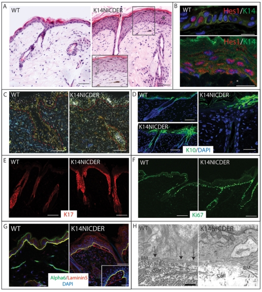
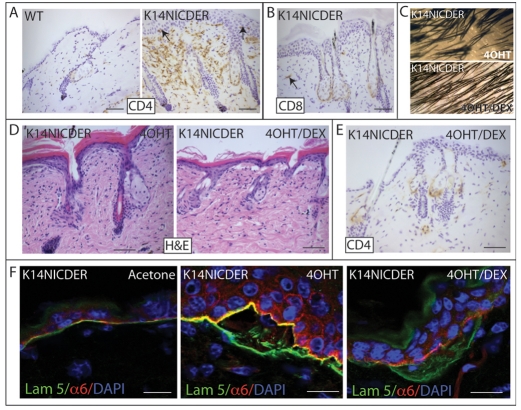
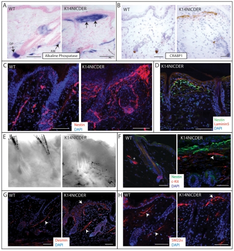
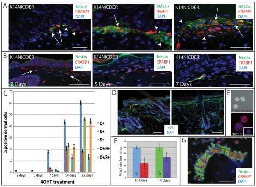
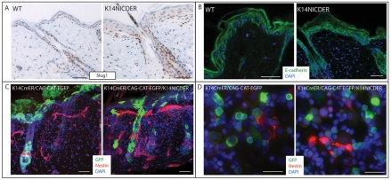
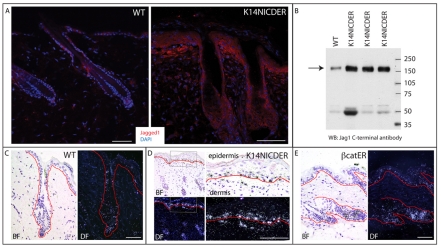
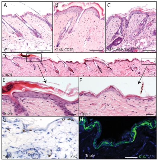
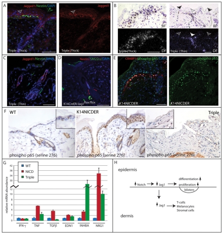
Notch signalling regulates epidermal differentiation and tumour formation via non-cell autonomous mechanisms that are incompletely understood. This study shows that epidermal Notch activation via a 4-hydroxy-tamoxifen-inducible transgene caused epidermal thickening, focal detachment from the underlying dermis and hair clumping. In addition, there was dermal accumulation of T lymphocytes and stromal cells, some of which localised to the blisters at the epidermal-dermal boundary. The T cell infiltrate was responsible for hair clumping but not for other Notch phenotypes. Notch-induced stromal cells were heterogeneous, expressing markers of neural crest, melanocytes, smooth muscle and peripheral nerve. Although Slug1 expression was expanded in the epidermis, the stromal cells did not arise through epithelial-mesenchymal transition. Epidermal Notch activation resulted in upregulation of jagged 1 in both epidermis and dermis. When Notch was activated in the absence of epidermal jagged 1, jagged 1 was not upregulated in the dermis, and epidermal thickening, blister formation, accumulation of T cells and stromal cells were inhibited. Gene expression profiling revealed that epidermal Notch activation resulted in upregulation of several growth factors and cytokines, including TNFα, the expression of which was dependent on epidermal jagged 1. We conclude that jagged 1 is a key mediator of non-cell autonomous Notch signalling in skin.


Figures

Fig. 1.
Fig. 1.
Characterisation of K14NICDER transgenic mice. (A) Haematoxylin and Eosin-stained back skin sections of 4OHT-treated wild-type (WT) and K14NICDER littermates. Inset shows a higher magnification view of epidermis and underlying dermis. Epidermal layers are indicated. B, basal; S, spinous; G, granular; C, cornified. (B) Back skin of 4OHT-treated wild-type (WT) and K14NICDER littermates stained with antibodies to Hes1 (red) and keratin 14 (green). Asterisk marks a Hes1-positive dermal cell. (C) RNA in situ hybridisation with radiolabelled antisense Hes1 probe on back skin sections from 4OHT-treated wild type (WT) and K14NICDER mice. Epidermal-dermal boundary is indicated by a broken red line. (D-G) Back skin of 4OHT-treated wild-type (WT) and K14NICDER littermates mice stained with antibodies to keratin 10 (green, D), keratin 17 (red, E), Ki67 (green, F), α6 integrin subunit (green, G) and laminin 5 (red, G). (H) Transmission electron micrographs of the epidermal basement membrane zone in wild type (WT) and K14NICDER littermates. Arrows indicate hemidesmosomes. BM, basement membrane; Coll, collagen fibres. (B,D,G) Sections were DAPI counterstained (blue). Mice were 4OHT-treated for 14 days (C-F), 15 days (B) or 21 days (A,G-H). Scale bars: 1 μm in H; 10 μm in B,G (inset); 25 μm in D (right panel), G (left and right panels); 50 μm in A,C,D (left panels), E,F.
Fig. 2.
Fig. 2.
Notch-induced skin inflammation. (A,B) Back skin sections of 4OHT-treated wild-type (WT) and K14NICDER littermates stained with antibodies to CD4 (A) and CD8 (B) (brown). Arrows indicate T cells in epidermis and dermis. (C) Macroscopic phenotype of K14NICDER transgenic mice treated with 4OHT or 4OHT + dexamethasone (4OHT/DEX). (D-F) Back skin sections of K14NICDER mice treated with acetone, 4OHT or 4OHT and dexamethasone (4OHT/DEX). Sections were stained with Haematoxylin and Eosin (D) or labeled with antibodies to CD4 (E), α6 integrin subunit (red, F) and laminin 5 (green, F). Nuclei were counterstained with Haematoxylin (A,B,E) or DAPI (blue, F). Mice were injected with dexamethasone or saline for 24 days and treated with 4OHT or acetone for 21 days. Scale bars: 50 μm in A,B,D,E; 10 μm in F.
Fig. 3.
Fig. 3.
Epidermal Notch activity induces accumulation of stromal cells in the upper dermis. (A-H) 4 μm (A,B,D,F-H) or 150 μm (C,E) back skin sections of 4OHT-treated wild-type (WT) and K14NICDER littermates were analysed. (A) Alkaline phosphatase activity (blue). Locations of dermal papilla (DP) and arrector pili muscle (AM) are indicated with arrows. Arrows in the right-hand panel show stromal cell accumulation. (B-D,F-H) Immunolabelling with antibodies to Crabp1 (B, brown), nestin (C, red; D, green; F, green), laminin 5 (D, red), Kit (F, red), desmin (G, red) and SM22α (H, red). Arrowheads in G,H indicate arrector pili muscle; arrows in G,H indicate stromal cells at the epidermal/dermal junction. Asterisk in F indicates a Kit-positive mast cell. (E) Brightfield image showing dermal melanocytes in K14NICDER transgenic skin. Sections were counterstained with Fast Red (A), Haematoxylin (B) or DAPI (C,D,F-H, blue). Mice were treated with 4OHT for 14 days. Scale bars: 50 μm.
Fig. 4.
Fig. 4.
Kinetics of appearance of Notch-induced stromal cells. (A,B,D) Back skin sections of 4OHT-treated K14NICDER mice stained with antibodies to nestin (A, green or red; B, green), Crabp1 (A,B; red), Sm22α (A, green) and p75 (D, green). (A) Arrows indicate double-labeled cells; arrowheads indicate single-labeled cells. (B) Arrows indicate Crabp1-positive cells. (C) Percentage Crabp1 (C), nestin (N), Sm22α (S) single-positive cells and Crabp1/nestin and Crabp1/SM22α double-positive cells at the epidermal-dermal boundary (n≥15 samples). (E) Skin-derived neurospheres 7 days after seeding, viewed by phase contrast (top) or anti-nestin labelling (bottom, red) and counterstained with DAPI (blue). Inset: labelling with secondary antibody alone. (F) Percentage sphere formation by cells from wild-type (WT) and K14NICDER (TG) littermates treated with 4OHT for 10 or 14 days. (G) Cytospin preparation of dermal cells isolated from 4OHT-treated K14NICDER skin stained with antibodies to nestin (green) and Crabp1 (red). Sections and cells were counterstained with DAPI (blue; A,B,D,E,G). Mice were treated with 4OHT for 2 days (B), 5 days (B), 7 days (B), 10 days (A, left panel; G) or 14 days (A, middle and right panels; D). Scale bars: 25 μm in A,B,G; 50 μm in D. Data are mean±s.e.m.
Fig. 5.
Fig. 5.
Nestin-positive dermal cells are not of epidermal origin. (A,B) Back skin sections of 4OHT-treated wild-type (WT) and K14NICDER littermates stained with antibodies to Slug 1 (A, brown) and E-cadherin (B, green). (C,D) Back skin sections (150 μm) (C) and cytospin preparations (D) of cells isolated from 4OHT-treated K14CreER/CAG-CAT-EGFP mice and K14CreER/CAG-CAT-EGFP/K14NICDER mice stained with antibodies to GFP (green) and nestin (red) with DAPI nuclear counterstain (blue). Mice were 4OHT-treated for 14 days (A-D). Scale bars: 20 μm in D; 50 μm in A-C.
Fig. 6.
Fig. 6.
Notch induces Jag1 in the epidermis and dermis. (A,C-E) Back skin of wild-type (A,C), K14NICDER (A,D) and K14-ΔNβcateninER (βcatER; E) mice. (A) Jagged 1 (red) immunolabelling with DAPI counterstain (blue). (B) Western blot of protein lysates from skin of wild-type (WT) and K14NICDER mice probed with anti-jagged 1. Each lane contains protein from a different mouse. Arrow indicates position of jagged 1 protein. Lower molecular mass bands are nonspecific and serve as loading controls. Molecular mass markers (kDa) are indicated. (C-E) RNA in situ hybridisation using jagged 1 radiolabelled antisense probe. Corresponding brightfield (BF) and darkfield (DF) panels show same field. Red lines mark the epidermal/dermal boundary. The right-hand panels in D are higher magnification views of the boxed region in the left-hand panels. Mice were treated with 4OHT for 21 days (A,B); 10 days (C,D) or 7 days (E). Scale bars: 50 μm.
Fig. 7.
Fig. 7.
Jag1 is required for Notch-induced skin phenotype. (A-F) Haematoxylin and Eosin-stained sections of back skin of wild type (A, WT), K14NICDER (B), K14CreER/Jag1flox/flox (C) and K14CreER/Jag1flox/flox/K14NICDER (D-F, triple) mice treated with 4OHT for 10 days. In triple transgenics, areas of normal (F) and thickened (E) epidermis were found. (E,F) Higher magnifications views of boxed regions in D. (G,H) Back skin sections of triple transgenics in areas of normal thickness stained with antibodies to Ki67 (G, brown) and K10 (H, green). Sections were counterstained with Haematoxylin (G) or DAPI (H). Scale bars: 100 μm in A-D; 25 μm in E-H.
Fig. 8.
Fig. 8.
Relationship between jagged 1 expression and Notch-induced stromal cells. (A-F) K14CreER/Jag1flox/flox/K14NICDER (triple) skin with either increased (thick) or normal (thin) epidermal thickness (A-C) and K14CreER/Jag1flox/flox skin (D) were 4OHT-treated for 10 days. (E,F) Wild-type (WT), K14NICDER and triple (thin) skin was 4OHT-treated for 14 days. (A,C-F) Sections were stained with antibodies to nestin (A,C, green; D, red), jagged 1 (A,C, red), SM22α (D, green), phosphorylated p65 (serine 276; E, green; F, brown) and Crabp1 (E, red) with DAPI (A,C-E, blue) or Haematoxylin (F) counterstain. Both panels in A and E show same field. Arrow in A marks jagged 1 staining. Insets in F show higher magnification views. (B) RNA in situ hybridisation using a radiolabelled jagged 1 antisense probe. Corresponding brightfield (BF, top) and darkfield (DF, bottom) panels show same field. In B, black arrows indicate interfollicular epidermis and the white arrows indicate hair follicle infundibulum. Scale bars: 50 μm. (G) Quantitative polymerase chain reaction of cDNA from 4OHT-treated wild-type (WT), K14NICDER (NICD) and K14CreER/Jag1flox/flox/K14NICDER (Triple) epidermis. Results (mean±s.e.m.) are expressed relative to wild type (=1) for each TAQman probe. (H) Schematic summary of results. Epidermal Notch activation results in increased expression of jagged 1 in epidermis and dermis, which contributes to the epidermal changes and accumulation of cells in the dermis.

References

    1. Aho S. (2004). Soluble form of Jagged1: unique product of epithelial keratinocytes and a regulator of keratinocyte differentiation. J. Cell Biochem. 92, 1271-1281 - PubMed
    1. Ambler C. A., Watt F. M. (2007). Expression of Notch pathway genes in mammalian epidermis and modulation by beta-catenin. Dev. Dyn. 236, 1595-1601 - PubMed
    1. Amoh Y., Li L., Katsuoka K., Penman S., Hoffman R. M. (2005). Multipotent Nestin-positive, keratin-negative hair-follicle bulge stem cells can form neurons. Proc. Natl. Acad. Sci. USA 102, 5530-5534 - PMC - PubMed
    1. Blanpain C., Lowry W. E., Pasolli H. A., Fuchs E. (2006). Canonical notch signaling functions as a commitment switch in the epidermal lineage. Genes Dev. 20, 3022-3035 - PMC - PubMed
    1. Botchkarev V. A., Eichmuller S., Johansson O., Paus R. (1997). Hair cycle-dependent plasticity of skin and hair follicle innervation in normal murine skin. J. Comp. Neurol. 386, 379-395 - PubMed

Publication types

MeSH terms