. 2010 Nov 1;123(Pt 21):3734-44.
doi: 10.1242/jcs.070466. Epub 2010 Oct 12.

Polypurine-repeat-containing RNAs: a novel class of long non-coding RNA in mammalian cells

Affiliations

Polypurine-repeat-containing RNAs: a novel class of long non-coding RNA in mammalian cells

Ruiping Zheng et al. J Cell Sci. .

Abstract

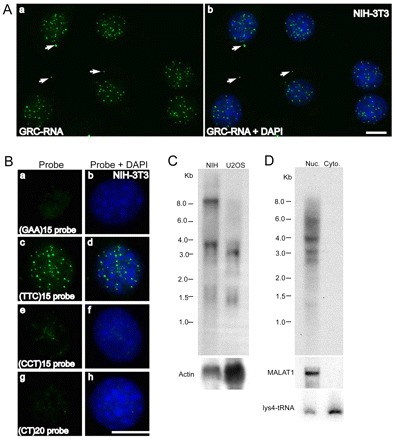
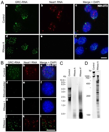
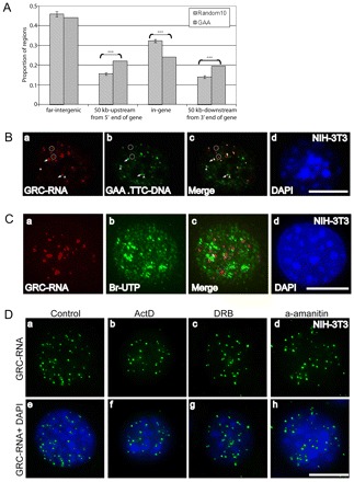
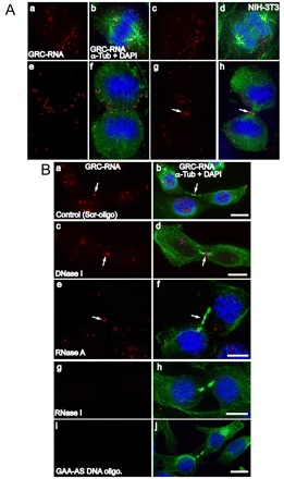
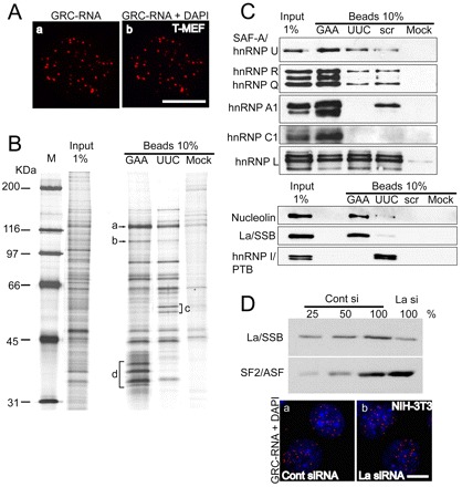
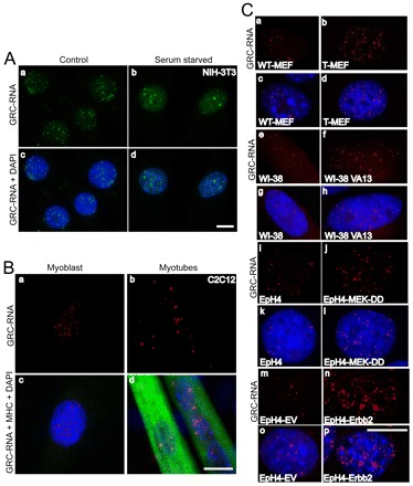
In higher eukaryotic cells, long non-protein-coding RNAs (lncRNAs) have been implicated in a wide array of cellular functions. Cell- or tissue-specific expression of lncRNA genes encoded in the mammalian genome is thought to contribute to the complex gene networks needed to regulate cellular function. Here, we have identified a novel species of polypurine triplet repeat-rich lncRNAs, designated as GAA repeat-containing RNAs (GRC-RNAs), that localize to numerous punctate foci in the mammalian interphase nuclei. GRC-RNAs consist of a heterogeneous population of RNAs, ranging in size from ~1.5 kb to ~4 kb and localize to subnuclear domains, several of which associate with GAA.TTC-repeat-containing genomic regions. GRC-RNAs are components of the nuclear matrix and interact with various nuclear matrix-associated proteins. In mitotic cells, GRC-RNAs form distinct cytoplasmic foci and, in telophase and G1 cells, localize to the midbody, a structure involved in accurate cell division. Differentiation of tissue culture cells leads to a decrease in the number of GRC-RNA nuclear foci, albeit with an increase in size as compared with proliferating cells. Conversely, the number of GRC-RNA foci increases during cellular transformation. We propose that nuclear GRC-RNAs represent a novel family of mammalian lncRNAs that might play crucial roles in the cell nucleus.


Figures

Fig. 1.
Fig. 1.
GRC-RNA is a member of the nrRNA family of lncRNAs. (A) RNA-FISH using an AK082015 probe (green) in NIH-3T3 cells revealed the punctate nuclear localization of GRC-RNA. In addition to nuclear foci, GRC-RNA also localized to a few cytoplasmic foci (arrows). Scale bar: 10 μm. (B) RNA-FISH using FITC-labeled (GAA)15 (a,b), (TTC)15 (c,d), (CCT)15 (e,f) and (CT)20 (g,h) single-stranded oligonucleotide probes in NIH-3T3 cells revealed that only the (TTC)15 probe hybridized to nuclear-enriched GRC-RNA (c,d). DNA was counterstained with DAPI (blue). Scale bar: 10 μm. (C) Northern blot using a 32P-labeled (TTC)15 probe from total RNA revealed that GRC-RNAs represent heterogeneous transcripts. Actin RNA was used as a loading control. (D) Northern blot using a 32P-labeled (TTC)15 probe in nuclear and cytoplasmic RNA fractions from EpH4 cells revealed nuclear enrichment of GRC-RNA. MALAT1 and lysine tRNA were used as markers for nuclear and cytoplasmic RNA fractionation, respectively.
Fig. 2.
Fig. 2.
GRC-RNA nuclear foci are sensitive to RNase but not DNase I treatment. (A) Co-RNA-FISH in NIH-3T3 cells for GRC-RNA (green; a,d) and Neat1 RNA (red; b,e) revealed that, unlike Neat1 RNA (e), GRC-RNA foci were insensitive to RNase A treatment (d). Merged images are shown in c and f, with chromatin stained with DAPI in blue. Scale bar: 10 μm. (B) Co-RNA-FISH in NIH-3T3 cells for GRC-RNA (green; a,d,g,j) and Neat1 RNA (red; b,e,h,k) revealed that both GRC-RNA and Neat1 RNA were degraded by Riboshredder (d–f) and RNase I (g–i) treatments, but not by DNase I (j–l) treatment. Chromatin was stained with DAPI in blue. Merged images are shown in c, f, i and l. Scale bar: 10 μm. (C) Northern blot using a 32P-labeled (TTC)15 probe from nuclear RNA of NIH-3T3 cells revealed that RNase A treatment resulted in the cleavage of GRC-RNA into smaller fragments. Longer exposure of the RNase-A-treated RNA blot (RNase A*) revealed the presence of RNase-A-resistant GRC-RNA. (D) Control and RNase-A-treated nuclear RNA were run on an acrylamide gel and hybridized with a 32P-labeled (TTC)15 probe.
Fig. 3.
Fig. 3.
GAA-repeats are preferentially enriched in the 5′ and 3′ regulatory regions of genes. (A) GAA-repeats are preferentially enriched in the 5′ and 3′ regulatory regions of genes. Based on the Refseq gene definition from the UCSC genome browser, the mouse genome is divided into four parts: the gene region (from start to end), 50 kb upstream of the gene 5′ end, 50 kb downstream of the gene 3′ end, and others called ‘far-intergenic regions’. The distribution of 1142 GAA-enriched regions (with at least 7 continuous GAAs and at least 300 bp of A/G stretch) in each of these four parts is shown by the gray bar. The significance of enrichment of GAA-repeats in each part is estimated based on the distribution of randomly selected genomic DNA regions (10 times) with same amount in each chromosome and same length distribution as the GAA fragments. (***P<0.001). The P-values are: 50 kb up-gene=0.00007; in gene=0.00002; and 50 kb down-gene=0.0006. (B) GRC-RNA foci associate with GAA.TTC-repeat DNA elements: RNA-FISH using a (TTC)15 (red; a,c) probe followed by DNA-FISH using a (GAA)15 probe (green; b,c) revealed association of GRC-RNA with GAA.TTC-repeat DNA elements in the genome. Arrows show a few of the representative GRC-RNA foci that colocalize with GAA.TTC-repeats; open circles represent the GRC-RNA foci that do not associate with GAA.TTC-repeats; arrowheads represent a few of the GAA.TTC-repeat DNA regions that do not associate with GRC-RNA. (C) GRC-RNA foci do not represent highly active transcription sites: RNA-FISH using a (TTC)15 (red; a,c) probe coupled with in vivo Br-UTP incorporation assays (5 minutes, Br-UTP pulse; green; b,c) revealed weak to no significant overlap between GRC-RNA foci and actively transcribing sites (c). (D) GRC-RNA foci contains stable nuclear RNAs: RNA-FISH for GRC-RNA in control (a,e) and RNA polymerase II inhibitor-treated NIH-3T3 cells [(actinomycin D for 1 hour; 5 μg/ml; b,f), (DRB for 3 hours; 32 μg/ml: c,g), (α-amanitin for 6 hours; 50 μg/ml: d,h)] showed a similar number of nuclear foci. Chromatin was stained with DAPI (blue). Scale bars: 10 μm.
Fig. 4.
Fig. 4.
GRC-RNAs form cytoplasmic foci in mitotic cells and localize to the midbody during cell division. (A) Co-RNA-FISH for GRC-RNA (red) and immunostaining for α-tubulin (green) in mitotic NIH-3T3 cells [(metaphase; a,b), (anaphase; c,d), (early telophase; e,f), (late telophase; g,h)] revealed the presence of GRC-RNA in cytoplasmic foci. The arrows in g and h show the presence of GRC-RNA in the midbody. (B) Co-RNA FISH for GRC-RNA (red) and immunostaining for α-tubulin (green) was conducted in early G1 cells that were treated with scrambled DNA oligonucleotide (control; a,b), DNase I (c,d), RNase A (e,f), RNase I (g,h) or GAA-antisense DNA oligonucleotides (i,j). GRC-RNA showed midbody localization (arrows) in control, DNase-I- and RNase-A-treated cells. However, RNase I treatment (g,h) and GAA-antisense DNA oligonucleotide transfection (i,j) resulted in the loss of GRC-RNA signal. The chromatin is stained with DAPI (blue). Scale bars: 10 μm.
Fig. 5.
Fig. 5.
GRC-RNA is associated with the nuclear matrix. (A) GRC-RNA-FISH in nuclear matrix preparations from transformed MEFs revealed the presence of GRC-RNA nuclear foci (a,b). The chromatin was stained with DAPI (blue). The absence of DAPI staining in b is due to DNase I treatment, which degrades most of the nuclear DNA. (B) Isolation of the GRC-RNA binding protein complex. RNA affinity chromatography was performed as described in the Materials and Methods, using affinity matrices containing different RNA sequences [(GAA)15, (UUC)15 and scrambled (scr) RNA oligonucleotides]. For identification, specific bands that showed enrichment were excised, digested with trypsin and subjected to mass spectrometry. a-d represent some of the bands that were excised and analyzed by mass spectrometry (a: hnRNP U; b: nucleolin; c: hnRNP I; d: hnRNP A1, A2/B1, hnRNP C1). (C) Western blot analysis of GAA- and UUC-RNA affinity-purified samples using antibodies against proteins that were identified by mass spectrometry. (D) Depletion of La/SSB in NIH-3T3 cells does not influence the nuclear distribution of GRC-RNA. Immunoblot analysis using La-antibody in cell extracts revealed >75% depletion of La protein in La-siRNA-treated cell extracts. GRC-RNA FISH (red) in control (a) and La/SSB-siRNA-treated (b) NIH-3T3 cells showed similar distributions of GRC-RNA. The chromatin is stained with DAPI. Scale bars: 10 μm.
Fig. 6.
Fig. 6.
GRC-RNA nuclear foci number is altered upon serum starvation and during cellular transformation. (A) GRC-RNA-FISH (green) in untreated (a,c) and serum-starved (3 days: b,d) NIH-3T3 cells reveal a reduced number of GRC-RNA foci in serum-deprived cells. Scale bar: 10 μm. (B) GRC-RNA-FISH in myoblasts (a,c) and myotubes (b,d; differentiated by incubating the myoblasts in serum-free media) revealed a reduced number, but prominent, GRC-RNA foci in myotubes. Note that the myogenesis marker, myosin heavy chain (MHC) specifically stains differentiated myotubes (d) and not proliferating myoblasts (c). Chromatin was stained with DAPI. Scale bar: 10 μm. (C) GRC-RNA-FISH (red) in WT-MEF (a,c), transformed MEF (b,d), WI38 (e,g), WI38 VA13 (f,h), WT-EpH4 (i,k), empty vector (EV)-transfected EpH4 (m,o), transformed constitutively active MEKDD (j-l) or Erbb2 (n,p) EpH4 cell lines revealed increased numbers of GRC-RNA foci in the transformed highly proliferating cell lines (SV-40 T-antigen-transformed MEF, WI38 VA13 human fibroblast, EpH4-MEKDD and EpH4-Erbb2) compared with their wild-type counterparts (WT-MEF, WI38 primary fibroblast and EpH4 and EpH4-EV mammary cells). DNA was counterstained with DAPI. Scale bar: 10 μm.

References

    1. Alexandre S., Rast C., Nguyen-Ba G., Vasseur P. (2000). Detection of apoptosis induced by topoisomerase inhibitors and serum deprivation in syrian hamster embryo cells. Exp. Cell Res. 255, 30-39. - PubMed
    1. Amaral P. P., Dinger M. E., Mercer T. R., Mattick J. S. (2008). The eukaryotic genome as an RNA machine. Science 319, 1787-1789. - PubMed
    1. Bidichandani S. I., Ashizawa T., Patel P. I. (1998). The GAA triplet-repeat expansion in Friedreich ataxia interferes with transcription and may be associated with an unusual DNA structure. Am. J. Hum. Genet. 62, 111-121. - PMC - PubMed
    1. Braidotti G., Baubec T., Pauler F., Seidl C., Smrzka O., Stricker S., Yotova I., Barlow D. P. (2004). The Air noncoding RNA: an imprinted cis-silencing transcript. Cold Spring Harbor Symp. Quant. Biol. 69, 55-66. - PMC - PubMed
    1. Brenet F., Dussault N., Borch J., Ferracci G., Delfino C., Roepstorff P., Miquelis R., Ouafik L. (2005). Mammalian peptidylglycine alpha-amidating monooxygenase mRNA expression can be modulated by the La autoantigen. Mol. Cell. Biol. 25, 7505-7521. - PMC - PubMed

Publication types