. 2011 Feb;60(2):177-88.
doi: 10.1136/gut.2009.202283. Epub 2010 Oct 12.

SHIP deficiency causes Crohn's disease-like ileitis

Affiliations

SHIP deficiency causes Crohn's disease-like ileitis

William G Kerr et al. Gut. 2011 Feb.

Abstract

Background: Inflammatory bowel disease (IBD) can arise from genetic mutations that compromise intestinal epithelial cell integrity or immune regulation. SHIP has previously been shown to play a pivotal role in limiting the number of immunoregulatory cells and their function.

Aim: To determine whether SHIP plays a pivotal role in control of immune tolerance in the gut mucosa.

Methods: Gastrointestinal pathology was assessed in three separate strains of SHIP-deficient mice and their respective wild-type (WT) littermates. Gastrointestinal pathology was analysed in SHIP-deficient hosts reconstituted with WT haematopoietic cell grafts, and WT hosts reconstituted with SHIP-deficient haematopoietic cell grafts including whole splenocytes, purified T cells or natural killer (NK) cells. Major immune cell populations were also analysed in the small intestine of SHIP-deficient mice and WT controls.

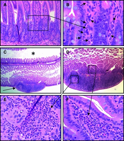
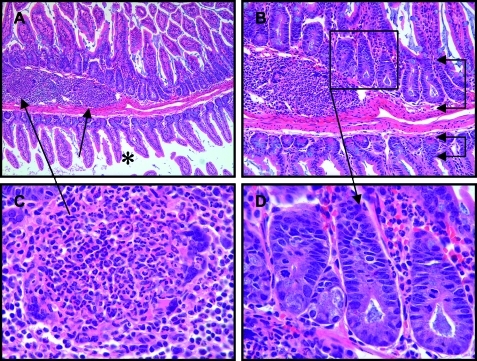
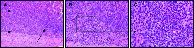
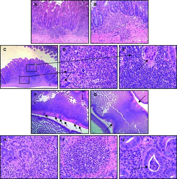
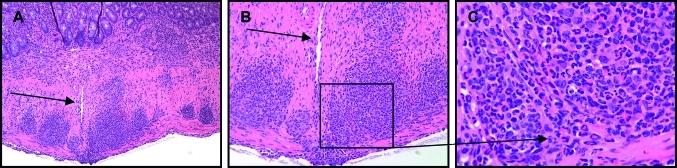
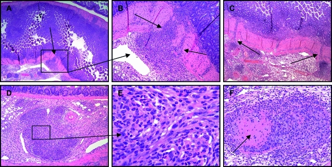
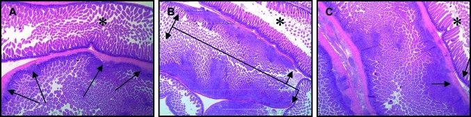
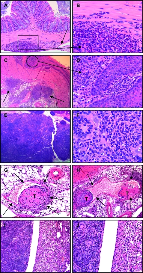
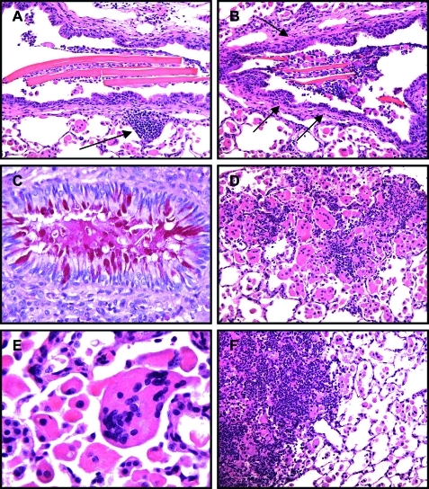
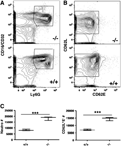
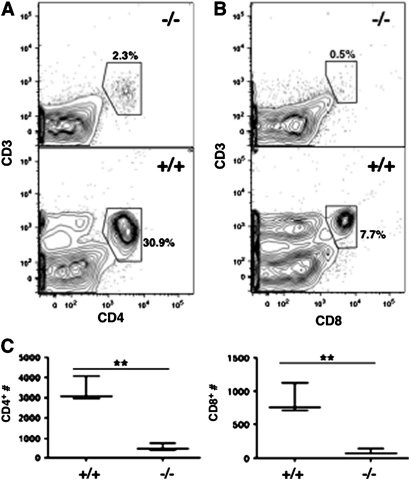
Results: SHIP-deficient mice developed segmental, transmural pyo-granulomatous ilietis that recapitulated classical features of Crohn's disease enteric pathology. Analysis of haematopoietic chimeras showed that WT bone marrow reconstitution of SHIP⁻/⁻ hosts corrects ileitis. Reconstitution with SHIP⁻/⁻ splenocytes transferred ileitis to WT hosts. Adoptive transfer of purified SHIP⁻/⁻ T cells or NK cells to WT hosts did not transfer ileitis. There was a paucity of both CD4 and CD8 T cells in the small intestines of SHIP-deficient mice; however, neutrophil numbers were significantly increased.

Conclusions: SHIP plays a pivotal role in immune function in the intestine; further scrutiny of this pathway in IBD patients is warranted. It is proposed that SHIP-deficient ileitis results from a local deficit in mucosal T cell immunity that promotes a damaging granulocyte-monocyte inflammation of the distal ileum.


Conflict of interest statement

Competing interests: None.

Figures

Figure 1
Figure 1
Grade 1 Crohn's disease (CD)-like ileitis of SHIP-deficient mice. Longitudinal sections through Swiss rolls of small intestine from SHIP-deficient mice with early manifestations of CD-like disease, interpreted as grade 1, with mild predominantly polymorphonuclear (PMN) leucocyte infiltration of the lamina propria (B, arrows). This PMN infiltration of the ileum was typically present in the mucosa overlying and within lymph nodules (C–F). Adjacent ileum and more proximal small intestine (C, asterisk) were unaffected with normal villous mucosal architecture. Higher magnifications show a predominantly PMN leucocyte infiltration of the villous lamina propria (F), and within the underlying lymph nodule, in this case accompanied by histiocytes and multinucleated giant cells (E).
Figure 2
Figure 2
Grade 2 Crohn's disease-like ileitis of SHIP-deficient mice was comprised of a moderate, mixed, predominantly polymorphonuclear leucocyte infiltration of the ileum lamina propria (D) and underlying lymph nodule (C), with attendant crypt hyperplasia (B, upper bracketed arrows) with elongation, crowding, and numerous mitotic figures (D), compared to the unaffected mucosa of the adjacent small intestine (A, asterisk) with normal villous architecture and typical crypt depth (B, lower bracketed arrows).
Figure 3
Figure 3
Grade 3 Crohn's disease-like ileitis of SHIP-deficient mice was comprised of a marked, mixed, often predominantly polymorphonuclear (PMN) inflammatory cell infiltration of the ileum with expansion of the mucosa and submucosa (A, bracketed arrows), loss of normal villous architecture, and crypt elongation (A, arrow). Higher magnifications of the affected ileum (B, C) showed a mixed, predominantly PMN leucocyte infiltration.
Figure 4
Figure 4
Grade 4 Crohn's disease-like ileitis of SHIP-deficient mice was comprised of a marked, mixed inflammatory cell infiltration of the ileum extending through the mucosa, submucosa, and into the tunica muscularis (A, B, C, F, G). Such inflammatory cell infiltrations varied in composition, were in some areas of affected ileum predominantly polymorphonuclear (D), while in other areas were predominantly mononuclear with lymphocytes, histiocytes and numerous multi-nucleated giant cells present (B, E). Grade 4 ileitis was typically sharply delimited aggregates of pyo-granulomatous inflammatory cell infiltrations, at times in rosary-bead arrays (F, arrows), that caused thickening of the bowel wall (G, arrows) and lumen narrowing. Multinucleated giant cells (H), and pyo-granulomas (I) were frequently present in affected segments, but crypt abscesses (J, arrow) were less frequently observed.
Figure 5
Figure 5
Grade 5 Crohn's disease-like ileitis of SHIP-deficient mice consisted of marked, mixed, transmural inflammatory cell infiltrations that extended to the serosa, some of which due to bowel wall thickening and stricture formation had developed fissures that penetrated into and ended blindly within the tunica muscularis (A; B, arrow).
Figure 6
Figure 6
Most SHIP-deficient mice presented with grade 6 Crohn's disease-like ileitis which consisted of marked, mixed, transmural inflammatory cell infiltrations that extended by way of fissures through the ileum serosa into the mesentery (A–C, arrows), the inflammation of which was comprised of mixed inflammatory cells and fibroblasts in aggregates and whirls (E), and pyo-granuloma, some with central necrosis (F, arrow).
Figure 7
Figure 7
Segmental nature of Crohn's disease-like ileitis of SHIP-deficient mice comprised of either discrete, discontinuous foci of inflammation (A, arrows), or broad bands of inflammation (B, bracketed arrows), each with expansion of the mucosa and submucosa, and thickening of the tunica muscularis (C, left arrow) compared to neighbouring unaffected segments (C, right arrow; A, B, C, asterisks) with normal villous architecture and bowel wall thickness.
Figure 8
Figure 8
Inflammation extended beyond the ileum in a few SHIP-deficient mice where mixed leucocyte infiltrations of the mesentery infiltrated into the colon wall (A, arrow; B), or the juncture of the oesophagus and stomach (C, arrows; D). Draining mesenteric lymph nodes were frequently enlarged and consisted of mixed inflammatory cells with numerous multinucleate cells (E, F). Rarely, inflammatory cellular thrombi (T) were present within mesenteric veins (G, H, arrows), and as emboli may have lodged in pulmonary microvasculature resulting in a granulomatous pulmonary consolidation comprised of mixed inflammatory cells with numerous multinucleate cells (I, J, left) that contrasted sharply with the eosinophilic crystalline pneumonia of neighbouring lobes (I, J, right) typical of SHIP-deficient mice.
Figure 9
Figure 9
SHIP-deficient mice develop an eosinophilic crystalline pneumonia that consists of large crystals in bronchiolar airways, foci of mixed inflammatory cell infiltrations (A, arrow), bronchiolar subepithelial fibrosis (B, arrows), hypertrophy and mucous metaplasia of bronchiolar epithelium (C), numerous alveolar macrophages and multinucleate cells (D, E), and infiltrating leucocytes resulting in patchy pulmonary consolidation (F, left).
Figure 10
Figure 10
Neutrophilia in SHIP-deficient small intestine. FACS detection of CD16/32+Ly6G+ neutrophils (A) and their CD62L/E surface staining (B) in small intestine of SHIP-deficient mice and wild-type (WT) littermates. (C) Absolute total CD16/32+Ly6G+ neutrophils and CD62L+ neutrophils in the small intestine of SHIP-deficient and WT mice as indicated. ***p<0.001.
Figure 11
Figure 11
Profound T cell deficit in SHIP-deficient small intestine. FACS detection of CD3+CD4+ (A) and CD3+CD8+ (B) T cells in the small intestine of SHIP-deficient mice and their wild-type (WT) littermates. (C) Absolute CD4 and CD8 T cell numbers in the small intestine of SHIP-deficient and WT mice as indicated. **p<0.01.

Comment in

References

    1. Sartor RB. Mechanisms of disease: pathogenesis of Crohn's disease and ulcerative colitis. Nat Clin Pract Gastroenterol Hepatol 2006;3:390–407 - PubMed
    1. Xavier RJ, Podolsky DK. Unravelling the pathogenesis of inflammatory bowel disease. Nature 2007;448:427–34 - PubMed
    1. Packey CD, Sartor RB. Interplay of commensal and pathogenic bacteria, genetic mutations, and immunoregulatory defects in the pathogenesis of inflammatory bowel diseases. J Intern Med 2008;263:597–606 - PubMed
    1. Cho JH. The genetics and immunopathogenesis of inflammatory bowel disease. Nat Rev Immunol 2008;8:458–66 - PubMed
    1. Wirtz S, Neurath MF. Mouse models of inflammatory bowel disease. Adv Drug Deliv Rev 2007;59:1073–83 - PubMed

Publication types

MeSH terms

Substances