. 2010 Dec 17;285(51):39597-608.
doi: 10.1074/jbc.M110.129718. Epub 2010 Oct 12.

Muscle wasting in aged, sarcopenic rats is associated with enhanced activity of the ubiquitin proteasome pathway

Affiliations

Muscle wasting in aged, sarcopenic rats is associated with enhanced activity of the ubiquitin proteasome pathway

Mikael Altun et al. J Biol Chem. .

Abstract

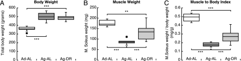
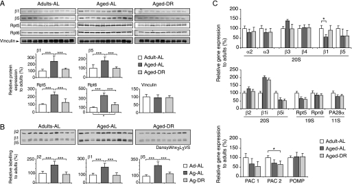
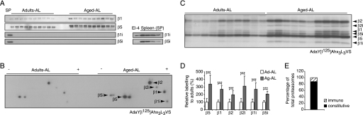
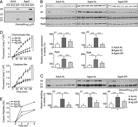
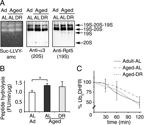
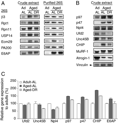
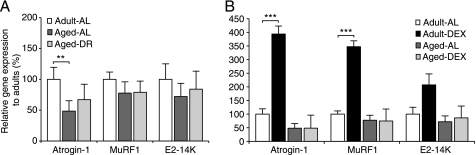
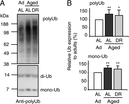
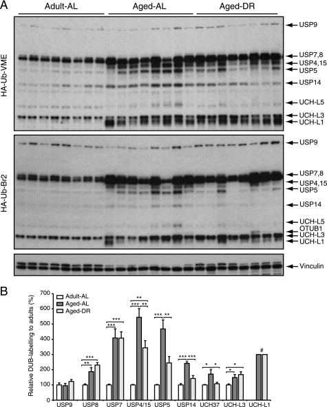
Among the hallmarks of aged organisms are an accumulation of misfolded proteins and a reduction in skeletal muscle mass ("sarcopenia"). We have examined the effects of aging and dietary restriction (which retards many age-related changes) on components of the ubiquitin proteasome system (UPS) in muscle. The hindlimb muscles of aged (30 months old) rats showed a marked loss of muscle mass and contained 2-3-fold higher levels of 26S proteasomes than those of adult (4 months old) controls. 26S proteasomes purified from muscles of aged and adult rats showed a similar capacity to degrade peptides, proteins, and an ubiquitylated substrate, but differed in levels of proteasome-associated proteins (e.g. the ubiquitin ligase E6AP and deubiquitylating enzyme USP14). Also, the activities of many other deubiquitylating enzymes were greatly enhanced in the aged muscles. Nevertheless, their content of polyubiquitylated proteins was higher than in adult animals. The aged muscles contained higher levels of the ubiquitin ligase CHIP, involved in eliminating misfolded proteins, and MuRF1, which ubiquitylates myofibrillar proteins. These muscles differed from ones rapidly atrophying due to disease, fasting, or disuse in that Atrogin-1/MAFbx expression was low and not inducible by glucocorticoids. Thus, the muscles of aged rats showed many adaptations indicating enhanced proteolysis by the UPS, which may enhance their capacity to eliminate misfolded proteins and seems to contribute to the sarcopenia. Accordingly, dietary restriction decreased or prevented the aging-associated increases in proteasomes and other UPS components and reduced muscle wasting.


Figures

FIGURE 1.
FIGURE 1.
Aged rats (30 months old) have much less muscle mass, but greater body weights than adult (4 months old), and dietary restriction decreases this loss of muscle. A, boxplots illustrate the body weights of 10 adults (Ad-AL) and 10 aged rats fed AL (Ag-AL) or 9 aged rats maintained on dietary restriction (Ag-DR), as described under “Materials and Methods.” B, soleus muscle weight (mg) at 4 and 30 (AL and DR) months. C, ratio between soleus weights (mg) and body weights (g). Box limits represent upper and lower quartile values and are separated by the median (crossbar within box). Maximum and minimum values, which are not defined as outliers (circles), are illustrated using error bars. Outliers are defined as values deviating from the quartile borders by more than 1.5 times the interquartile distance. ***, p < 0.001.
FIGURE 2.
FIGURE 2.
Proteasome content increases in muscles of aged but not in rats on dietary restriction. A, levels of the 20S subunits, β1 and β5, and the 19S components, Rpt5 and Rpt6, were measured by immunoblotting with Vinculin as loading control. B, 20S proteasomes were covalently modified in crude extracts with the active site-directed probe dansyl-Ahx3L3VS followed by immunoblotting with anti-dansyl antibody. Each lane in panels A or B represents 25 μg of crude extract from gastrocnemius muscles of 4-month-old (n = 10) and 30-month-old AL (n = 10) and DR (n = 9) rats. Densitometry data and statistical analysis of protein levels (A) and proteasome activity (B) are plotted relative to the 4-month-old AL group (set to 100%). Error bars indicate standard deviation. C, changes in mRNA levels of several proteasome subunits as well as factors involved in proteasome assembly were assayed by qPCR. ***, p < 0.001.
FIGURE 3.
FIGURE 3.
In aged muscles, immunoproteasomes are increased, but immunosubunits (β1i, β2i, and β5i) are still much less abundant than the standard catalytic subunits. A, representative immunoblot of the standard catalytic subunits of the proteasome, β1 (caspase-like activity), β5 (chrymotrypsin-like activity), and their homologs β1i and β5i in gastrocnemius muscle extracts prepared from rats. As a control, equal amounts of rat splenocytes (SP) were used to detect β1i and β5i adult and aged. B and C, gastrocnemius muscle extracts prepared from 4-month- and 30-month-old rats were incubated with the active site-directed probe AdaY[I125]Ahx3L3VS and analyzed by two-dimensional (B) and one-dimensional (C) gel electrophoresis. Each assay was performed in triplicates and a representative result is shown. D, densitometric quantification. E, distribution of 20S species as percent of total 20S proteasomes. ***, p < 0.001.
FIGURE 4.
FIGURE 4.
Proteasome activity increases with age and this change is prevented by dietary restriction. A, to enrich for 26S proteasomes, muscle extracts were ultracentrifuged for 1 h at 100,000 × g to remove the microsomal fraction, and the supernatant (1hS) spun for 5 h at 100,000 × g. Proteasomes were detected only in the 5-h pellet (5hP) by immunoblotting (top) and using the active site probe dansyl-Ahx3L3VS (bottom) as described in the legend to Fig. 2; a representative result is shown. B and C, proteasome-enriched pellets were prepared from seven animals of each condition and proteasome content (B) and reactivity with the active site probe (C) were quantified. D, fluorogenic substrates were used to measure caspase-like (Suc-YVAD-AMC) and chymotrypsin-like (Suc-LLVY-AMC) activities. Error bars represent standard deviation. E, degradation of casein assessed by the appearance of casein fragments using tandem mass spectrometry. Proteasome-enriched fractions from 4-month (Ad-AL), 30-month (Ag-AL), or 30-month-old rat on DR (Ag-DR) were incubated with casein for different times at 37 °C, followed by LC-MS/MS analysis (see “Materials and Methods”). One of two independent experiments is shown. *, p < 0.05; **, p < 0.01; ***, p < 0.001.
FIGURE 5.
FIGURE 5.
26S proteasomes isolated from aged and adult muscles show similar size distributions and similar abilities to hydrolyze peptide substrates and ubiquitin conjugates. A, gastrocnemius muscles from individual animals were pooled (60 mg each; n = 7) and 26S proteasomes were isolated using the UBL affinity technique (7). Purified proteasomes were analyzed on native PAGE by peptidase activity (Suc-LLVY-AMC overlay assay) and immunoblot. B, the specific activity of purified 26S proteasomes was determined using Suc-LLVY-AMC and expressed relative to the activity of adult proteasomes. The averaged results from three different purifications are shown. C, to assay the degradation of a ubiquitylated protein, 50 nm Ub5DHFR was incubated with 2 nm 26S for 0, 30, 60, and 120 min. The amount of Ub5DHFR was determined by immunoblot and quantified by densitometry. The panel presents averages of two independent measurements, each analyzed by two sets of immunoblots. Error bars are SD. *, p < 0.05.
FIGURE 6.
FIGURE 6.
Aged and adult muscles differ in the proteins associated with the 26S proteasome and their contents of ubiquitin ligases and the p97/VCP complex samples. 60 mg of gastrocnemius muscles from 7 animals in each group were pooled and homogenized. Equal amounts of crude extracts (40 μg/lane) and isolated proteasomes (0.4 μg/lane) were analyzed by immunoblot. A, β3 is a 20S subunit, Rpt1 is found in the 19S base, and Rpn11 in the 19S lid. USP14, Ecm29, and PA200 are proteasome-associated proteins and E6AP is a ubiquitin ligase. B, levels of p97 and its cofactor p47 were increased in the aged muscle, whereas its associated ubiquitin ligase Ufd2 and the myofibril assembly factor Unc45B were decreased. CHIP, an E3 that ubiquitylates unfolded proteins, also increased in muscle with aging but less in animals on DR. Atrogin-1/MAFbx levels decreased in aged muscle, whereas the MuRF1 content doubled. Vinculin served as loading control for panels A and B. C, mRNA levels of several proteins assayed in panels A and B determined by qPCR.
FIGURE 7.
FIGURE 7.
Expression of the ubiquitin ligases, Atrogin-1/MAFbx and MuRF1, and the E2, E2–14K, does not increase in muscles of aged animals and cannot be induced by dexamethasone. Real time PCR analysis of mRNAs encoding Atrogin-1/MAFbx and MuRF1, and the E2 ubiquitin conjugating enzyme E2-14K. A, muscle expression in 4-month and 30-month-old rats fed AL (AL) or DR. B, expression in muscle in 4-month and 30-month-old AL rats treated with dexamethasone and age-matched controls. Individual samples were run in triplicates and expressed relative to adult control samples. Error bars are S.E. **, p < 0.01; ***, p < 0.001.
FIGURE 8.
FIGURE 8.
In aged (sarcopenic) muscle levels of free ubiquitin and ubiquitylated proteins are increased. Equal amounts of crude extracts derived from pooled muscles from 7 animals per group (40 μg/lane; see Fig. 6) were analyzed by immunoblot for poly- and monoubiquitin content. Densitometric analysis of seven is presented to the right, *, p < 0.05; **, p < 0.01.
FIGURE 9.
FIGURE 9.
In aged (sarcopenic) muscle, deubiquitylating activities are increased. Deubiquitylating enzyme activity profiling using two ubiquitin-based probes (HA-Ub-VME and HA-Ub-Br2) that target complementary DUB subsets. The identity of the DUBs was determined by comparison with labeled DUBs identified in EL-4 cell extract; for details see supplemental Fig. S1 (35). Error bars are SD. *, p < 0.05; **, p < 0.01; ***, p < 0.001; # indicates that levels in adults were too low to be quantified.

References

    1. Evans W. J. (1995) J. Gerontol. A Biol. Sci. Med. Sci. 50, 5–8 - PubMed
    1. Lecker S. H., Goldberg A. L., Mitch W. E. (2006) J. Am. Soc. Nephrol. 17, 1807–1819 - PubMed
    1. Rock K. L., Gramm C., Rothstein L., Clark K., Stein R., Dick L., Hwang D., Goldberg A. L. (1994) Cell 78, 761–771 - PubMed
    1. Voges D., Zwickl P., Baumeister W. (1999) Annu. Rev. Biochem. 68, 1015–1068 - PubMed
    1. Ciechanover A. (2005) Nat. Rev. Mol. Cell Biol. 6, 79–87 - PubMed

Publication types