. 2011 Jan;10(1):M110.004036.
doi: 10.1074/mcp.M110.004036. Epub 2010 Oct 12.

Quantitative protein and mRNA profiling shows selective post-transcriptional control of protein expression by vasopressin in kidney cells

Affiliations

Quantitative protein and mRNA profiling shows selective post-transcriptional control of protein expression by vasopressin in kidney cells

Sookkasem Khositseth et al. Mol Cell Proteomics. 2011 Jan.

Abstract

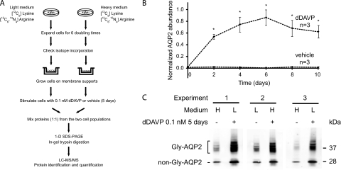
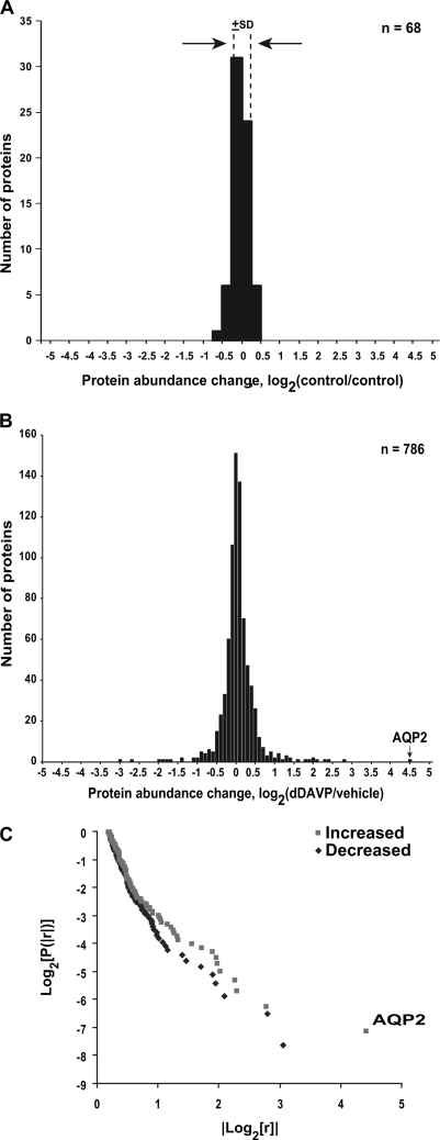
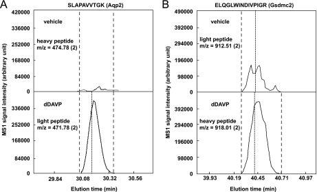
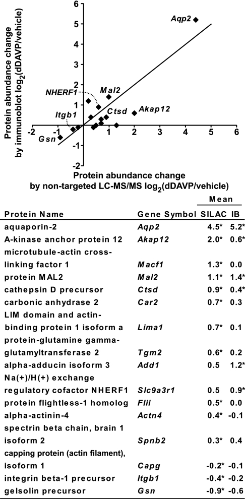
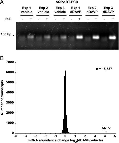
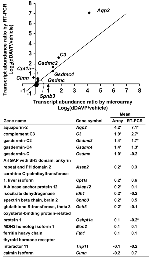
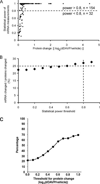
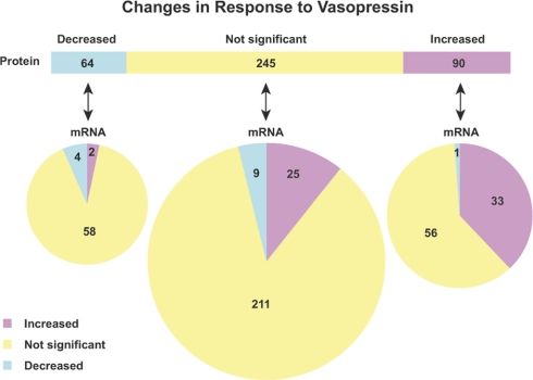
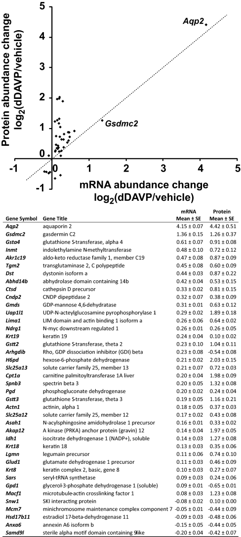
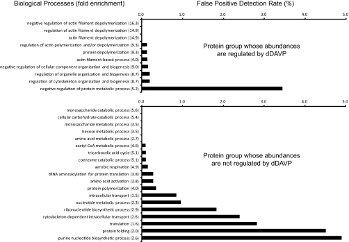
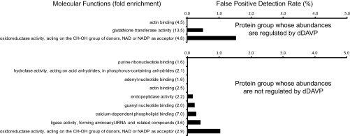
Previous studies in yeast have supported the view that post-transcriptional regulation of protein abundances may be more important than previously believed. Here we ask the question: "In a physiological regulatory process (the response of mammalian kidney cells to the hormone vasopressin), what fraction of the expressed proteome undergoes a change in abundance and what fraction of the regulated proteins have corresponding changes in mRNA levels?" In humans and other mammals, vasopressin fulfills a vital homeostatic role (viz. regulation of renal water excretion) by regulating the water channel aquaporin-2 in collecting duct cells. To address the question posed, we utilized large-scale quantitative protein mass spectrometry (LC-MS/MS) employing stable isotopic labeling in cultured mpkCCD cells ('SILAC') coupled with transcriptomic profiling using oligonucleotide expression arrays (Affymetrix). Preliminary studies analyzing two nominally identical control samples by SILAC LC-MS/MS yielded a relative S.D. of 13% (for ratios), establishing the precision of the SILAC approach in our hands. We quantified nearly 3000 proteins with nontargeted SILAC LC-MS/MS, comparing vasopressin- versus vehicle-treated samples. Of these proteins 786 of them were quantified in each of 3 experiments, allowing statistical analysis and 188 of these showed significant vasopressin-induced changes in abundance, including aquaporin-2 (20-fold increase). Among the proteins with statistically significant abundance changes, a large fraction (at least one-third) was found to lack changes in the corresponding mRNA species (despite sufficient statistical power), indicating that post-transcriptional regulation of protein abundance plays an important role in the vasopressin response. Bioinformatic analysis of the regulated proteins (versus all transcripts) shows enrichment of glutathione S-transferase isoforms as well as proteins involved in organization of the actin cytoskeleton. The latter suggests that long-term regulatory processes may contribute to actomyosin-dependent trafficking of the water channel aquaporin-2. The results provide impetus for increased focus on translational regulation and regulation of protein degradation in physiological control in mammalian epithelial cells.


Figures

Fig. 1.
Fig. 1.
Quantitative SILAC LC-MS/MS protein mass spectrometry. A, A flowchart of protocol for SILAC and LC-MS/MS. B, Time course of changes in aquaporin-2 (AQP2) protein abundance in mpkCCD cells in response to dDAVP. The mpkCCD cells were grown on membrane supports until confluence before they were exposed to 0.1 nm dDAVP or vehicle starting on day zero. Twenty μg protein samples collected at different time points were used for semiquantitative immunoblotting with an aquaporin-2 antibody. Values are mean ±S.E. Asterisks denote significant differences in aquaporin-2 abundances between dDAVP- and vehicle exposed cells for the same day (p < 0.01). Individual immunoblots are shown in the Supplemental Fig. S2. C, Steady-state aquaporin-2 protein abundance in mpkCCD cells used for LC-MS/MS quantification. The mpkCCD cells labeled with either heavy (H) or light (L) amino acids were grown on permeable supports until confluence before exposure to either 0.1 nm dDAVP or vehicle for 5 days. Proteins were extracted and used for immunoblotting with an aquaporin-2 antibody that detects both glycosylated (Gly-AQP2) and nonglycosylated aquaporin-2 (non-Gly-AQP2). 20 μg protein were loaded in each lane.
Fig. 2.
Fig. 2.
SILAC LC-MS/MS protein quantification. A, Variability of SILAC method. Distribution of protein abundance ratios for nominally identically treated cells (control versus control) except for labeling with SILAC light (L) and heavy (H) amino acids. The standard deviation of log2(L/H) values was ± 0.18 (dashed lines). Bin size = 0.25. B, Distribution of protein log2(D/C) values among all proteins quantified, where D is signal obtained in presence of dDAVP (5 day) and C is signal obtained in presence of vehicle. The value corresponding to aquaporin-2 (AQP2) abundance change is indicated. Bin size = 0.1. C, Log-log plot of cumulative probability versus absolute value of ratio for all mean values from Supplemental Table S4 in which log2(D/C) is greater than or equal to 0.2, showing “long-tailed distribution.” AQP2 change is labeled.
Fig. 3.
Fig. 3.
Typical reconstructed SILAC LC-MS/MS chromatograms for A, aquaporin-2 and B, gasdermin C2 peptides. Examples of corresponding heavy- and light-labeled aquaporin-2 (Aqp2, NP_033829) and gasdermin C2 (Gsdmc2, NP_808580) peptides are shown. Quantification was carried out using the trapezoidal rule for numerical integration (QUIL program, see Experimental Procedures). Dashed lines define the range within which the peptide intensity was integrated. Dotted lines indicate the time point when a peptide sequence was identified. The m/z (charge) of the peptides are indicated. Similar results were obtained for all three sample pairs, despite alternating heavy and light labeling.
Fig. 4.
Fig. 4.
Comparison of SILAC LC-MS/MS quantification with semiquantitative immunoblotting. Immunoblots are shown in the Supplemental Fig. S3. Log2 values of the protein abundance ratios for paired dDAVP- and vehicle-exposed cells are shown on the right [mean ± S.E., * p < 0.05 versus log2(1)]. IB, immunoblot. The RefSeq accession numbers are: Actn4, NP_068695; Add1, NP_001095914; Akap12, NP_112462; Aqp2, NP_033829; Capg, NP_031625; Car2, NP_033931; Ctsd, NP_034113; Flii, NP_071292; Gsn, NP_666232; Itgb1, NP_034708; Lima1, NP_075550; Macf1, NP_033730; Mal2, NP_849251; Nherf1, NP_036160; Spnb2, NP_787030; and Tgm2, NP_033399.
Fig. 5.
Fig. 5.
Transcript changes in mpkCCD cells in response to vasopressin analog dDAVP. A, Aquaporin-2 mRNA abundance changes in mpkCCD cells in response to dDAVP. Polarized mpkCCD cells grown on membrane supports were exposed to dDAVP or vehicle for 5 days. Total RNA was extracted and used for reverse transcription and polymerase chain reaction analysis (RT-PCR): 0.5 μg of total RNA per sample; 30 cycles of PCR. -, no RT reaction control; V, vehicle; D, 0.1 nm dDAVP. B, Distribution of mRNA abundance changes in response to dDAVP among all transcripts quantified. The bar corresponding to aquaporin-2 response is indicated. The results were obtained from Affymetrix GeneChip Mouse Genome 430 2.0 Arrays.
Fig. 6.
Fig. 6.
Comparison of transcript quantification by Affymetrix microarray with real time RT-PCR. A 200 ng aliquot total RNA from dDAVP- and vehicle-exposed mpkCCD cells was used for quantification of transcript abundances with real time RT-PCR. *, statistically significant versus no change i.e. log2(dDAVP/vehicle) = 0 (p < 0.05, n = 3). The RefSeq accession numbers are: Akap12, NM_031185; Aqp2, NM_009699; Asap2, NM_001004364; C3, NM_009778; Clmn, NM_001040682; Cpt1a, NM_013495; Fth1, NM_010239; Gsdmc1, NM_031378; Gsdmc2, NM_177912; Gsdmc4, XM_001474104; Gstt3, NM_133994; Idh1, NM_001111320; Mon2, NM_153395; Osbpl1a, NM_207530; Spnb3, NM_021287; and Trip11, XM_001001171.
Fig. 7.
Fig. 7.
Comparison of protein changes with transcript changes. A, Statistical power coefficients (vertical axis) obtained when testing for mRNA abundance differences that are percentage-wise equivalent to the measured protein abundance changes (horizontal axis) for the same gene. Data shown include only proteins whose abundances were significantly altered in response to dDAVP as quantified by SILAC LC-MS/MS. B, Percent of the regulated proteins that show corresponding changes in transcript abundances as a function of statistical power threshold. For example, 27% of the regulated proteins have significant changes in the corresponding mRNAs when limited to comparisons with statistical power greater than 0.8. C, Percent of regulated proteins that show corresponding changes in transcript abundance as a function of magnitude of protein abundance change threshold.
Fig. 8.
Fig. 8.
Relationship between protein changes and transcript changes in response to vasopressin analog dDAVP. A total of 399 proteins that have mRNA measurements with statistical powers greater than 0.8 were summarized. Proteins (top bar) are grouped with regard to their responses to dDAVP. Each protein group was then subgrouped with regard to corresponding mRNA changes (bottom pies).
Fig. 9.
Fig. 9.
Summary of genes for which changes in protein abundance were accompanied by changes in transcript abundance in response to dDAVP. Values are mean ± S.E. of log2 values of dDAVP/vehicle abundance ratios.
Fig. 10.
Fig. 10.
Gene Ontology Biological Process analysis for proteins that change in abundances in response to dDAVP. Top, 188 proteins that changed in abundances (p < 0.05) in response to dDAVP were used for Gene Ontology Biological Process analysis using the DAVID bioinformatic suite (http://david.abcc.ncifcrf.gov). Enrichment of Biological Process terms in these significantly changed proteins were statistically compared with all transcripts expressed in the mpkCCD cells by Fisher exact test. All GO terms listed were significantly enriched (p < 0.05). Fold enrichment is given in parentheses. Bars represent the estimated false positive detection rate. Bottom, 598 proteins that did not change in abundances were analyzed the same way, providing a control for the regulated group.
Fig. 11.
Fig. 11.
Gene Ontology Molecular Function analysis for proteins that change in abundances in response to dDAVP. Top, 188 proteins that changed in abundance (p < 0.05) in response to dDAVP were used for Gene Ontology Molecular Function analysis using the DAVID bioinformatic suite (http://david.abcc.ncifcrf.gov). Enrichment of Molecular Function terms in these significantly changed proteins were statistically compared with all transcripts expressed in the mpkCCD cells by Fisher exact test (All GO terms listed were significantly enriched at p < 0.05.). Fold enrichment is given in parentheses. Bars represent the estimated false positive detection rate. Bottom, 598 proteins that did not change in abundance were analyzed the same way, providing a control for the regulated group.
Fig. 12.
Fig. 12.
Comparison of regulated-proteins with those found in two prior proteomic studies of long-term vasopressin effects. Prior studies that have addressed protein abundance changes in response to long-term exposure to vasopressin in native collecting duct cells used either two-dimensional gels with fluorescent dye labeling (42) or ICAT LC-MS/MS (43) for quantification. Integers represent the number of proteins found to be significantly changed in abundance in each subsection of the Venn diagram (total for this study, 188 proteins).

References

    1. Gygi S. P., Rochon Y., Franza B. R., Aebersold R. (1999) Correlation between protein and mRNA abundance in yeast. Mol. Cell. Biol. 19, 1720–1730 - PMC - PubMed
    1. Ideker T., Thorsson V., Ranish J. A., Christmas R., Buhler J., Eng J. K., Bumgarner R., Goodlett D. R., Aebersold R., Hood L. (2001) Integrated genomic and proteomic analyses of a systematically perturbed metabolic network. Science 292, 929–934 - PubMed
    1. Washburn M. P., Koller A., Oshiro G., Ulaszek R. R., Plouffe D., Deciu C., Winzeler E., Yates J. R., 3rd. (2003) Protein pathway and complex clustering of correlated mRNA and protein expression analyses in Saccharomyces cerevisiae. Proc. Natl. Acad. Sci. U.S.A. 100, 3107–3112 - PMC - PubMed
    1. Nielsen S., Frøkiaer J., Marples D., Kwon T. H., Agre P., Knepper M. A. (2002) Aquaporins in the kidney: from molecules to medicine. Physiol. Rev. 82, 205–244 - PubMed
    1. Wall S. M., Han J. S., Chou C. L., Knepper M. A. (1992) Kinetics of urea and water permeability activation by vasopressin in rat terminal IMCD. Am. J. Physiol.. 262, F989–F998 - PubMed

Publication types

MeSH terms