. 2010 Oct;120(10):3668-72.
doi: 10.1172/JCI43699.

Defective migration of neuroendocrine GnRH cells in human arrhinencephalic conditions

Affiliations

Defective migration of neuroendocrine GnRH cells in human arrhinencephalic conditions

Luis Teixeira et al. J Clin Invest. 2010 Oct.

Abstract

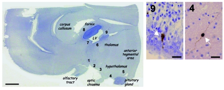
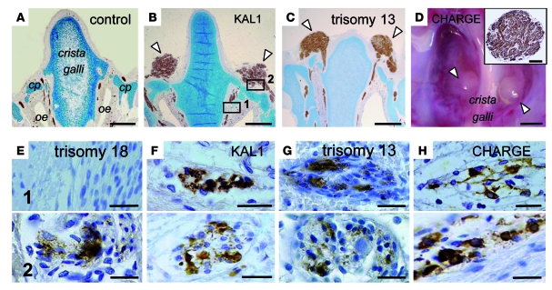
Patients with Kallmann syndrome (KS) have hypogonadotropic hypogonadism caused by a deficiency of gonadotropin-releasing hormone (GnRH) and a defective sense of smell related to olfactory bulb aplasia. Based on the findings in a fetus affected by the X chromosome–linked form of the disease, it has been suggested that hypogonadism in KS results from the failed embryonic migration of neuroendocrine GnRH1 cells from the nasal epithelium to the forebrain. We asked whether this singular observation might extend to other developmental disorders that also include arrhinencephaly. We therefore studied the location of GnRH1 cells in fetuses affected by different arrhinencephalic disorders, specifically X-linked KS, CHARGE syndrome, trisomy 13, and trisomy 18, using immunohistochemistry. Few or no neuroendocrine GnRH1 cells were detected in the preoptic and hypothalamic regions of all arrhinencephalic fetuses, whereas large numbers of these cells were present in control fetuses. In all arrhinencephalic fetuses, many GnRH1 cells were present in the frontonasal region, the first part of their migratory path, as were interrupted olfactory nerve fibers that formed bilateral neuromas. Our findings define a pathological sequence whereby a lack of migration of neuroendocrine GnRH cells stems from the primary embryonic failure of peripheral olfactory structures. This can occur either alone, as in isolated KS, or as part of a pleiotropic disease, such as CHARGE syndrome, trisomy 13, and trisomy 18.


Figures

Figure 1
Figure 1. Distribution of GnRH1-immunoreactive cells in a normal fetal brain.
Left: Mayer hemalun staining of a sagittal section of the forebrain from a 24-week-old control fetus (rostral region at left). Arabic numerals denote the locations of GnRH1-immunoreactive cells: 1–5 are typical of neuroendocrine GnRH1 cells, whereas 6–9 presumably correspond to some misrouted GnRH1 neurons. Right: detailed microscopic views of regions 4 and 9, showing GnRH1 neurons (arrows). LV, lateral ventricle. Scale bars: 3 mm (left); 50 μm (right).
Figure 2
Figure 2. Neuromas and accumulation of GnRH1-immunoreactive cells in the frontonasal region of arrhinencephalic fetuses.
(AC) Coronal sections of the frontonasal region in the KAL1 fetus and 23-week-old trisomy 13 and control fetuses, immunostained with an anti–βIII-tubulin antibody. The brains have been removed. In the KAL1 and trisomy 13 fetuses, bilateral spherical structures corresponding to neuromas (arrowheads) are visible on the dorsal aspect of the cribriform plate (cp) stained with Alcian blue. oe, olfactory epithelium. (D) Endocranial view of the cribriform plate region of the ethmoid bone in the CHARGE fetus. Abnormal spherical structures (arrowheads) were present on both sides of the crista galli. Inset: section from one of the spherical structures. The entangled nerve fibers that make up the neuroma were stained using an anti–βIII-tubulin antibody. (EH) Coronal sections corresponding to the boxed regions in B from KAL1, CHARGE, trisomy 13, and trisomy 18 fetuses. Sections were immunostained using an anti-GnRH1 antibody. In all fetuses, GnRH1 cells were seen in the upper nasal region (region 1) and/or on the dorsal aspect of the cribriform plate (region 2). Note the presence of GnRH1 cells only on the dorsal aspect of the cribriform plate for the 35-week-old trisomy 18 fetus. Scale bars: 1 mm (AD); 200 μm (D, inset); 30 μm (EH).
Figure 3
Figure 3. Distribution of GnRH1-immunoreactive cells in the forebrains and frontonasal regions of the 4 arrhinencephalic fetuses and a control fetus.
Red dots denote GnRH1 cells. Green lines indicate the path of the olfactory and terminal nerve fibers. The intracerebral fluid compartment is shaded gray. In the control fetus, GnRH1 cells were present in the preoptic and hypothalamic areas and were not detected in the frontonasal region. In addition, scattered, presumably misrouted GnRH1 neurons were found at several periventricular locations in the forebrain. In the arrhinencephalic fetuses, GnRH1 cells accumulated along the broken path of the olfactory and terminal nerve fibers that do not make contact with the forebrain. Few or no neuroendocrine cells were found in the preoptic/hypothalamic region. Scattered, periventricular GnRH1 neurons were also detected in the trisomy 13 and trisomy 18 fetuses. Scale bars: 1 cm. ob, olfactory bulb; h, hypothalamus; t, thalamus.

References

    1. Plant TM. Hypothalamic control of the pituitary-gonadal axis in higher primates: key advances over the last two decades. J Neuroendocrinol. 2008;20(6):719–726. doi: 10.1111/j.1365-2826.2008.01708.x. - DOI - PubMed
    1. Schwanzel-Fukuda M, Pfaff DW. Origin of luteinizing hormone-releasing hormone neurons. Nature. 1989;338(6211):161–164. doi: 10.1038/338161a0. - DOI - PubMed
    1. Schwanzel-Fukuda M, Crossin KL, Pfaff DW, Bouloux PMG, Hardelin JP, Petit C. Migration of Luteinizing Hormone-Releasing Hormone (LHRH) neurons in early human embryos. J Comp Neurol. 1996;366(3):547–557. doi: 10.1002/(SICI)1096-9861(19960311)366:3<547::AID-CNE12>3.0.CO;2-M. - DOI - PubMed
    1. Dodé C, Hardelin JP. Kallmann syndrome. Eur J Hum Genet. 2009;17(2):139–146. doi: 10.1038/ejhg.2008.206. - DOI - PMC - PubMed
    1. Franco B, et al. A gene deleted in Kallmann’s syndrome shares homology with neural cell adhesion and axonal path-finding molecules. Nature. 1991;353(6344):529–536. doi: 10.1038/353529a0. - DOI - PubMed

Publication types

Substances