

doi: 10.1364/OE.18.019413.
Automatic segmentation of seven retinal layers in SDOCT images congruent with expert manual segmentation
Affiliations
- PMID: 20940837
- PMCID: PMC3408910
- DOI: 10.1364/OE.18.019413
Item in Clipboard
Automatic segmentation of seven retinal layers in SDOCT images congruent with expert manual segmentation
Opt Express.
.
Abstract
Segmentation of anatomical and pathological structures in ophthalmic images is crucial for the diagnosis and study of ocular diseases. However, manual segmentation is often a time-consuming and subjective process. This paper presents an automatic approach for segmenting retinal layers in Spectral Domain Optical Coherence Tomography images using graph theory and dynamic programming. Results show that this method accurately segments eight retinal layer boundaries in normal adult eyes more closely to an expert grader as compared to a second expert grader.
Figures
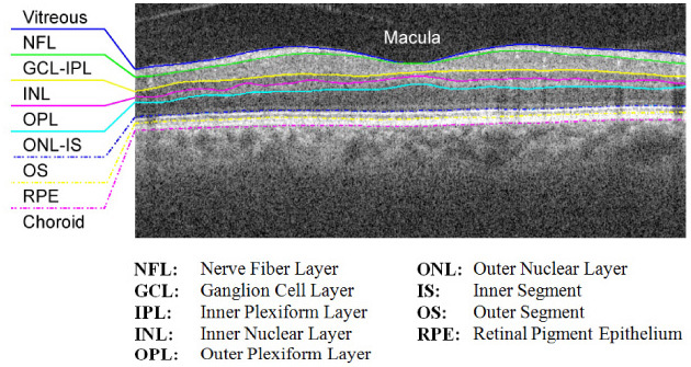
Target retinal layers of a cross-sectional SDOCT image (B-scan) centered at the macula.
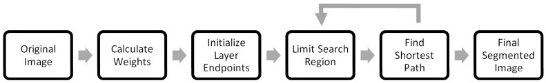
A generalized layer segmentation algorithm schematic.

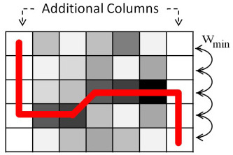
Example graph weights (adjacency matrix) for three connected nodes.
An example segmentation using automatic endpoint initialization.
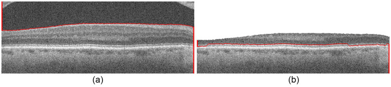
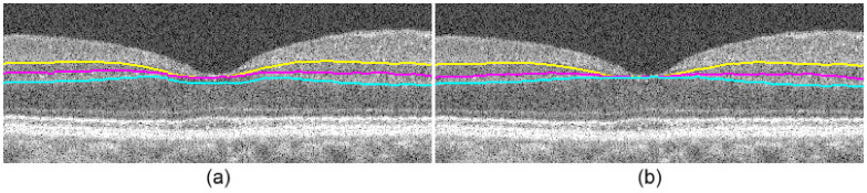
Segmentation of Fig. 6b using Dijkstra’s algorithm, automatic endpoint initialization, and search space limitation. (a) The vitreous-NFL layer boundary segmented. (b) The pilot IS-OS layer boundary segmented.
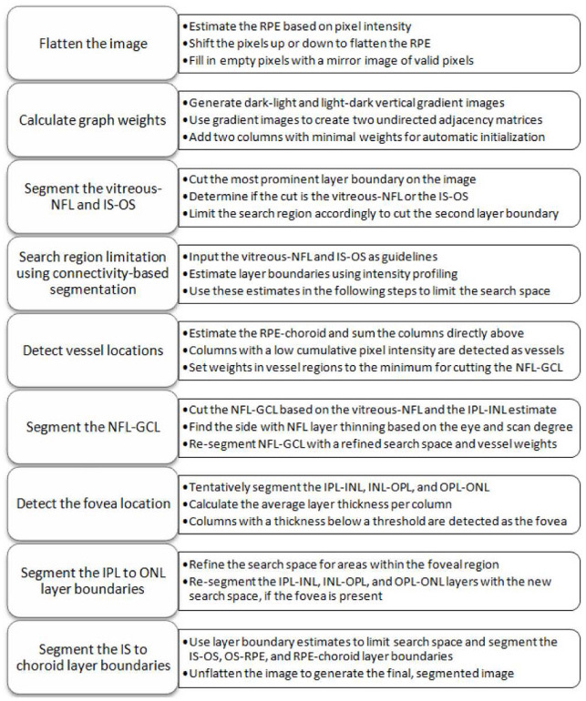
Eight retinal layer boundary segmentation algorithm schematic for SDOCT images.
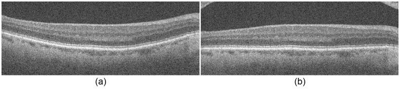
Image flattening. (a) The original retinal SDOCT image. (b) The flattened image.
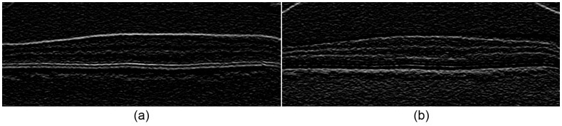
Gradient images used for calculating graph weights for Fig. 6b. (a) Dark-to-light image for segmenting a darker layer above a lighter layer. (b) Light-to-dark image for segmenting a lighter layer above a darker layer.
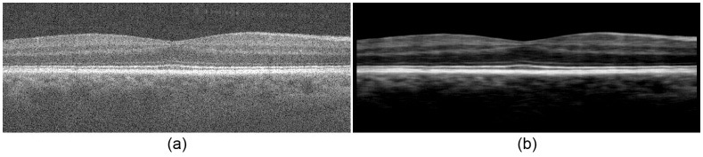
Contrast enhancement. (a) A flattened retinal SDOCT image. (b) The contrast-enhanced image.
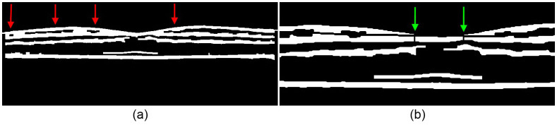
(a) A binary mask of the filtered image in Fig. 9b. The red arrows mark the location of the holes corresponding to the GCL-IPL complex. (b) A zoomed in binary mask with disconnected layers achieved by interpolating the lower boundaries of corresponding holes from Fig. 10a. The green arrows point to vertical breaks used to separate clusters that were not disconnected through interpolation.
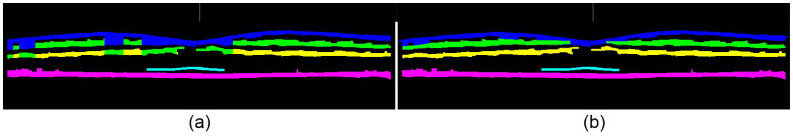
Retinal layer assignments, where blue = NFL, green = IPL, yellow = OPL, cyan = IS-OS, magenta = RPE. (a) Column-wise layer assignments of the mask in Fig. 10a. Note the conflicts in the top three layer assignments. (b) Cluster layer assignments of the mask in Fig. 10b.
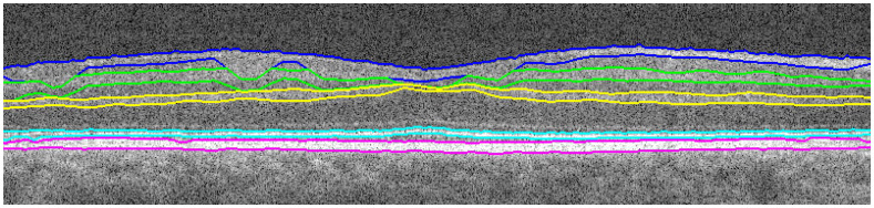
Connectivity-based segmentation of retinal layers using the layer assignments from Fig. 11b.
Vessel detection. (a) NFL-GCL without vessel detection. (b) NFL-GCL with vessel detection.

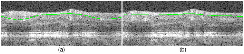
NFL-GCL segmentation. (a) NFL-GCL before correction. (b) NFL-GCL after correction.
Fovea correction. (a) Segmented image before correction. (b) Segmented image after correction.
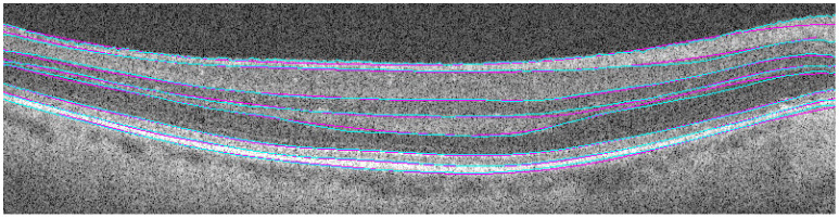
Comparison of automatic (cyan) versus manual (magenta) segmentation.
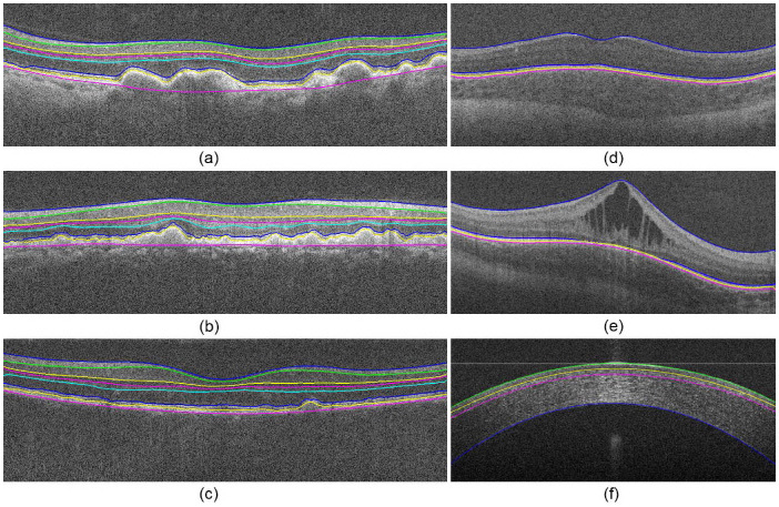
Segmentation of anatomical and pathological images of the eye. (a-c) Level 3 aged-macular degeneration retinas with drusen. (d) Pediatric retina. (e) Pediatric retina with edema. (f) Cornea.
References
-
- Schuman S. G., Koreishi A. F., Farsiu S., Jung S., Izatt J. A., Toth C. A., “Photoreceptor Layer Thinning over Drusen in Eyes with Age-Related Macular Degeneration Imaged In Vivo with Spectral-Domain Optical Coherence Tomography,” Ophthalmology 116, 488–496(2009). 10.1016/j.ophtha.2008.10.006 - DOI - PMC - PubMed
Publication types
MeSH terms
Grants and funding
LinkOut - more resources
Full Text Sources
Other Literature Sources
Medical
