. 2010 Sep 30;6(9):e1001128.
doi: 10.1371/journal.ppat.1001128.

Pathogenic bacteria target NEDD8-conjugated cullins to hijack host-cell signaling pathways

Affiliations

Pathogenic bacteria target NEDD8-conjugated cullins to hijack host-cell signaling pathways

Grégory Jubelin et al. PLoS Pathog. .

Abstract

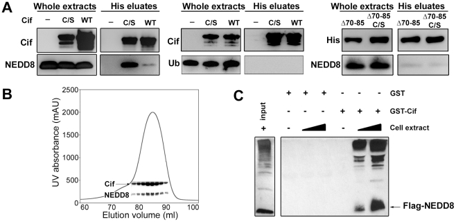
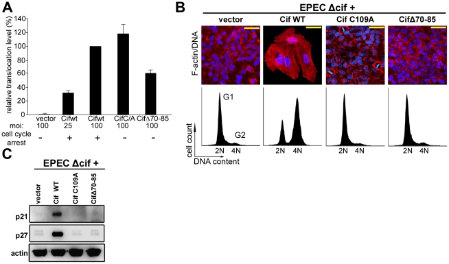
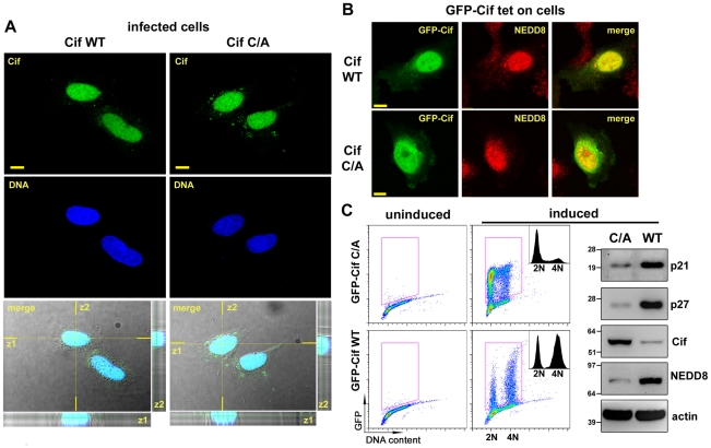
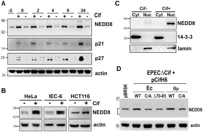
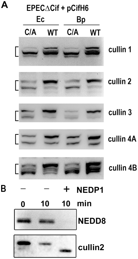
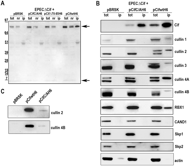
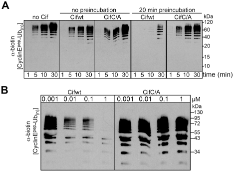
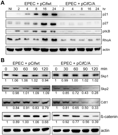
The cycle inhibiting factors (Cif), produced by pathogenic bacteria isolated from vertebrates and invertebrates, belong to a family of molecules called cyclomodulins that interfere with the eukaryotic cell cycle. Cif blocks the cell cycle at both the G₁/S and G₂/M transitions by inducing the stabilization of cyclin-dependent kinase inhibitors p21(waf1) and p27(kip1). Using yeast two-hybrid screens, we identified the ubiquitin-like protein NEDD8 as a target of Cif. Cif co-compartmentalized with NEDD8 in the host cell nucleus and induced accumulation of NEDD8-conjugated cullins. This accumulation occurred early after cell infection and correlated with that of p21 and p27. Co-immunoprecipitation revealed that Cif interacted with cullin-RING ubiquitin ligase complexes (CRLs) through binding with the neddylated forms of cullins 1, 2, 3, 4A and 4B subunits of CRL. Using an in vitro ubiquitylation assay, we demonstrate that Cif directly inhibits the neddylated CUL1-associated ubiquitin ligase activity. Consistent with this inhibition and the interaction of Cif with several neddylated cullins, we further observed that Cif modulates the cellular half-lives of various CRL targets, which might contribute to the pathogenic potential of diverse bacteria.


Conflict of interest statement

The authors have declared that no competing interests exist.

Figures

Figure 1
Figure 1. Cif interacts in vitro with the ubiquitin-like protein NEDD8.
(A) Hexa-histidine tagged wild-type Cif (WT), Cif cysteine 109 mutant Cys-to-Ser (C/S), Cif deleted for amino-acids 70-85 mutant (Δ70-85) or Cif Δ70-85 Cys-to-Ser mutant(Δ70-65 C/S) and NEDD8 or ubiquitin (Ub) proteins were co-expressed in bacteria, and then His-Cif proteins were purified from bacterial lysates using nickel affinity. Whole extracts and eluted fractions were probed with indicated antibodies. (B) Wild-type His-Cif and NEDD8 were co-expressed in bacteria and His-Cif was purified using nickel affinity and gel filtration chromatography. The peak in the chromatograph corresponds to the His-Cif complex. NEDD8 and the corresponding fractions were visualized using SDS-PAGE stained with Coomassie blue. (C) Glutathione sepharose matrix was mixed with 100 µg of GST or GST-Cif Cys-to-Ser mutant purified proteins and then incubated with a lysate of HEK293T cells expressing a FLAG-NEDD8 construct. 100 µg or 250 µg of cells extract were used to obtain a GST constructs/cell extract ratio of 1∶1 and 1∶2.5 respectively. Cells extract (input) and eluted fractions were immunoblotted with anti-FLAG antibodies. FLAG-NEDD8 fusion protein is indicated with an arrow.
Figure 2
Figure 2. Cytopathic activities of wild-type Cif, Cif C/A and CifΔ70-85 mutants.
(A) Translocation efficiency of Cif wild-type and mutant proteins were assessed as follow: HeLa cells were loaded with CCF2/AM substrate and were infected 1 h 30 with E22Δcif hosting plasmids expressing the different Cif-TEM1 fusions and intracellular β-lactamase activity was detected by measuring cleavage of the CCF2/AM substrate. Translocation level is represented as percentages relative to translocation of the Cif WT-TEM protein (see materials and methods). Experiments were performed in triplicate and error bars represent standard errors of the mean. The multiplicity of infection (moi) and corresponding cell cycle arrest phenotype are indicated. Note that infection at moi of 25 with low level of translocation of wild-type Cif was sufficient to induce cell cycle arrest. (B) G1/S synchronized HeLa cells were infected 1.5 h with E22Δcif expressing the different Cif proteins, washed and incubated with antibiotic for 20-72 h. Upper panels: F-actin was labeled with phalloidin-rhodamine (red) and DNA with DAPI (blue) 72 h post-infection. Bars represent 50 µm. Lower panels: cell cycle distribution was analyzed by flow cytometry 20 h post-infection. 2N (G1) and 4N (G2) populations are indicated. (C) HeLa cells were infected as in B, washed and incubated with antibiotics for 24 h. Cell protein extracts were probed with anti-p21, anti-p27 and anti-actin antibodies.
Figure 3
Figure 3. Cif is sorted to the host cell nucleus and co-compartmentalizes with NEDD8.
(A) HeLa cells were infected 2 h with EPECΔcif expressing wild-type Cif (WT) or the CifC109A (C/A) mutant, washed and further incubated 3 h with antibiotics. Cif was then visualized by indirect immunofluorescence and confocal imaging, with anti-Cif antibodies (green) and TO-PRO-3 to stain DNA (dark blue). Single optical XY slices and the corresponding Z slices of the image stacks are shown. In the merged images, coincident fluorescent sources appear cyan. Bars represent 10 µm. (B) Stable HeLa TetOn cells expressing GFP-Cif (WT) or GFP-CifC109A (C/A) were induced for 24 h with doxycycline. GFP-Cif was visualized by GFP fluorescence acquisition (green) and NEDD8 was detected by indirect immunofluorescence with anti-NEDD8 antibodies (red). In the merged images, coincident fluorescent sources appear orange-yellow. Bars represent 10 µm. (C) HeLa TetOn cells were induced or not with doxycycline, then DNA was stained with propidium iodide and DNA/GFP content was analyzed by flow cytometry. GFP-positive cells were gated as shown in the DNA content/GFP fluorescence 2D plots to generate the cell cycle histograms (insets). 2N and 4N DNA content are indicated. Induced cell protein lysates were probed with anti-p21, anti-p27, anti-Cif, anti-NEDD8 and anti-actin antibodies. Molecular weights in kDa are indicated.
Figure 4
Figure 4. Infection with Cif-producing EPEC induces the accumulation of neddylated proteins.
(A) HeLa cells were infected (starting at -2 h) 2 h with wild-type EPEC (+) or a Cif-deleted mutant EPEC (-), washed and further incubated with antibiotics up to 24 h. Cell protein extracts were probed with anti-NEDD8, anti-p21, anti-p27 and anti-actin antibodies. Molecular weights in kDa are indicated. (B) HeLa, IEC-6 or HCT116 cells were infected as in A and further incubated 6 h with antibiotics. Cell protein extracts were probed with anti-NEDD8 and anti-actin antibodies. (C) HeLa cells were infected as in A, washed and further incubated 6 h with antibiotics. Cytoplasmic (Cyt) and nuclear (Nuc) extracts were probed with anti-NEDD8, anti-14-3-3 (cytoplasmic control) and anti-lamin (nuclear control) antibodies. (D) HeLa cells were infected 3 h with EPECΔcif strain carrying the empty vector (pBRSK) or plasmids coding for His-tagged wild-type (WT), cysteine mutant (C/A) or Δ70-85 mutant of Cif from E. coli (Ec) or from B. pseudomallei (Bp) and further incubated 3 h with antibiotics. Cell protein extracts were probed with anti-NEDD8 and anti-actin antibodies. The brackets encompass the 73-81 kDa region.
Figure 5
Figure 5. Cif induces the accumulation of NEDD8-conjugated cullins in host cells.
(A) HeLa cells were infected 3 h with EPECΔcif strain carrying plasmids coding for His-tagged wild-type (WT) or cysteine mutant (C/A) from E. coli (Ec) or from B. pseudomallei (Bp) and further incubated 3 h with antibiotics. Cell protein extracts were probed with anti-cullins (cullin 1 to cullin 4B) antibodies. The brackets encompass the 73-81 kDa regions. (B) Protein extracts from HeLa cells infected with EPEC expressing wild-type Cif were incubated for 10 min in presence (+) or absence (-) of NEDP1 and probed with anti-NEDD8 and anti-cullin2 antibodies.
Figure 6
Figure 6. Cif interacts with NEDD8-conjugated cullins.
HeLa cells were infected 3 h with EPECΔcif carrying the empty vector (pBRSK) or plasmids expressing His-tagged E. coli Cif cysteine mutant (C/A), 70-85 deleted mutant (Δ70-85) or wild-type (WT) and further incubated 3 h with antibiotics. Cell extracts were immunoprecipitated with anti-His antibodies. (A) Total (tot), non-retained (nr) and immunoprecipitated (ip) protein fractions were probed with anti-NEDD8 antibodies. Arrows indicate neddylated cullins (top) and monomeric NEDD8 (bottom). Molecular weights in kDa are indicated. (B) Total and immunoprecipitated protein fractions were probed with anti-Cif, anti-cullins (CUL1 to CUL4B), anti-RBX1, anti-CAND1, anti-Skp1, anti-Skp2 and anti-actin antibodies. (C) High loads of immunoprecipitated fractions were probed with anti-cullins (CUL2 and CUL4B) antibodies.
Figure 7
Figure 7. Cif decreases neddylated SCF-mediated substrate polyubiquitylation in vitro.
(A) Anti-biotin western blot showing time courses of polyubiquitylation of a biotin-labeled cyclin E phosphopeptide by NEDD8-modified SCFFbw7ΔD in the presence or absence of purified His-tagged wild type Cif (Cifwt) or Cif catalytic Cys-to-Ala (CifC/A) mutant. Reactions were performed by adding Cif straight to the reaction (no preincubation), or after 20 min room temperature incubation of NEDD8-modified SCFFbw7(263-C) with Cif prior to initiating the reaction by adding all other proteins for polyubiquitylation activity (20 min preincubation). (B) Anti-biotin western blot showing titration of the indicated amount of Cifwt or CifC/A with 200 nM SCFFbw7ΔD, prior to 10-min long polyubiquitylation reactions for a biotin-labeled cyclin E phosphopeptide. In all these reactions, the indicated version of Cif was preincubated with NEDD8-modified SCFFbw7ΔD for 1 h prior to reaction initiation.
Figure 8
Figure 8. Cif induces stabilization of various targets of cullin-RING ubiquitin ligases.
(A) HeLa cells were infected (starting at 0 h) for 2 h with EPECΔcif carrying a plasmid encoding wild-type (WT) or cysteine mutant (C/A) Cif and further incubated for indicated times with antibiotics. Cell protein extracts were probed with anti-p21, p27, phospho-IκB, RhoA or actin antibodies. NI: non-infected. (B) HeLa cells were infected for 2 h with EPECΔcif carrying the plasmid expressing cysteine mutant (pCifC/A) or wild-type Cif (pCifwt) and then (starting at 0 min) incubated for the indicated time with cycloheximide. Cell protein extracts were probed with anti-Skp1, Skp2, Cdt1, β-catenin or actin antibodies. The relative amount of protein (relative to 0 min and normalized to actin level) is indicated below each western blot.

Similar articles

Cited by

References

    1. Samba-Louaka A, Taieb F, Nougayrede JP, Oswald E. Cif type III effector protein: a smart hijacker of the host cell cycle. Future Microbiol. 2009;4:867–877. - PubMed
    1. Nougayrede JP, Taieb F, De Rycke J, Oswald E. Cyclomodulins: bacterial effectors that modulate the eukaryotic cell cycle. Trends Microbiol. 2005;13:103–110. - PubMed
    1. Oswald E, Nougayrede JP, Taieb F, Sugai M. Bacterial toxins that modulate host cell-cycle progression. Curr Opin Microbiol. 2005;8:83–91. - PubMed
    1. Marches O, Ledger TN, Boury M, Ohara M, Tu X, et al. Enteropathogenic and enterohaemorrhagic Escherichia coli deliver a novel effector called Cif, which blocks cell cycle G2/M transition. Mol Microbiol. 2003;50:1553–1567. - PubMed
    1. Yao Q, Cui J, Zhu Y, Wang G, Hu L, et al. A bacterial type III effector family uses the papain-like hydrolytic activity to arrest the host cell cycle. Proc Natl Acad Sci U S A. 2009;106:3716–3721. - PMC - PubMed

Publication types

MeSH terms