. 2010 Oct 7;6(6):599-612.
doi: 10.7150/ijbs.6.599.

Aberrant cytoplasm localization and protein stability of SIRT1 is regulated by PI3K/IGF-1R signaling in human cancer cells

Affiliations

Aberrant cytoplasm localization and protein stability of SIRT1 is regulated by PI3K/IGF-1R signaling in human cancer cells

Vanessa Byles et al. Int J Biol Sci. .

Abstract

SIRT1, an NAD-dependent histone/protein deacetylase, has classically been thought of as a nuclear protein. In this study, we demonstrate that SIRT1 is mainly localized in the nucleus of normal cells, but is predominantly localized in the cytoplasm of the cancer / transformed cells we tested. We found this predominant cytoplasmic localization of SIRT1 is regulated by elevated mitotic activity and PI3K/IGF-1R signaling in cancer cells. We show that aberrant cytoplasmic localization of SIRT1 is due to increased protein stability and is regulated by PI3K/IGF-1R signaling. In addition, we determined that SIRT1 is required for PI3K-mediated cancer cell growth. Our study represents the first identification that aberrant cytoplasm localization is one of the specific alternations to SIRT1 that occur in cancer cells, and PI3K/IGF-1R signaling plays an important role in the regulation of cytoplasmic SIRT1 stability. Our findings suggest that the over-expressed cytoplasmic SIRT1 in cancer cells may greatly contribute to its cancer-specific function by working downstream of the PI3K/IGF-1R signaling pathway.

Keywords: PI3K/IGF-1R; SIRT1; cancer cells; cytoplasm localization; protein stability.


Conflict of interest statement

CONFLICTS OF INTEREST: The authors declare that they have no conflicts of interest

Figures

Fig 1
Fig 1
Localization patterns of SIRT1 in cancer and normal cells. A. Cytoplasmic and nuclear fractions of DU145 and PC3 cells and prostate cancer tissues were isolated, the concentration of nuclear and cytoplasmic protein was measured by Bradford assay, the same amount of nuclear and cytoplasmic protein was subjected to SDS gel, and immunoblots were performed with anti-SIRT1, α-tubulin and LaminA/C antibodies. B. Cytoplasmic and nuclear proteins were isolated from DU145 cells transfected with RNA interference of SIRT1 (siSIRT1) or vector control (siVector). Equal amounts of nuclear and cytoplasmic protein were loaded, and immunoblots were performed with the same antibodies used in Fig.1A. C. The nuclear and cytoplasmic fractions of normal prostate cell PZ-HPV-7, normal lung cell MRC5 and normal prostate tissues were prepared. The same amount of nuclear and cytoplasmic protein was loaded, and immunoblots were performed with the same antibodies as Fig.1A. D. The nuclear and cytoplasmic fractions of primary and transformed human fibroblast cells BR3 and BR3neo, normal prostate cell PZ-HPV-7 and prostate cancer cell line DU145 were prepared. Equal amounts of nuclear and cytoplasmic protein were loaded and immunoblots were performed with the same antibodies as Fig.1A.
Fig 2
Fig 2
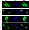
Mitotic signaling regulates cytoplasmic localization of SIRT1. DU145 cells were cultured in the chamber slide with DMEM+10% FBS. The immunofluorescence was performed with anti-SIRT1 antibody (sc 74504, Santa Cruz 1:1000, RT 1hr) and Alexa Fluor 488 anti-mouse (Invitrogen, 1:200, RT 1hr). Nuclear protein was stained with DAPI (1:10,000). A. DU145 cells were cultured with DMEM+10% FBS. B. The DU145 cells that were cultured in the DMEM+10% FBS were shifted to serum-free DMEM for 48 hr. C. After culturing the DU145 cells in serum-free medium for 48hr, the medium was replaced with DMEM+10% FBS for 24hr. D. DU145 cells that were cultured in DMEM+10%FBS were treated with hydroxyurea for 24 hr.
Fig 3
Fig 3
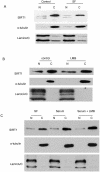
Translocation does not play a dominant role in SIRT1 cytoplasm localization. A. The DU145 cells that were cultured in the DMEM+10% FBS were shifted to serum-free DMEM for 24 hr. The nuclear and cytoplasmic proteins were fractionated and equal amounts of nuclear and cytoplasmic protein were loaded, and immunoblots were performed with anti-SIRT1, Lamin A/C and tubulin antibodies. B. The DU145 cells were treated with either vehicle or 10nM LMB for two hours. The nuclear and cytoplasmic proteins were fractionated and equal amounts of nuclear and cytoplasmic protein were loaded and immunoblots were performed with anti-SIRT1, Lamin A/C and tubulin antibodies. C. The DU145 cells that were cultured in the serum-free DMEM were shifted to DMEM+10% FBS with or without LMB for 2hr. The nuclear and cytoplasmic proteins were fractionated and the same amounts of nuclear and cytoplasmic protein were loaded and immunoblots were performed with anti-SIRT1, Lamin A/C and tubulin antibodies.
Fig 4
Fig 4
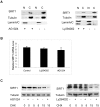
PI3K and IGF-1R regulates the protein stability of cytoplasmic SIRT1. A. DU145 cells were treated with or without AG1024 (4µM) (left panel) or Ly294002 (20µM) (right panel) for 12hr. The nuclear and cytoplasmic proteins were prepared and equal amounts of nuclear and cytoplasmic protein were loaded to SDS gel. The immunoblot was performed with anti-SIRT1, tubulin and LaminA/C antibodies. B. Transcript levels of SIRT1 was measured by quantitative RT-PCR analysis of RNA extracted from the cells treated with or without AG1024 (4µM) or Ly294002 (20µM) for 12hr. Transcript levels are expressed relative to β-actin transcripts. C. DU145 cells were pretreated with or without AG1024 (4µM) (left panel) or Ly294002 (20µM) (right panel) for 30minutes, then treated with cycloheximide (10μg/ml). The nuclear and cytoplasmic proteins were isolated after treatment with cycloheximide for 0, 5 and 15hr and the same amounts of nuclear and cytoplasmic proteins were subjected to SDS gel. The immunoblot was performed with anti-SIRT1 and anti-tubulin antibodies.
Fig 5
Fig 5
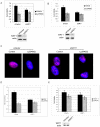
SIRT1 silencing attenuates PI3K inhibition-mediated prostate cancer cell growth arrest. Prostate cancer cell lines PC3 (5A) and LNCaP (5B) in which SIRT1 had been knocked down by siRNA expression (siSIRT1) or empty-vector-transfected cells (siVector) were exposed to the Ly292002 (20µM).Viable cells were quantitated at 72hr by MTS assay. The error bars represent the SEM. The SIRT1 knockdown efficiency was shown in 5A and 5B lower panel. C. The SIRT1 knockdown (siSIRT1) and empty-vector control (siVector) PC3 cells were treated with or without Ly292002 (20µM) for 24hr, then exposed to the BrdU (20uM) for another 24hr. The immunostaining was performed with anti-BrdU antibody (NMM1645458) and anti-mouse 594 secondary antibody. The images were taken with Nikon deconvolution wide-field Epifluorescence system. 100x. D. The SIRT1 knockdown (siSIRT1) and empty-vector control (siVector) PC3 cells were treated with or without Ly292002 (20µM) for 24hr, then exposed to the BrdU (4uM) for 24hr. The cells were fixed, immunostainned with anti-BrdU antibody and peroxidase conjugate secondary antibody; the peroxidase substrate was added and plates were read at 450nM. The error bars represent the SEM. E. MTS analysis for normal prostate cells PZ-HPV-7 cells. The experiment conditions are the same as 5A and 5B. The SIRT1 knockdown efficiency was shown in 5E lower panel.
Fig S1
Fig S1
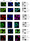
SIRT1 localization determined by both Immunofluorescence and immunobolt analysis. DU145 cells were cultured in the chamber slide with DMEM+10% FBS for the immunoflurorescence analysis (left panel). A. Anti-SIRT1 (C05187, Epitomics, 1:100, RT 1hr) and Alexa Fluor 488 goat anti-rabbit (Invitrogen, 200x, RT 1hr). B. Anti-hSir2 (856, 1:500, RT 1hr) and Alexa Fluor 488 goat anti-rabbit (Invitrogen, 200x, RT 1hr). C. Anti-SIRT1 (SC-19857, Santa Cruz, 1:50. RT 1hr) and Alexa Fluor 546 donkey anti-goat (Invitrogen, 100x, RT 1hr). D. Anti-SIRT1 (07-131, Milipore, 1:50, RT 1hr) and Alexa Fluor 488 goat anti-rabbit (Invitrogen, 200x, RT 1hr). E. Anti-SIRT1 (sc-74504, Santa Cruz, 1:1000, RT 1hr) and Alexa Fluor 488 goat anti-mouse (Invitrogen, 200x, RT 1hr). F. Anti-SIRT1 (05-707, upstate, 1:1000, RT 1hr) and Alexa Fluor 594 goat anti-mouse (Invitrogen, 100x, RT 1hr). Cytoplasmic and nuclear fractions of DU145 cells were isolated and same amount of nuclear and cytoplasmic protein were subjected to SDS gels, and immunoblots were performed (right panel). A. Anti-SIRT1 (C05187, Epitomics, 1:500, RT 1hr). B. Anti-hSir2 (856, 1:1000, RT 1hr). C. Anti-SIRT1 (SC-19857, Santa Cruz, 1:200, 4oC O/N). D. Anti-SIRT1 (07-131, Milipore, 1:500, 4oC O/N). E. Anti-SIRT1 (sc-74504, Santa Cruz, 1:1000, RT 1hr). F. Anti-SIRT1 (05-707, upstate, 1:1000, RT 1hr).

Similar articles

Cited by

References

    1. Saunders L.R, Verdin E. Sirtuins: critical regulators at the crossroads between cancer and aging. Oncogene. 2007;26(37):5489–504. - PubMed
    1. Yamamoto H, Schoonjans K, Auwerx J. Sirtuin functions in health and disease. Mol Endocrinol. 2007;21(8):1745–55. - PubMed
    1. Zeng L, Chen R, Liang F. et al.Silent information regulator, Sirtuin 1, and age-related diseases. Geriatr Gerontol Int. 2009;9(1):7–15. - PubMed
    1. Donmez G, Guarente L. Aging and disease: connections to sirtuins. Aging Cell. 2010;9(2):285–90. - PubMed
    1. Haigis M.C, Sinclair D.A. Mammalian sirtuins: biological insights and disease relevance. Annu Rev Pathol. 2010;5:253–95. - PMC - PubMed

Publication types