Review
. 2010 Sep 30;6(9):e1001032.
doi: 10.1371/journal.ppat.1001032.

Sequestration and tissue accumulation of human malaria parasites: can we learn anything from rodent models of malaria?

Affiliations
Review

Sequestration and tissue accumulation of human malaria parasites: can we learn anything from rodent models of malaria?

Blandine Franke-Fayard et al. PLoS Pathog. .

Abstract

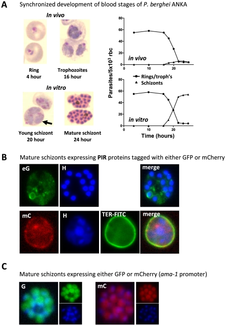
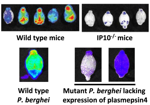
The sequestration of Plasmodium falciparum-infected red blood cells (irbcs) in the microvasculature of organs is associated with severe disease; correspondingly, the molecular basis of irbc adherence is an active area of study. In contrast to P. falciparum, much less is known about sequestration in other Plasmodium parasites, including those species that are used as models to study severe malaria. Here, we review the cytoadherence properties of irbcs of the rodent parasite Plasmodium berghei ANKA, where schizonts demonstrate a clear sequestration phenotype. Real-time in vivo imaging of transgenic P. berghei parasites in rodents has revealed a CD36-dependent sequestration in lungs and adipose tissue. In the absence of direct orthologs of the P. falciparum proteins that mediate binding to human CD36, the P. berghei proteins and/or mechanisms of rodent CD36 binding are as yet unknown. In addition to CD36-dependent schizont sequestration, irbcs accumulate during severe disease in different tissues, including the brain. The role of sequestration is discussed in the context of disease as are the general (dis)similarities of P. berghei and P. falciparum sequestration.


Conflict of interest statement

The authors have declared that no competing interests exist.

Figures

Figure 1
Figure 1. P. berghei ANKA asexual blood stage development and expression of proteins in mature schizonts.
(A) In vivo and in vitro development of rings, trophozoites, and schizonts during one cycle of synchronized development. In mice, rings and trophozoites do not sequester but schizonts disappear from the peripheral circulation (upper graph). In vitro schizogony takes place between 18 and 24 hours after invasion of the red blood cell (lower graph). The arrow indicates a multiply infected red blood cell containing three trophozoite-stage parasites; above this cell is a 20-hour schizont (graphs adapted from and . (B) Live mature schizonts of two transgenic lines expressing two different fluorescently tagged PIR proteins either tagged with GFP (eG; PB200064.00.0) or mCherry (mC; PB200026.00.0). These proteins are exported into the cytoplasm of the erythrocyte nucleus stained with Hoechst (H; blue), red blood cell membrane surface protein stained in mC parasites (TER-FITC; green) (J. Braks and B. Franke-Fayard, unpublished data). (C) Live mature schizonts that express GFP and mCherry in the cytoplasm of the merozoites (J. Braks and B. Franke-Fayard, unpublished data).
Figure 2
Figure 2. Imaging of transgenic P. berghei ANKA parasites in vivo and ex vivo.
CD36-mediated sequestration of schizonts in adipose tissue and lungs (adapted from PNAS, 2005 [35]). (A, B) Distribution of transgenic P. berghei ANKA parasites, expressing GFP::luciferase fusion protein (ama-1 promoter, see Box 3). Parasites are visible in lungs, spleen, and adipose tissue in wild-type mice, and principally in the blood circulation and accumulated in the spleen in CD36 knock-out mice. In wild-type mice infected with a non-sequestering K173 line, schizonts are also mainly found in the peripheral blood circulation and accumulated in the spleen (1: adipose tissue; 2: spleen; 3 liver; 4: lungs; 5: heart; 6: kidney; 7: brain). (C) Sequestration of transgenic P. berghei ANKA parasites in microvasculature of adipose tissue (upper panel with under phase contrast and lower panel with GFP-positive schizonts indicated by arrows).
Figure 3
Figure 3. Imaging of transgenic P. berghei ANKA parasites in brains of mice ex vivo.
Matched sets of experiments with P. berghei ANKA infections in ECM-sensitive mice (i.e., wild-type mice) or knock-out mice (i.e., IP10−/−). Knock-out mice do not develop cerebral pathology and this corresponds to a strong reduction in irbc accumulation as compared to infections in wild-type mice (adapted from [41]). Similar examples of a lack of irbc accumulation can be observed in the brains of mice treated with antibodies against host molecules (e.g., anti-LTβ mAB and anti-CD25 mAB; see [42], [104]). Parasites express GFP::luciferase fusion protein under the control of the eef1a promoter, see Box 3). Also, mice infected with a P. berghei ANKA mutant that has had the gene encoding plasmepsin 4 removed do not develop cerebral complications, and again there is a strong reduction of irbc accumulation in the brain of these infected animals (adapted from [43]).

Similar articles

Cited by

References

    1. Rogerson SJ, Hviid L, Duffy PE, Leke RF, Taylor DW. Malaria in pregnancy: pathogenesis and immunity. Lancet Infect Dis. 2007;7:105–117. - PubMed
    1. Desai M, ter Kuile FO, Nosten F, McGready R, Asamoa K, et al. Epidemiology and burden of malaria in pregnancy. Lancet Infect Dis. 2007;7:93–104. - PubMed
    1. Beeson JG, Duffy PE. The immunology and pathogenesis of malaria during pregnancy. Curr Top Microbiol Immunol. 2005;297:187–227. - PubMed
    1. Mackintosh CL, Beeson JG, Marsh K. Clinical features and pathogenesis of severe malaria. Trends Parasitol. 2004;20:597–603. - PubMed
    1. Rasti N, Wahlgren M, Chen Q. Molecular aspects of malaria pathogenesis. FEMS Immunol Med Microbiol. 2004;41:9–26. - PubMed

Publication types