. 2010 Oct;120(10):3663-7.
doi: 10.1172/jci42771.

Dual function of MyD88 in RAS signaling and inflammation, leading to mouse and human cell transformation

Affiliations

Dual function of MyD88 in RAS signaling and inflammation, leading to mouse and human cell transformation

Isabelle Coste et al. J Clin Invest. 2010 Oct.

Abstract

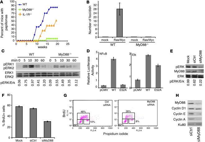
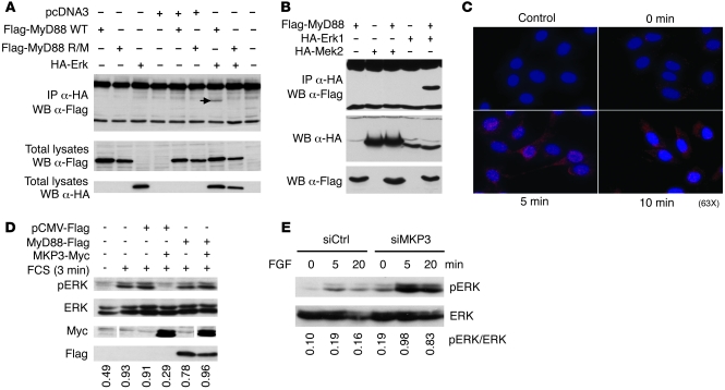
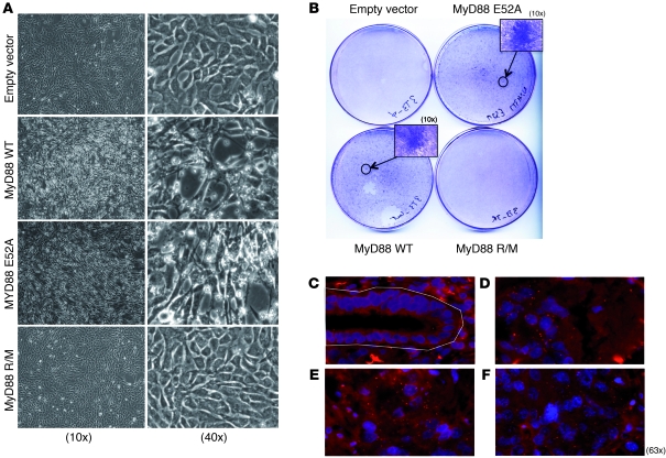
Accumulating evidence points to inflammation as a promoter of carcinogenesis. MyD88 is an adaptor molecule in TLR and IL-1R signaling that was recently implicated in tumorigenesis through proinflammatory mechanisms. Here we have shown that MyD88 is also required in a cell-autonomous fashion for RAS-mediated carcinogenesis in mice in vivo and for MAPK activation and transformation in vitro. Mechanistically, MyD88 bound to the key MAPK, Erk, and prevented its inactivation by its phosphatase, MKP3, thereby amplifying the activation of the canonical RAS pathway. The relevance of this mechanism to human neoplasia was suggested by the finding that MyD88 was overexpressed and interacted with activated Erk in primary human cancer tissues. Collectively, these results show that in addition to its role in inflammation, MyD88 plays what we believe to be a crucial direct role in RAS signaling, cell-cycle control, and cell transformation.


Figures

Figure 1
Figure 1. MyD88 is required for Ras transformation, full Erk activation, and cell cycle.
(A) Incidence of skin papilloma in WT, MyD88–/–, and IL-1R–/– mice (n = 20 per group) subjected to a 2-stage carcinogenesis (DMBA and TPA). (B) Quantification of foci developed in WT and MyD88–/– MEFs transfected with Rasv12 and Myc. (C) Erk phosphorylation is reduced in MyD88–/– MEFs treated with FGF for the indicated times. Lower panel shows total Erk on the same blots. (D) Luciferase activity from NF-κB luciferase or Elk luciferase in HeLa cells overexpressing WT MyD88, MyD88-E52A, or control plasmid. Values were normalized based on Renilla luminescence. (EH) A375 melanoma cell line was transfected with control (siCtrl) or MyD88-specific siRNA (siMyD88). (E) Western blot shows reduced constitutive Erk phosphorylation in MyD88 siRNA–treated cells. (F) Decreased percentage of cells incorporating BrdU after a 1-hour pulse. (G) BrdU/propidium iodide profile showing a block in the G1/S transition of the cell cycle. Boxes (clockwise starting from lower left) represent populations in G0/G1, S, and G2/M, respectively. (H) Western blot showing reduced expression of cyclins A and E, but not of cyclin D, in MyD88 siRNA–treated cells. Ku80 is a housekeeping protein used as a gel-loading control. Bar graph data represent mean ± SEM.
Figure 2
Figure 2. MyD88 forms a functional complex with Erk and protects it from dephosphorylation by MKP3.
(A) MyD88 interacts with Erk via a conserved motif in its death domain. HEK293T cells were transfected with a plasmid encoding HA-Erk and/or Flag-MyD88 (WT), and Flag-MyD88 R/M. Lysates were immunoprecipitated with anti-HA antibody and immunoblotted with anti-Flag antibody (upper panel). Only WT MyD88 interacted with Erk (arrow). (B) MyD88 does not bind to MEK. HEK293 cells were cotransfected with Flag-MyD88 construct and with HA-Erk1 or HA-Mek2. Lysates were immunoprecipitated with anti-HA and immunoblotted with anti-Flag antibody. (C) Interaction between endogenous MyD88 and p-Erk upon cell activation revealed by proximity ligation assay (red dots). B16F10 melanoma cells were starved overnight (0 minutes), then activated with FCS for 5 or 10 minutes. Cells were stained with antibodies to MyD88 and p-Erk followed by the appropriate DNA-linked secondary antibodies according to the Duolink protocol. Control: labeling in absence of primary antibodies. (D) MyD88 inhibits p-Erk dephosphorylation by MKP3. MyD88-Flag, pCMV-Flag, and MKP3-Myc were transfected into HeLa cells as indicated. Cells were starved, then activated with FCS. Western blot analysis was performed on total cell extracts using antibodies to p-Erk, Erk, Flag, and Myc. (E) MKP3 knockdown restores Erk activation in MyD88–/– MEFs. MyD88-deficient MEFs were transfected with control siRNA (siCtrl) or MKP3-specific siRNA (siMKP3), starved, then activated for the indicated times with FGF. Upper panel is a Western blot revealed with anti–p-Erk; lower panel with anti–total Erk.
Figure 3
Figure 3. MyD88 induces Erk-dependent transformation of fibroblasts in vitro and interacts with p-Erk in primary human cancers.
(A) NIH 3T3 fibroblasts were transfected with Myc and either empty vector, MyD88 WT, MyD88 R/M, or MyD88-E52A. Antibiotic-selected MyD88 WT– and MyD88-E52A–, but not MyD88 R/M–transfected cells started showing signs of microscopic morphological transformation after 10 days of culture and (B) focus formation 3 weeks after culture. (CF) MyD88 and p-Erk interaction in primary lung tumor tissue revealed by proximity ligation assay (red dots). Paraffin sections were stained with antibodies to MyD88 and p-Erk followed by the appropriate DNA-linked secondary antibodies according to the Duolink protocol. Shown are normal peritumoral epithelium (within the white outline) (C) and glandular tumor structures (DF) from the same section.

References

    1. West AP, Koblansky AA, Ghosh S. Recognition and signaling by toll-like receptors. Annu Rev Cell Dev Biol. 2006;22:409–437. doi: 10.1146/annurev.cellbio.21.122303.115827. - DOI - PubMed
    1. Lee MS, Kim YJ. Signaling pathways downstream of pattern-recognition receptors and their cross talk. Annu Rev Biochem. 2007;76:447–480. doi: 10.1146/annurev.biochem.76.060605.122847. - DOI - PubMed
    1. Clevers H. At the crossroads of inflammation and cancer. Cell. 2004;118(6):671–674. doi: 10.1016/j.cell.2004.09.005. - DOI - PubMed
    1. Rakoff-Nahoum S, Medzhitov R. Regulation of spontaneous intestinal tumorigenesis through the adaptor protein MyD88. Science. 2007;317(5834):124–127. - PubMed
    1. Swann JB, et al. Demonstration of inflammation-induced cancer and cancer immunoediting during primary tumorigenesis. Proc Natl Acad Sci U S A. 2008;105(2):652–656. doi: 10.1073/pnas.0708594105. - DOI - PMC - PubMed

Publication types

Substances