

Development of electrically conductive oligo(polyethylene glycol) fumarate-polypyrrole hydrogels for nerve regeneration
- PMID: 20942380
- PMCID: PMC3947846
- DOI: 10.1021/bm100526a
Development of electrically conductive oligo(polyethylene glycol) fumarate-polypyrrole hydrogels for nerve regeneration
Abstract
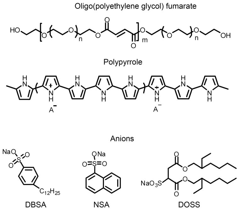
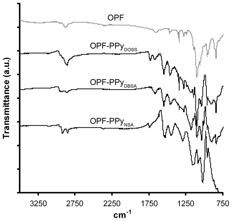
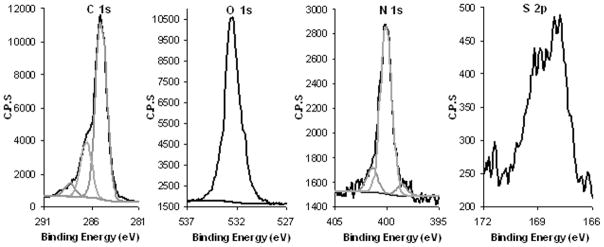
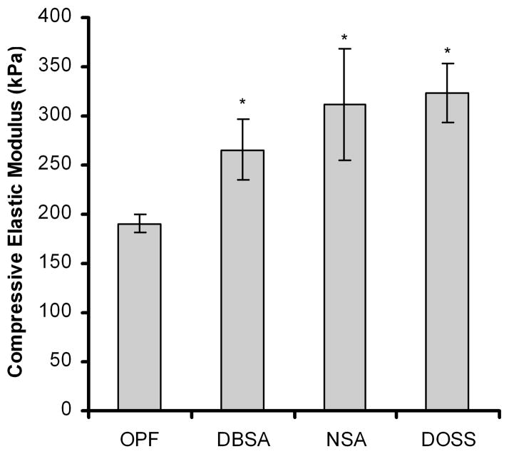
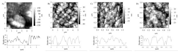
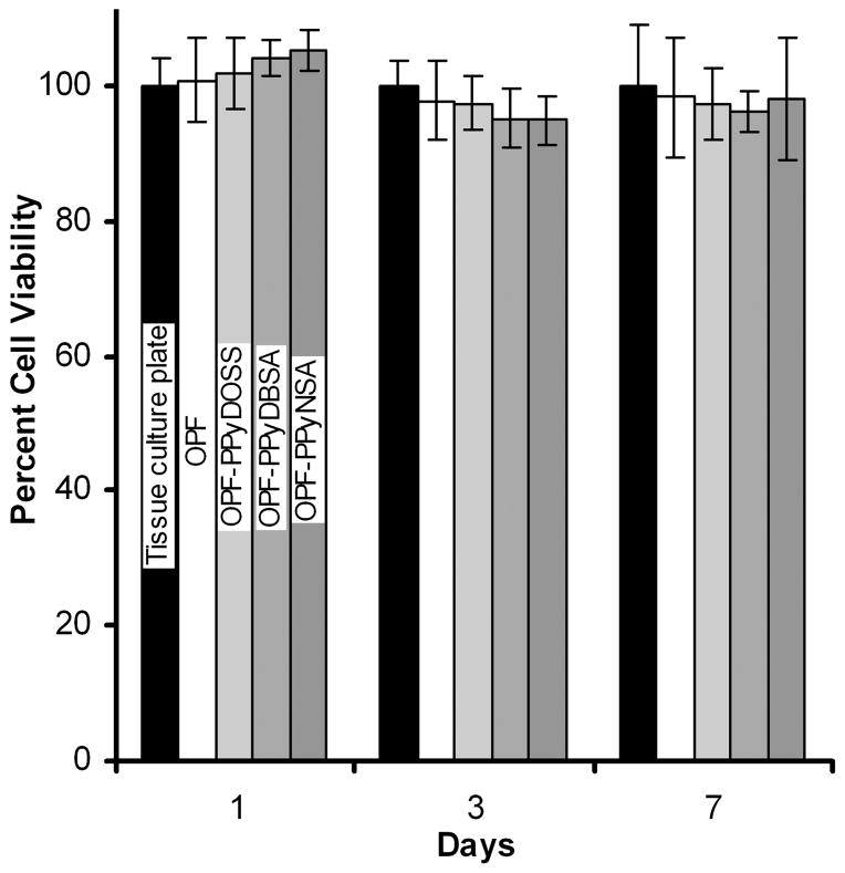
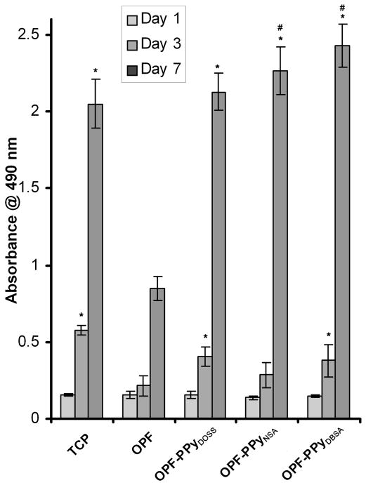
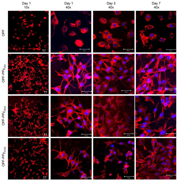
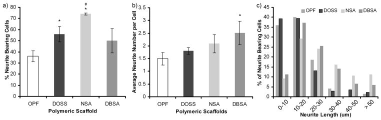
Electrically conductive hydrogel composites consisting of oligo(polyethylene glycol) fumarate (OPF) and polypyrrole (PPy) were developed for applications in nerve regeneration. OPF-PPy scaffolds were synthesized using three different anions: naphthalene-2-sulfonic acid sodium salt (NSA), dodecylbenzenesulfonic acid sodium salt (DBSA), and dioctyl sulfosuccinate sodium salt (DOSS). Scaffolds were characterized by ATR-FTIR, XPS, AFM, dynamic mechanical analysis, electrical resistivity measurements, and swelling experiments. OPF-PPy scaffolds were shown to consist of up to 25 mol % polypyrrole with a compressive modulus ranging from 265 to 323 kPa and a sheet resistance ranging from 6 to 30 × 10(3) Ohms/square. In vitro studies using PC12 cells showed OPF-PPy materials had no cytotoxicity and PC12 cells showed distinctly better cell attachment and an increase in the percent of neurite bearing cells on OPF-PPy materials compared to OPF. The neurite lengths of PC12 cells were significantly higher on OPF-PPyNSA and OPF-PPyDBSA. These results show that electrically conductive OPF-PPy hydrogels are promising candidates for future applications in nerve regeneration.
Conflict of interest statement
A provisional patent has been filed on oligo(polyethylene glycol) fumarate-polypyrrole based materials, and this technology has been licensed to BonWrx.
Figures
Similar articles
-
The development of electrically conductive polycaprolactone fumarate-polypyrrole composite materials for nerve regeneration.Biomaterials. 2010 Aug;31(23):5916-26. doi: 10.1016/j.biomaterials.2010.04.012. Epub 2010 May 21. Biomaterials. 2010. PMID: 20483452 Free PMC article.
-
In situ synthesis of robust conductive cellulose/polypyrrole composite aerogels and their potential application in nerve regeneration.Angew Chem Int Ed Engl. 2014 May 19;53(21):5380-4. doi: 10.1002/anie.201402751. Epub 2014 Apr 7. Angew Chem Int Ed Engl. 2014. PMID: 24711342
-
Material properties and electrical stimulation regimens of polycaprolactone fumarate-polypyrrole scaffolds as potential conductive nerve conduits.Acta Biomater. 2011 Mar;7(3):944-53. doi: 10.1016/j.actbio.2010.10.013. Epub 2010 Oct 20. Acta Biomater. 2011. PMID: 20965280 Free PMC article.
-
Injectable OPF/graphene oxide hydrogels provide mechanical support and enhance cell electrical signaling after implantation into myocardial infarct.Theranostics. 2018 May 12;8(12):3317-3330. doi: 10.7150/thno.25504. eCollection 2018. Theranostics. 2018. PMID: 29930732 Free PMC article.
-
In vivo bone and soft tissue response to injectable, biodegradable oligo(poly(ethylene glycol) fumarate) hydrogels.Biomaterials. 2003 Aug;24(19):3201-11. doi: 10.1016/s0142-9612(03)00168-6. Biomaterials. 2003. PMID: 12763447
Cited by
-
Conducting Polymers, Hydrogels and Their Composites: Preparation, Properties and Bioapplications.Polymers (Basel). 2019 Feb 17;11(2):350. doi: 10.3390/polym11020350. Polymers (Basel). 2019. PMID: 30960334 Free PMC article. Review.
-
Organic electrode coatings for next-generation neural interfaces.Front Neuroeng. 2014 May 27;7:15. doi: 10.3389/fneng.2014.00015. eCollection 2014. Front Neuroeng. 2014. PMID: 24904405 Free PMC article. Review.
-
Peripheral Nerve Regeneration Strategies: Electrically Stimulating Polymer Based Nerve Growth Conduits.Crit Rev Biomed Eng. 2015;43(2-3):131-59. doi: 10.1615/CritRevBiomedEng.2015014015. Crit Rev Biomed Eng. 2015. PMID: 27278739 Free PMC article.
-
Combined Optimized Effect of a Highly Self-Organized Nanosubstrate and an Electric Field on Osteoblast Bone Cells Activity.Biomed Res Int. 2019 Mar 21;2019:7574635. doi: 10.1155/2019/7574635. eCollection 2019. Biomed Res Int. 2019. PMID: 31016196 Free PMC article.
-
Injectable hydrogels in central nervous system: Unique and novel platforms for promoting extracellular matrix remodeling and tissue engineering.Mater Today Bio. 2023 Mar 22;20:100614. doi: 10.1016/j.mtbio.2023.100614. eCollection 2023 Jun. Mater Today Bio. 2023. PMID: 37008830 Free PMC article. Review.
References
-
- Dahlin L, Johansson F, Lindwall C, Kanje M. Int Rev Neurobiol. 2009;87:507–30. - PubMed
-
- Colen KL, Choi M, Chiu DT. Plast Reconstr Surg. 2009;124:e386–94. - PubMed
-
- Siemionow M, Brzezicki G. Int Rev Neurobiol. 2009;87:141–72. - PubMed
-
- Schmidt CE, Leach JB. Annu Rev Biomed Eng. 2003;5:293–347. - PubMed
-
- Cao H, Liu T, Chew SY. Adv Drug Deliv Rev. 2009;61:1055–1064. - PubMed
Publication types
MeSH terms
Substances
Grants and funding
LinkOut - more resources
Full Text Sources
Other Literature Sources
Miscellaneous
