

Reconstitution of gloeobacter rhodopsin with echinenone: role of the 4-keto group
- PMID: 20942439
- PMCID: PMC2995442
- DOI: 10.1021/bi1014166
Reconstitution of gloeobacter rhodopsin with echinenone: role of the 4-keto group
Abstract
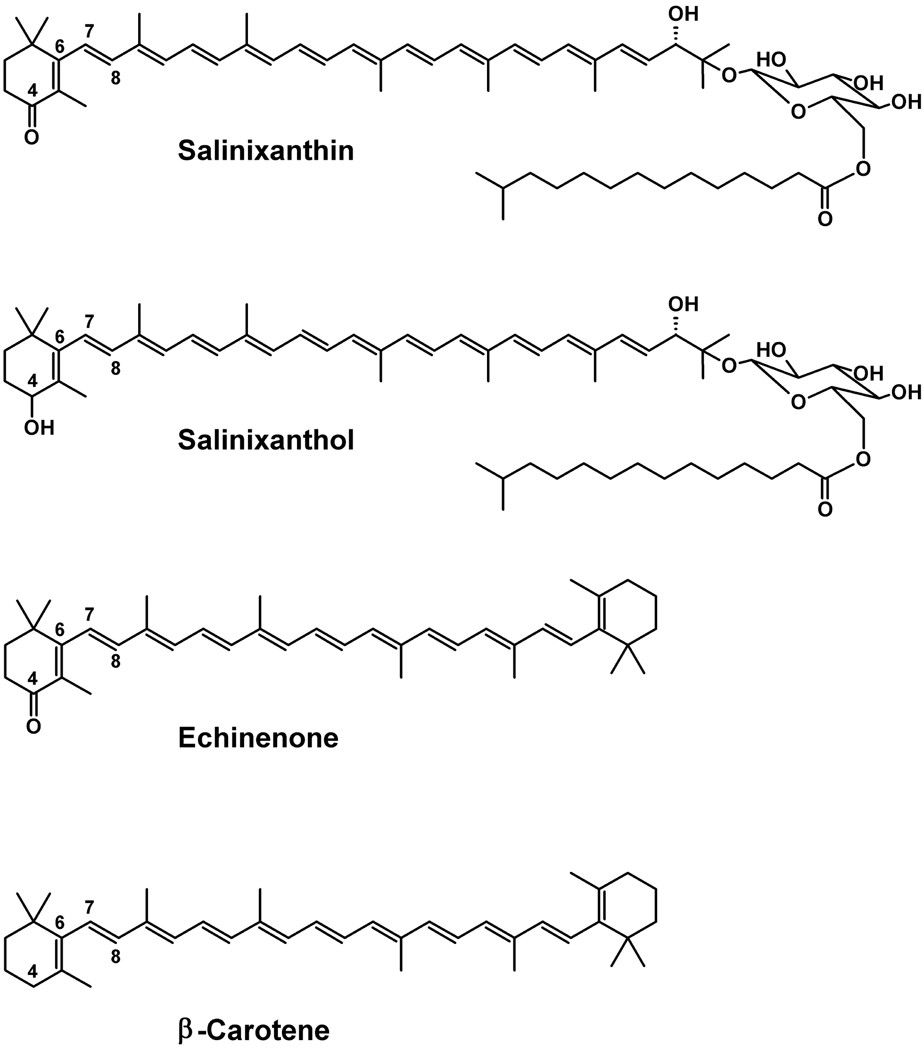
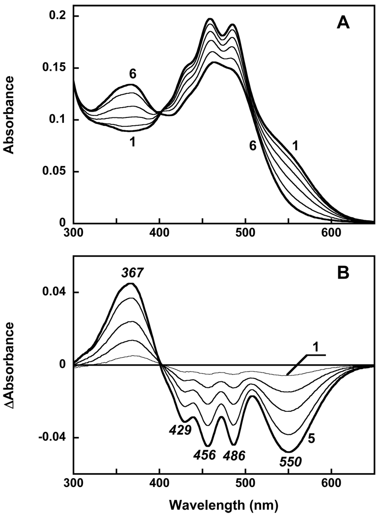
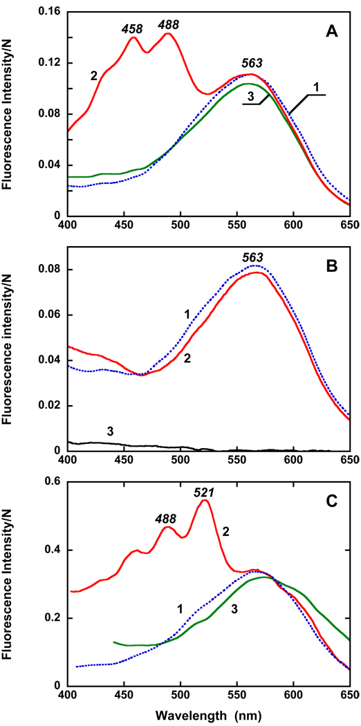
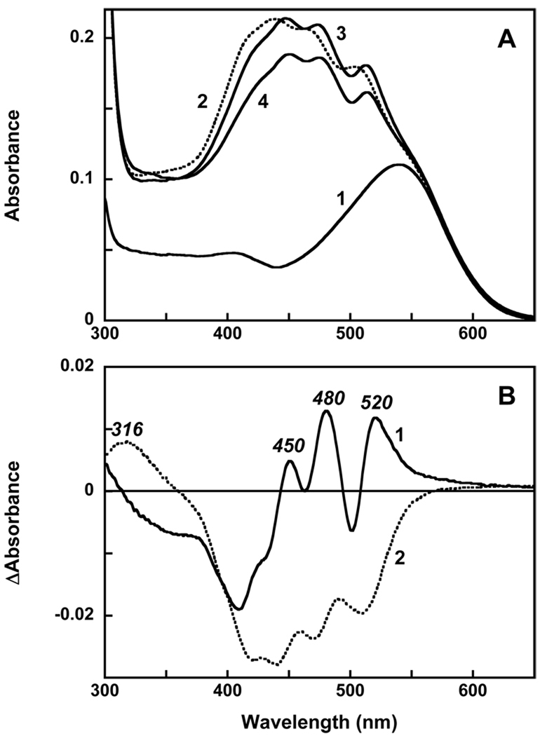
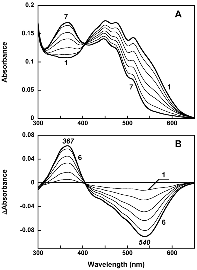
In previous work, we reconstituted salinixanthin, the C(40)-carotenoid acyl glycoside that serves as a light-harvesting antenna to the light-driven proton pump xanthorhodopsin, into a different protein, gloeobacter rhodopsin expressed in Escherichia coli, and demonstrated that it transfers energy to the retinal chromophore [Imasheva, E. S., et al. (2009) Biochemistry 48, 10948]. The key to binding of salinixanthin was the accommodation of its ring near the retinal β-ionone ring. Here we examine two questions. Do any of the native Gloeobacter carotenoids bind to gloeobacter rhodopsin, and does the 4-keto group of the ring play a role in binding? There is no salinixanthin in Gloeobacter violaceous, but a simpler carotenoid, echinenone, also with a 4-keto group but lacking the acyl glycoside, is present in addition to β-carotene and oscillol. We show that β-carotene does not bind to gloeobacter rhodopsin, but its 4-keto derivative, echinenone, does and functions as a light-harvesting antenna. This indicates that the 4-keto group is critical for carotenoid binding. Further evidence of this is the fact that salinixanthol, an analogue of salinixanthin in which the 4-keto group is reduced to hydroxyl, does not bind and is not engaged in energy transfer. According to the crystal structure of xanthorhodopsin, the ring of salinixanthin in the binding site is turned out of the plane of the polyene conjugated chain. A similar conformation is expected for echinenone in the gloeobacter rhodopsin. We suggest that the 4-keto group in salinixanthin and echinenone allows for the twisted conformation of the ring around the C6-C7 bond and probably is engaged in an interaction that locks the carotenoid in the binding site.
Figures
Similar articles
-
Reconstitution of Gloeobacter violaceus rhodopsin with a light-harvesting carotenoid antenna.Biochemistry. 2009 Nov 24;48(46):10948-55. doi: 10.1021/bi901552x. Biochemistry. 2009. PMID: 19842712 Free PMC article.
-
Removal and reconstitution of the carotenoid antenna of xanthorhodopsin.J Membr Biol. 2011 Jan;239(1-2):95-104. doi: 10.1007/s00232-010-9322-x. Epub 2010 Nov 21. J Membr Biol. 2011. PMID: 21104180 Free PMC article.
-
Retinal β-ionone ring-salinixanthin interactions in xanthorhodopsin: a study using artificial pigments.Biochemistry. 2013 Feb 19;52(7):1290-301. doi: 10.1021/bi301318n. Epub 2013 Feb 7. Biochemistry. 2013. PMID: 23331279
-
Xanthorhodopsin: Proton pump with a carotenoid antenna.Cell Mol Life Sci. 2007 Sep;64(18):2323-8. doi: 10.1007/s00018-007-7167-y. Cell Mol Life Sci. 2007. PMID: 17571211 Free PMC article. Review.
-
Light-driven ion-translocating rhodopsins in marine bacteria.Trends Microbiol. 2015 Feb;23(2):91-8. doi: 10.1016/j.tim.2014.10.009. Trends Microbiol. 2015. PMID: 25432080 Review.
Cited by
-
Genomic and phenotypic attributes of novel salinivibrios from stromatolites, sediment and water from a high altitude lake.BMC Genomics. 2014 Jun 13;15:473. doi: 10.1186/1471-2164-15-473. BMC Genomics. 2014. PMID: 24927949 Free PMC article.
-
Engineering a carotenoid-binding site in Dokdonia sp. PRO95 Na+-translocating rhodopsin by a single amino acid substitution.Photosynth Res. 2018 May;136(2):161-169. doi: 10.1007/s11120-017-0453-0. Epub 2017 Oct 5. Photosynth Res. 2018. PMID: 28983723
-
Carbon fixation and rhodopsin systems in microbial mats from hypersaline lakes Brava and Tebenquiche, Salar de Atacama, Chile.PLoS One. 2021 Feb 9;16(2):e0246656. doi: 10.1371/journal.pone.0246656. eCollection 2021. PLoS One. 2021. PMID: 33561170 Free PMC article.
-
Selective Choice of the Efficient Carotenoid Antenna by a Xanthorhodopsin: Controlling Factors for Binding and Excitation Energy Transfer.JACS Au. 2025 Jun 26;5(7):3070-3081. doi: 10.1021/jacsau.4c01243. eCollection 2025 Jul 28. JACS Au. 2025. PMID: 40747033 Free PMC article.
-
Integrated In Silico Analysis of Pathway Designs for Synthetic Photo-Electro-Autotrophy.PLoS One. 2016 Jun 23;11(6):e0157851. doi: 10.1371/journal.pone.0157851. eCollection 2016. PLoS One. 2016. PMID: 27336167 Free PMC article.
References
-
- Lanyi JK. Proton transfers in the bacteriorhodopsin photocycle. Biochim. Biophys. Acta. 2006;1757:1012–1018. - PubMed
-
- Béjà O, Aravind L, Koonin EV, Suzuki MT, Hadd A, Nguyen LP, Jovanovich SB, Gates CM, Feldman RA, Spudich JL, Spudich EN, DeLong EF. Bacterial rhodopsin: Evidence for a new type of phototrophy in the sea. Science. 2000;289:1902–1906. - PubMed
-
- Drachev LA, Frolov VN, Kaulen AD, Liberman EA, Ostroumov SA, Plakunova GV, Semenov AY, Skulachev VP. Reconstitution of biological molecular generators of electric current. Bacteriorhodopsin. J. Biol. Chem. 1976;251:7059–7065. - PubMed
-
- Michel H, Oesterhelt D. Light-induced changes of the pH gradient and the membrane potential in Halobacterium halobium. FEBS Lett. 1980;65:175–178. - PubMed
Publication types
MeSH terms
Substances
Grants and funding
LinkOut - more resources
Full Text Sources
Other Literature Sources
Miscellaneous
