. 2010 Oct 13:11:129.
doi: 10.1186/1471-2202-11-129.

Differential expression of a BMP4 reporter allele in anterior fungiform versus posterior circumvallate taste buds of mice

Affiliations

Differential expression of a BMP4 reporter allele in anterior fungiform versus posterior circumvallate taste buds of mice

Ha M Nguyen et al. BMC Neurosci. .

Abstract

Background: Bone Morphogenetic Protein 4 (BMP4) is a diffusible factor which regulates embryonic taste organ development. However, the role of BMP4 in taste buds of adult mice is unknown. We utilized transgenic mice with LacZ under the control of the BMP4 promoter to reveal the expression of BMP4 in the tongues of adult mice. Further we evaluate the pattern of BMP4 expression with that of markers of specific taste bud cell types and cell proliferation to define and compare the cell populations expressing BMP4 in anterior (fungiform papillae) and posterior (circumvallate papilla) tongue.

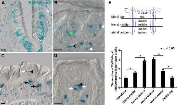
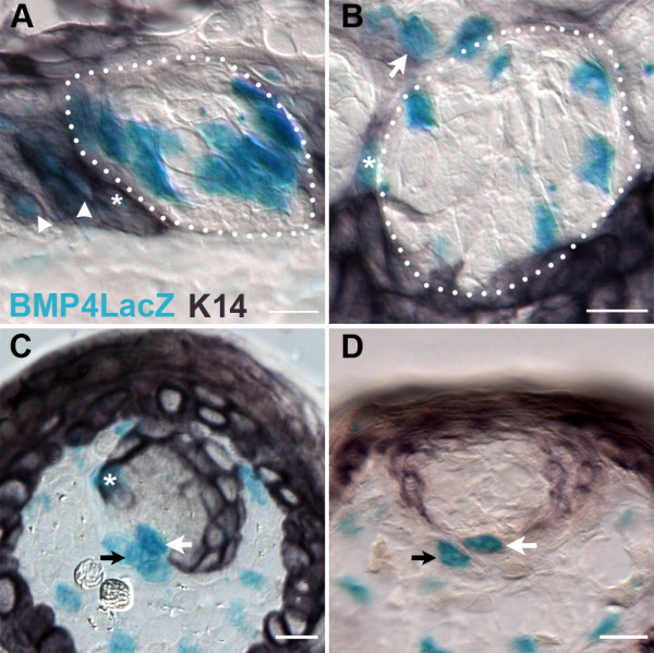
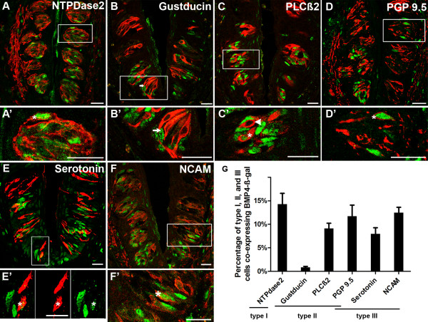
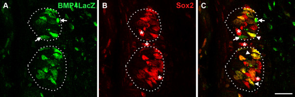
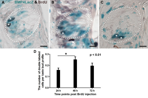
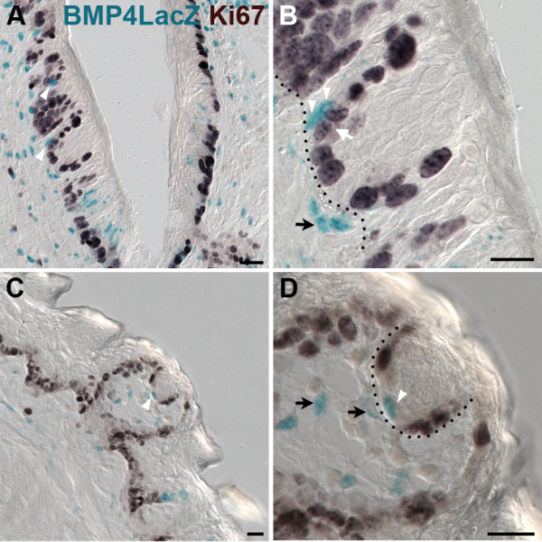
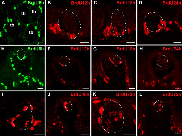
Results: BMP4 is expressed in adult fungiform and circumvallate papillae, i.e., lingual structures composed of non-taste epithelium and taste buds. Unexpectedly, we find both differences and similarities with respect to expression of BMP4-driven ß-galactosidase. In circumvallate papillae, many fusiform cells within taste buds are BMP4-ß-gal positive. Further, a low percentage of BMP4-expressing cells within circumvallate taste buds is immunopositive for markers of each of the three differentiated taste cell types (I, II and III). BMP4-positive intragemmal cells also expressed a putative marker of immature taste cells, Sox2, and consistent with this finding, intragemmal cells expressed BMP4-ß-gal within 24 hours after their final mitosis, as determined by BrdU birthdating. By contrast, in fungiform papillae, BMP4-ß-gal positive cells are never encountered within taste buds. However, in both circumvallate and fungiform papillae, BMP4-ß-gal expressing cells are located in the perigemmal region, comprising basal and edge epithelial cells adjacent to taste buds proper. This region houses the proliferative cell population that gives rise to adult taste cells. However, perigemmal BMP4-ß-gal cells appear mitotically silent in both fungiform and circumvallate taste papillae, as we do not find evidence of their active proliferation using cell cycle immunomarkers and BrdU birthdating.

Conclusion: Our data suggest that intragemmal BMP4-ß-gal cells in circumvallate papillae are immature taste cells which eventually differentiate into each of the 3 taste cell types, whereas perigemmal BMP4-ß-gal cells in both circumvallate and fungiform papillae may be slow cycling stem cells, or belong to the stem cell niche to regulate taste cell renewal from the proliferative cell population.


Figures

Figure 1
Figure 1
BMP4-ß-gal is expressed in both circumvallate and fungiform papillae of adult mice. A. In the circumvallate papilla, X-gal positive cells are evident in variable numbers within taste buds (intragemmal cells - one example outlined with white dashed line, and in papillary epithelium adjacent to taste buds (perigemmal cells, white asterisks), as well as in the subepithelial mesenchyme (white arrow). B. The heterogeneous nature of BMP4lacZ expression is evident at higher magnification. Some intragemmal fusiform cells express moderate levels of ß-galactosidase (green arrowhead), while others have higher levels (white arrowhead). Some basal epithelial cells are also strongly X-gal positive (black arrowhead), as are subepithelial lamina propria cells (white arrow). C. In fungiform papillae, BMP4-ß-gal is expressed in epithelial cells around fungiform taste buds (black arrowhead) and filiform papillae (black arrows), and is also evident in lamina propria beneath fungiform taste buds (white arrow). D. X-gal positive epithelial cells are common perigemmally in the epithelium (black arrowhead), as are positive mesenchymal cells (white arrows). E. A schematic diagram of circumvallate papillae is divided into six regions for each trench: lateral top, lateral middle, lateral bottom, medial bottom, medial middle, and medial top for each of the paired circumvallate trenches. The number of BMP4-ß-gal intragemmal cells per taste bud profile in the bottom of the papilla is significantly greater than in the middle part, and the middle part has significantly more BMP4-ß-gal cells than the top region (p < 0.01, Tukey comparison test); data indicate mean +/- standard error; n = 6 mice; at lease 50 taste buds were counted for each location. Scale bars: 20 μm.
Figure 2
Figure 2
BMP4-ß-gal is expressed in perigemmal cells of both circumvallate and fungiform papillae. A, B. X-gal positive cells in the circumvallate papilla comprise both extragemmal (A; white arrowheads) and perigemmal (A and B; white asterisks) K14-immunopositive cells. Some perigemmal BMP4-postivie cells are not K14-IR (B, white arrow). C, D. In fungiform papillae, perigemmal BMP4-LacZ expressing epithelial cells are K14-immunonegative (C, D; white arrows), or K14-IR (C, white asterisk). Black arrows indicate X-gal positive mesenchymal cells in close apposition to X-gal positive perigemmal epithelial cells. Scale bars: 20 μm.
Figure 3
Figure 3
BMP4-ß-gal is expressed in a subset of each differentiated taste cell type in circumvallate papillae. Double labeling of BMP4-ß-gal-IR (green nuclei) with differentiated taste cell immunomarkers (red) in circumvallate taste buds. A, A'. Double immunostaining for ß-gal and NTPDase2 (type I cells) reveals double labeled cells (arrowhead in A'). B, B'. In a double immunostained section for BMP4-ß-gal and Gustducin-IR (Type II cells), an arrow indicates one of several cells immunopositive for BMP4-ß-gal but not for gustducin. C, C'. Double staining for BMP4-ß-gal with PLCß2 (type II cells) showing both double labeled (white arrowhead) and singly labeled type II cells (white asterisk). D, D'. Double staining for BMP4-ß-gal with PGP9.5 (subset of type II and III cells) also reveals a double labeled cell (white arrowhead), as well as cells expressing one or the other immunomarker. E, E'. Double staining for BMP4-ß-gal with serotonin-IR (Type III cells) again shows single and double (white arrowhead) labeled taste cells. The boxed area shown in E, is shown in E' as a high magnification view of the merged and channels to better demonstrate double labeling F, F'. Double immunostaining for BMP4-ß-gal with NCAM-IR (type III cells) reveals double (arrowhead) and singly labeled taste cells. A',B',C',D',E', and F' are higher magnification of white boxes in A,B,C,D,E, and F respectively. Scale bars: 20 μm. G: Percentages of type I, II and III taste cells co-expressing BMP4-ß-gal in circumvallate taste buds (mean +/- standard error of the mean, n = 3 mice; 100 to 150 taste buds were counted for each taste cell type marker).
Figure 4
Figure 4
BMP4-ß-gal is expressed in taste cells expressing Sox2 in circumvallate papillae. BMP4-ß-gal-IR (A, green) is expressed in fusiform cells of circumvallate taste buds, and a large number of them are Sox2-IR (B, red). In C, white arrowheads indicate double-labeled cells. In B and C, white asterisks indicate intragemmal and perigemmal cells that are Sox2-IR only, while in A and C, white arrows point to BMP4-ß-gal-IR cells that are not Sox2-IR. Scale bar: 20 μm.
Figure 5
Figure 5
In circumvallate taste buds, intragemmal cells express BMP4-ß-gal within 24 hours of cell birth. Co-expression of BMP4-ß-gal (Xgal; blue) and BrdU (dark brown) was detected beginning at 24 hours after BrdU injection (A), and was still observed at 48 hours (B), and 72 hours (C). White asterisks indicate double-labeled intragemmal cells. An arrow in B points to a BrdU immunopositive perigemmal cell that does not express BMP4-ß-gal. D. The mean number of intragemmal cells labeled for both BrdU and BMP4-ß-gal is plotted with respect to time post-BrdU injection. The incidence of double labeled cells per bud profile peaks at 48 hrs. Taste buds were tallied throughout the circumvallate papilla, and only taste buds with a clear taste pore were included. (Mean +/- standard error, n = 3 mice for each time point, At least 200 taste buds were counted for each time point). Scale bars: 10 μm.
Figure 6
Figure 6
BMP4-ß-gal perigemmal cells in fungiform and circumvallate papillae are not actively cycling. BMP4-ß-gal expression (X-gal reaction; blue) and Ki-67 immunostaining (dark brown) in circumvallate (A,B) and fungiform papillae (C,D) reveals that cycling cells surround taste buds basolaterally, but that BMP4-ß-gal positive epithelial cells do not reside in this mitotic domain. In circumvallate papillae (A,B) and fungiform papillae (C,D), white arrowheads indicate BMP4-X-gal positive cells in the epithelium that are not Ki-67 positive, although X-gal positive epithelial cells are located near perigemmal Ki-67 immunopositive cells (e.g. white arrow in B). Black arrows indicate BMP4 expressing cells in adjacent mesenchyme, in close proximity to taste buds and BMP4-Xgal positive perigemmal epithelial cells. In B and D, the black dotted line delineates the basement membrane separating the epithelium and mesenchyme. Scale bars: 20 μm.
Figure 7
Figure 7
Proliferating and mitosing cells are basal epithelial and perigemmal cells in both fungiform and circumvallate taste papillae. Proliferating PCNA-IR cells (red) and mitosing pH3-IR cells (green) in the circumvallate (A) and fungiform papillae (B) are located perigemmally, and not inside taste buds (white dashed outlines). Scale bars: 50 μm.
Figure 8
Figure 8
Newly born taste cells enter taste buds in both circumvallate and fungiform papillae within 12-18 hours of birth. A,E. BrdU-IR cells are present in the basal epithelium and perigemmaly, around taste buds, at 6 hours post-injection in circumvallate papillae (A, transverse section) and fungiform papillae (E); tb = taste bud. B,F. At 12 hours post-injection, BrdU-IR cells are evident around taste buds, and some BrdU-IR cells are found inside taste buds in the circumvallate papilla (B circumvallate; F fungiform). C,G. In both circumvallate and fungiform papillae, BrdU-IR cells are present inside taste buds at 18 hours post-injection (C circumvallate; G fungiform). D,H. BrdU-IR cells reside in the basal compartment and along the inner limits of taste buds by 24 hours (D circumvallate; H fungiform). I,J,K,L. BrdU-IR cells are detected in more central regions of taste buds at 48 hours (I circumvallate; J fungiform) and 72 hours (K circumvallate; L fungiform), but are also still detected in the perigemmal zone (white arrows). Scale bars: 20 μm.

Similar articles

Cited by

References

    1. Pumplin DW, Getschman E, Boughter JD Jr, Yu C, Smith DV. Differential expression of carbohydrate blood-group antigens on rat taste-bud cells: relation to the functional marker alpha-gustducin. J Comp Neurol. 1999;415(2):230–239. doi: 10.1002/(SICI)1096-9861(19991213)415:2<230::AID-CNE7>3.0.CO;2-Y. - DOI - PubMed
    1. Lawton DM, Furness DN, Lindemann B, Hackney CM. Localization of the glutamate-aspartate transporter, GLAST, in rat taste buds. Eur J Neurosci. 2000;12(9):3163–3171. doi: 10.1046/j.1460-9568.2000.00207.x. - DOI - PubMed
    1. Bartel DL, Sullivan SL, Lavoie EG, Sevigny J, Finger TE. Nucleoside triphosphate diphosphohydrolase-2 is the ecto-ATPase of type I cells in taste buds. J Comp Neurol. 2006;497(1):1–12. doi: 10.1002/cne.20954. - DOI - PMC - PubMed
    1. Vandenbeuch A, Clapp TR, Kinnamon SC. Amiloride-sensitive channels in type I fungiform taste cells in mouse. BMC Neurosci. 2008;9:1. doi: 10.1186/1471-2202-9-1. - DOI - PMC - PubMed
    1. Bernhardt SJ, Naim M, Zehavi U, Lindemann B. Changes in IP3 and cytosolic Ca2+ in response to sugars and non-sugar sweeteners in transduction of sweet taste in the rat. J Physiol. 1996;490(Pt 2):325–336. - PMC - PubMed

Publication types

LinkOut - more resources