. 2010;12(5):R83.
doi: 10.1186/bcr2728. Epub 2010 Oct 13.

A novel approach for the generation of genetically modified mammary epithelial cell cultures yields new insights into TGFβ signaling in the mammary gland

Affiliations

A novel approach for the generation of genetically modified mammary epithelial cell cultures yields new insights into TGFβ signaling in the mammary gland

Ethan A Kohn et al. Breast Cancer Res. 2010.

Abstract

Introduction: Molecular dissection of the signaling pathways that underlie complex biological responses in the mammary epithelium is limited by the difficulty of propagating large numbers of mouse mammary epithelial cells, and by the inability of ribonucleic acid interference (RNAi)-based knockdown approaches to fully ablate gene function. Here we describe a method for the generation of conditionally immortalized mammary epithelial cells with defined genetic defects, and we show how such cells can be used to investigate complex signal transduction processes using the transforming growth factor beta (TGFβ/Smad pathway as an example.

Methods: We intercrossed the previously described H-2Kb-tsA58 transgenic mouse (Immortomouse) which expresses a temperature-sensitive mutant of the simian virus-40 large T-antigen (tsTAg), with mice of differing Smad genotypes. A panel of conditionally immortalized mammary epithelial cell (IMEC) cultures were derived from the virgin mammary glands of offspring of these crosses and used to assess the Smad dependency of different biological responses to TGFβ.

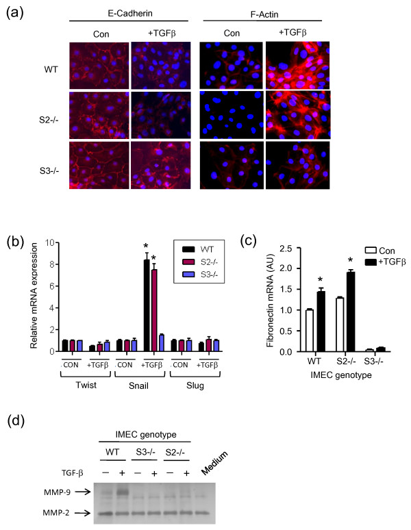
Results: IMECs could be propagated indefinitely at permissive temperatures and had a stable epithelial phenotype, resembling primary mammary epithelial cells with respect to several criteria, including responsiveness to TGFβ. Using this panel of cells, we demonstrated that Smad3, but not Smad2, is necessary for TGFβ-induced apoptotic, growth inhibitory and EMT responses, whereas either Smad can support TGFβ-induced invasion as long as a threshold level of total Smad is exceeded.

Conclusions: This work demonstrates the practicality and utility of generating conditionally immortalized mammary epithelial cell lines from genetically modified Immortomice for detailed investigation of complex signaling pathways in the mammary epithelium.


Figures

Figure 1
Figure 1
Using the Immortomouse in combination with genetically modified mice to generate conditionally immortalized cell cultures. General scheme for breeding strategy, generation and propagation of conditionally immortalized cells.
Figure 2
Figure 2
Morphology, proliferative capacity and intermediate filament expression of immortalized mammary epithelial cell cultures. (a) Western blot of tsTAg expression in immortalized mammary epithelial cells (IMECs) grown in the presence or absence of added IFNγ at permissive (33°C) or nonpermissive (37°C) temperatures as indicated. β-actin used as a loading control. (b) Phase contrast images of wildtype IMECs showing a cobblestone epithelial morphology under both permissive and nonpermissive conditions. White scale bar = 100 μm. (c) Proliferation of IMECs under the permissive and nonpermissive growth conditions, showing rapid loss of proliferative capacity under nonpermissive conditions. Results representative of two independent experiments. (d) Wildtype IMEC cultures were grown under nonpermissive conditions and the expression of intermediate filament proteins was assessed by immunofluorescence using pancytokeratin or vimentin antibodies. Swiss 3T3 fibroblasts were used as a positive control for vimentin expression. DAPI, 4',6-diamidino-2-phenylindole. (e) Flow cytometric analysis for expression of cytokeratins CK8 and CK14 in wildtype IMECs grown under nonpermissive conditions.
Figure 3
Figure 3
Functional characterization of immortalized mammary epithelial cell cultures. (a) Whole mount of a reconstituted mammary gland generated by transplantation of 2.5 × 10⁴ immortalized mammary epithelial cells (IMECs) into a cleared mammary fat pad. (b) PCR analysis of β-casein mRNA expression as an indicator of lactogenic differentiation. IMECs growing in two-dimensional culture under nonpermissive conditions were exposed to lactogenic hormones for 3 days prior to isolation of mRNA for analysis. RNA from HC11 cells treated in the same way and RNA isolated from a mid-pregnant mammary gland were used as positive controls. (c) Responsiveness to transforming growth factor beta (TGFβ)-mediated growth inhibition in IMECs and primary mammary epithelial cells (MECs) was determined under nonpermissive conditions by assessing incorporation of [3H]thymidine. Results are means of triplicate determinations. IMEC-A and IMEC-B are independently generated IMEC cultures derived from different mice, isolated several months apart. The responses of early (P20) and late (P64) passage cultures of IMEC-A were compared.
Figure 4
Figure 4
Characterization of immortalized mammary epithelial cell cultures with different Smad genotypes. (a) Western blot analysis of Smad2 and Smad3 expression in different immortalized mammary epithelial cell (IMEC) cultures. Smad2 was excised from IMECs that were homozygous for the conditional allele of Smad2 (Smad2fl/fl) by ex vivo exposure to adenovirus expressing Cre recombinase. Adenovirus expressing LacZ was used as a control for nonspecific effects of viral transduction. Mouse embryo fibroblasts (MEFs) derived from germline Smad2 knockout mice (S2KO) were used as a positive control for complete Smad2 deletion. β-actin used as a loading control. WT, wildtype. (b) Morphology of Smad null cultures under nonpermissive conditions. White scale bar = 100 μm. (c) Long-term growth curves for the various IMEC cultures grown under permissive conditions. Growth plotted as cumulative population doublings. Results are representative of two independent experiments. (d) Short-term growth curves for the various IMEC cultures after shifting to nonpermissive conditions. Results are the mean ± standard error of the mean for three independent experiments. (e) Western blot of Smad phosphorylation in response to transforming growth factor beta (TGFβ) (2 ng/ml for 30 minutes) in the IMECs of different genotypes. Note that the phospho-Smad3 antibody also recognizes phospho-Smad1, phospho-Smad5 and phospho-Smad8, which comprise the upper of the two bands on the phospho-Smad3 blot. (f) Quantitative RT-PCR assessment of TGFβ receptor type II (TGFBR2) mRNA. Data are normalized to the wildtype genotype. *P < 0.05 for expression difference between specified genotype and wildtype control (Student's t test). AU, arbitrary units.
Figure 5
Figure 5
Smad2 and Smad3 are differentially required for different TGFβ-mediated biological responses. Immortalized mammary epithelial cells (IMECs) of different Smad genotypes were shifted to nonpermissive temperatures and then assessed for their ability to respond to varying concentrations of transforming growth factor beta (TGFβ) in the following biological assays as described in Materials and methods: (a) growth inhibition, (b) apoptosis, (c) migration, and (d) invasion. For the growth inhibition and assays, all data are normalized to the control condition (no added TGFβ) for the respective genotype group. For migration and invasion, data are normalized to the wildtype (WT) IMECs, no added TGFβ control condition. LacZ indicates Smad2fl/fl cells that have been exposed to the LacZ control adenovirus and therefore have a wildtype complement of Smad2 and Smad3. Data are the mean ± standard deviation of three determinations. All experiments were repeated at least twice with essentially the same results. (a, b) *P < 0.05 for the difference between the indicated Smad genotype and wildtype control. (c, d) *P < 0.05 for the difference between specified pairwise comparisons.
Figure 6
Figure 6
Exogenous Smad2 or Smad3 expression restores TGFβ-mediated invasion in immortalized mammary epithelial cells lacking Smad. (a) Adenoviral expression of Smad2 in Smad2 null cells, or of Smad3 in Smad3 null cells, restores sensitivity to transforming growth factor beta (TGFβ)-induced invasion. (b) Adenoviral expression of Smad3 in Smad2 null cells, or of Smad2 in Smad3 null cells, largely restores sensitivity to TGFβ-induced invasion. All values normalized to wildtype (WT) control values. Results are the mean ± standard deviation of three determinations. *P < 0.05 for the difference between specified pairwise comparisons. Western blot analysis of Smad expression in adenovirally transduced immortalized mammary epithelial cells (IMECs) is shown for each experiment. β-actin used as a loading control. NS, not significant.
Figure 7
Figure 7
Smad dependency of induction of molecular markers of epithelial-to-mesenchymal transition and of MMP-9 by TGFβ. (a) Immunofluorescent staining for E-cadherin and F-actin following 48 hours with or without treatment with 2 ng/ml transforming growth factor beta (TGFβ). (b) Quantitative RT-PCR determination of mRNA levels for key transcriptional regulators of EMT under the same treatment conditions as (a). All data are normalized to the no TGFβ condition for each genotype. *P < 0.05 for the difference between the TGFβ-treated and control conditions. (c) Quantitative RT-PCR for fibronectin mRNA. Data normalized to the wildtype (WT) genotype, no TGFβ treatment. *P < 0.05 for the difference between the TGFβ-treated and control conditions. (d) Gelatin zymography of cell-conditioned medium following 48 hours with or without treatment with 2 ng/ml TGFβ. Medium indicates medium alone control; MMP-2 is present in the serum contained in the culture medium.

Similar articles

Cited by

References

    1. Massague J. TGFβ in Cancer. Cell. 2008;134:215–230. doi: 10.1016/j.cell.2008.07.001. - DOI - PMC - PubMed
    1. Derynck R, Miyazono K. The TGF-β Family. Cold Spring Harbor, NY: Cold Spring Harbor Laboratory Press; 2008. - PubMed
    1. Wakefield LM, Piek E, Bottinger EP. TGF-β signaling in mammary gland development and tumorigenesis. J Mammary Gland Biol Neoplasia. 2001;6:67–82. doi: 10.1023/A:1009568532177. - DOI - PubMed
    1. Flanders KC, Wakefield LM. Transforming growth factor-(β)s and mammary gland involution; functional roles and implications for cancer progression. J Mammary Gland Biol Neoplasia. 2009;14:131–144. doi: 10.1007/s10911-009-9122-z. - DOI - PMC - PubMed
    1. Tan AR, Alexe G, Reiss M. Transforming growth factor-β signaling: emerging stem cell target in metastatic breast cancer? Breast Cancer Res Treat. 2008;115:453–495. doi: 10.1007/s10549-008-0184-1. - DOI - PMC - PubMed

Publication types

MeSH terms

LinkOut - more resources