

Chemokine gene expression in lung CD8 T cells correlates with protective immunity in mice immunized intra-nasally with Adenovirus-85A
- PMID: 20942964
- PMCID: PMC2967494
- DOI: 10.1186/1755-8794-3-46
Chemokine gene expression in lung CD8 T cells correlates with protective immunity in mice immunized intra-nasally with Adenovirus-85A
Abstract
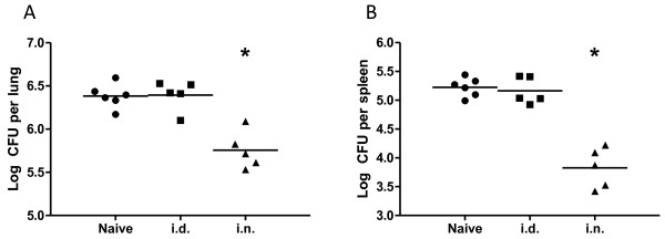
Background: Immunization of BALB/c mice with a recombinant adenovirus expressing Mycobacterium tuberculosis (M. tuberculosis) antigen 85A (Ad85A) protects against aerosol challenge with M. tuberculosis only when it is administered intra-nasally (i.n.). Immunization with Ad85A induces a lung-resident population of activated CD8 T cells that is antigen dependent, highly activated and mediates protection by early inhibition of M. tuberculosis growth. In order to determine why the i.n. route is so effective compared to parenteral immunization, we used microarray analysis to compare gene expression profiles of pulmonary and splenic CD8 T cells after i.n. or intra-dermal (i.d.) immunization.
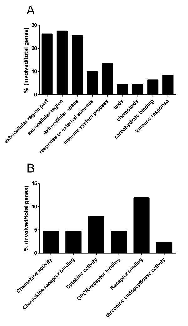
Method: Total RNA from CD8 T cells was isolated from lungs or spleens of mice immunized with Ad85A by the i.n. or i.d. route. The gene profiles generated from each condition were compared. Statistically significant (p ≤ 0.05) differentially expressed genes were analyzed to determine if they mapped to particular molecular functions, biological processes or pathways using Gene Ontology and Panther DB mapping tools.
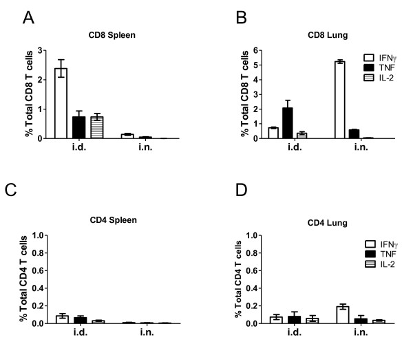
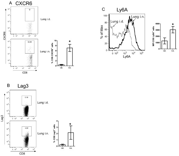
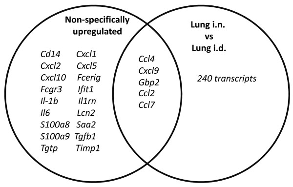
Results: CD8 T cells from lungs of i.n. immunized mice expressed a large number of chemokines chemotactic for resting and activated T cells as well as activation and survival genes. Lung lymphocytes from i.n. immunized mice also express the chemokine receptor gene Cxcr6, which is thought to aid long-term retention of antigen-responding T cells in the lungs. Expression of CXCR6 on CD8 T cells was confirmed by flow cytometry.
Conclusions: Our microarray analysis represents the first ex vivo study comparing gene expression profiles of CD8 T cells isolated from distinct sites after immunization with an adenoviral vector by different routes. It confirms earlier phenotypic data indicating that lung i.n. cells are more activated than lung i.d. CD8 T cells. The sustained expression of chemokines and activation genes enables CD8 T cells to remain in the lungs for extended periods after i.n. immunization. This may account for the early inhibition of M. tuberculosis growth observed in Ad85A i.n. immunized mice and explain the effectiveness of i.n. compared to parenteral immunization with this viral vector.
Figures
Similar articles
-
CXCR6 is a marker for protective antigen-specific cells in the lungs after intranasal immunization against Mycobacterium tuberculosis.Infect Immun. 2011 Aug;79(8):3328-37. doi: 10.1128/IAI.01133-10. Epub 2011 May 31. Infect Immun. 2011. PMID: 21628524 Free PMC article.
-
Immunization of mice with a recombinant adenovirus vaccine inhibits the early growth of Mycobacterium tuberculosis after infection.PLoS One. 2009 Dec 9;4(12):e8235. doi: 10.1371/journal.pone.0008235. PLoS One. 2009. PMID: 20011050 Free PMC article.
-
Mechanisms of mucosal and parenteral tuberculosis vaccinations: adenoviral-based mucosal immunization preferentially elicits sustained accumulation of immune protective CD4 and CD8 T cells within the airway lumen.J Immunol. 2005 Jun 15;174(12):7986-94. doi: 10.4049/jimmunol.174.12.7986. J Immunol. 2005. PMID: 15944305
-
Immunization with different formulations of Mycobacterium tuberculosis antigen 85A induces immune responses with different specificity and protective efficacy.Vaccine. 2013 Sep 23;31(41):4624-31. doi: 10.1016/j.vaccine.2013.07.040. Epub 2013 Jul 27. Vaccine. 2013. PMID: 23896422 Free PMC article.
-
[Novel vaccines against M. tuberculosis].Kekkaku. 2006 Dec;81(12):745-51. Kekkaku. 2006. PMID: 17240920 Review. Japanese.
Cited by
-
Gene Expression Profiling of Tuberculous Meningitis Co-infected with HIV.J Proteomics Bioinform. 2012 Sep;5(9):235-244. doi: 10.4172/jpb.1000243. Epub 2012 Sep 9. J Proteomics Bioinform. 2012. PMID: 27053842 Free PMC article.
-
Environmental effects on protection against Mycobacterium tuberculosis after immunization with Ad85A.Vaccine. 2013 Feb 4;31(7):1086-93. doi: 10.1016/j.vaccine.2012.12.024. Epub 2012 Dec 21. Vaccine. 2013. PMID: 23262169 Free PMC article.
-
The frequency and function of nucleoprotein-specific CD8+ T cells are critical for heterosubtypic immunity against influenza virus infection.J Virol. 2024 Aug 20;98(8):e0071124. doi: 10.1128/jvi.00711-24. Epub 2024 Jul 31. J Virol. 2024. PMID: 39082839 Free PMC article.
-
CXCR6-Deficiency Improves the Control of Pulmonary Mycobacterium tuberculosis and Influenza Infection Independent of T-Lymphocyte Recruitment to the Lungs.Front Immunol. 2019 Mar 7;10:339. doi: 10.3389/fimmu.2019.00339. eCollection 2019. Front Immunol. 2019. PMID: 30899256 Free PMC article.
-
Coinfection with Streptococcus pneumoniae negatively modulates the size and composition of the ongoing influenza-specific CD8⁺ T cell response.J Immunol. 2014 Nov 15;193(10):5076-87. doi: 10.4049/jimmunol.1400529. Epub 2014 Oct 13. J Immunol. 2014. PMID: 25311807 Free PMC article.
References
-
- Wang J, Thorson L, Stokes RW, Santosuosso M, Huygen K, Zganiacz A, Hitt M, Xing Z. Single mucosal, but not parenteral, immunization with recombinant adenoviral-based vaccine provides potent protection from pulmonary tuberculosis. J Immunol. 2004;173(10):6357–6365. - PubMed
Publication types
MeSH terms
Substances
Grants and funding
LinkOut - more resources
Full Text Sources
Other Literature Sources
Medical
Molecular Biology Databases
Research Materials
Miscellaneous
