. 2010 Dec;31(36):9422-30.
doi: 10.1016/j.biomaterials.2010.08.064. Epub 2010 Oct 12.

Phosphorylation-dependent mineral-type specificity for apatite-binding peptide sequences

Affiliations

Phosphorylation-dependent mineral-type specificity for apatite-binding peptide sequences

William N Addison et al. Biomaterials. 2010 Dec.

Abstract

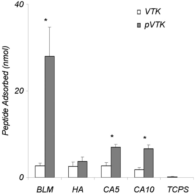
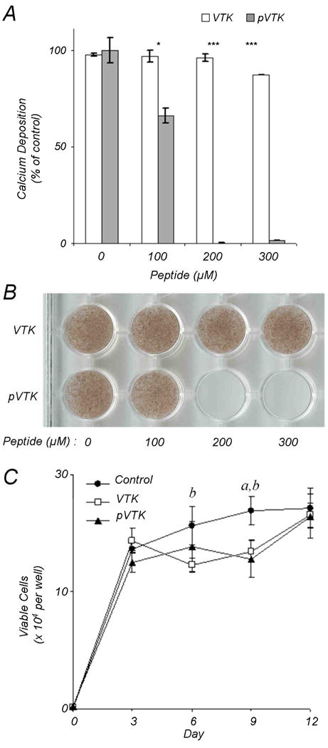
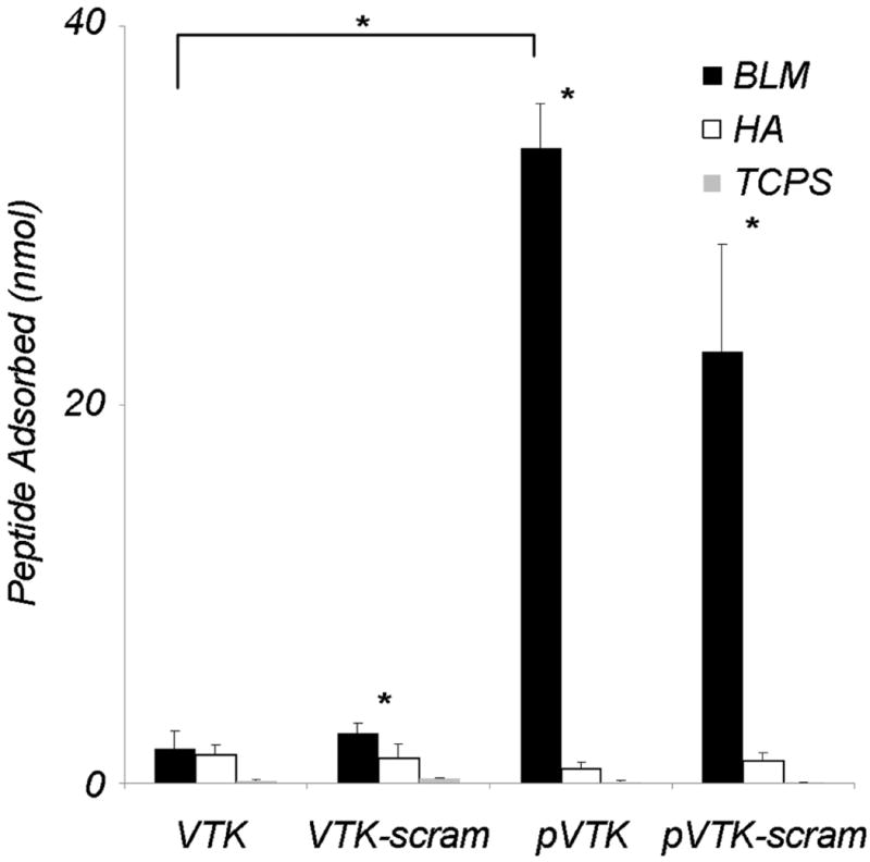
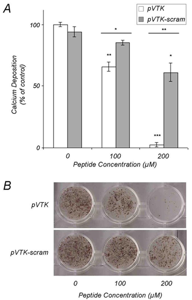
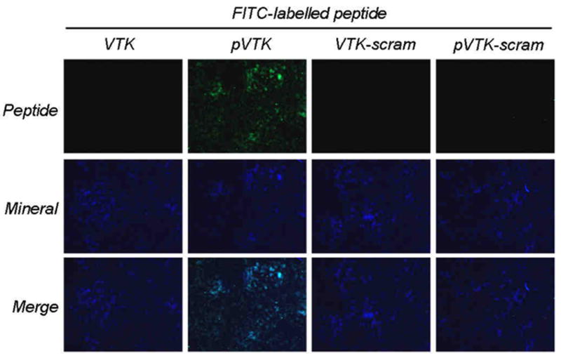
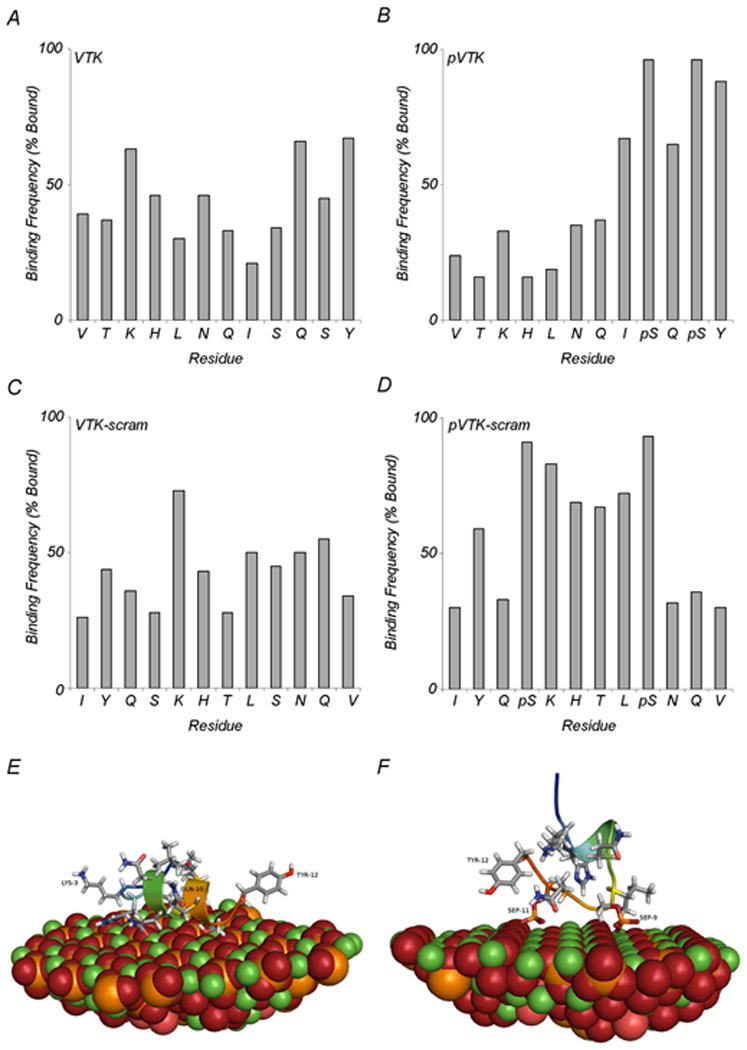
Apatite-binding peptides discovered by phage display provide an alternative design method for creating functional biomaterials for bone and tooth tissue repair. A limitation of this approach is the absence of display peptide phosphorylation--a post-translational modification important to mineral-binding proteins. To refine the material specificity of a recently identified apatite-binding peptide, and to determine critical design parameters (net charge, charge distribution, amino acid sequence and composition) controlling peptide affinity for mineral, we investigated the effects of phosphorylation and sequence scrambling on peptide adsorption to four different apatites (bone-like mineral, and three types of apatite containing initially 0, 5.6 and 10.5% carbonate). Phosphorylation of the VTKHLNQISQSY peptide (VTK peptide) led to a 10-fold increase in peptide adsorption (compared to nonphosphorylated peptide) to bone-like mineral, and a 2-fold increase in adsorption to the carbonated apatite, but there was no effect of phosphorylation on peptide affinity to pure hydroxyapatite (without carbonate). Sequence scrambling of the nonphosphorylated VTK peptide enhanced its specificity for the bone-like mineral, but scrambled phosphorylated VTK peptide (pVTK) did not significantly alter mineral-binding suggesting that despite the importance of sequence order and/or charge distribution to mineral-binding, the enhanced binding after phosphorylation exceeds any further enhancement by altered sequence order. Osteoblast culture mineralization was dose-dependently inhibited by pVTK and to a significantly lesser extent by scrambled pVTK, while the nonphosphorylated and scrambled forms had no effect, indicating that inhibition of osteoblast mineralization is dependent on both peptide sequence and charge. Computational modeling of peptide-mineral interactions indicated a favorable change in binding energy upon phosphorylation that was unaffected by scrambling. In conclusion, phosphorylation of serine residues increases peptide specificity for bone-like mineral, whose adsorption is determined primarily by sequence composition and net charge as opposed to sequence order. However, sequence order in addition to net charge modulates the mineralization of osteoblast cultures. The ability of such peptides to inhibit mineralization has potential utility in the management of pathologic calcification.


Figures

Fig. 1
Fig. 1
Adsorption of VTK and phosphorylated VTK (pVTK) peptides on bone-like mineral (BLM) layer, and pressed and sintered disks prepared from hydroxyapatite (HA), 5.6% carbonated apatite (CA5), and 10.5% carbonated apatite (CA10), and tissue culture polystyrene (TCPS). Phosphorylating serine residues in the VTK peptide increased peptide adsorption onto BLM 10-fold in comparison to nonphosphorylated VTK. While less, a significant increase in pVTK binding was also observed for the CA5 and CA10 substrates. Data are presented as means ± S.D. * denotes statistical differences between adsorption of VTK vs. pVTK on a given substrate (p < 0.05).
Fig. 2
Fig. 2
Mineralizing MC3T3-E1 osteoblast cultures were incubated with VTK and pVTK at the indicated concentrations for 12 days followed by mineral quantification by (A) calcium content determination expressed as a percentage of untreated control cultures, and (B) von Kossa (silver nitrate) staining for mineral. Data are presented as means ± S.D. *p < 0.05; ***p < 0.001 from Student's t-test for statistical differences between the two peptides at a given dose. (C) Cell proliferation in osteoblast cultures treated with, or without, 200 μM pVTK and VTK as measured by MTT assay. a denotes statistical significance between Control and VTK at the given time point. b denotes statistical significance between Control and pVTK at the given time point.
Fig. 3
Fig. 3
Adsorption of fluorescently-labelled VTK, phosphorylated VTK (pVTK), scrambled VTK (VTK-scram) and scrambled phosphorylated VTK (pVTK-scram) on bone-like mineral (BLM), hydroxyapatite (HA) and tissue culture polystyrene (TCPS). pVTK and pVTK-scram showed significantly higher adsorption to BLM than VTK. All peptides except VTK showed significantly higher binding to BLM than HA. Substrate specificity of the peptides was confirmed by absence of binding to TCPS. Bracket denotes a statistical difference between peptides on BLM (p < 0.05). Data are presented as means ± S.D. * denotes statistical differences between adsorption on BLM and HA for a given peptide (p < 0.01)
Fig. 4
Fig. 4
Effect of scrambling pVTK on inhibition of osteoblast culture mineralization. MC3T3-E1 osteoblast cultures were treated with the indicated concentrations of pVTK and scrambled pVTK (pVTK-scram) for 12 days followed by (A) quantification of mineralization by calcium content determination expressed as a percentage of untreated control cultures, and (B) von Kossa (silver nitrate) staining for visualization of mineral. Data are presented as means ± S.D. *p < 0.05; **p < 0.01; ***p < 0.001 from Student's t-test relative to the untreated control cultures or between the two peptides at a given dose (horizontal bars).
Fig. 5
Fig. 5
Binding of peptides to osteoblast culture mineral. Maximally mineralized (12-day) MC3T3-E1 osteoblast cultures were incubated with 150 μM fluorescently tagged peptides for 1 h and examined by fluorescence microscopy for peptides (green) and calcein blue-labelled mineral (blue). pVTK co-localized with mineral indicating peptide binding to apatite.
Fig. 6
Fig. 6
Molecular modeling of peptide binding to a high-calcium density (100) crystallographic face of hydroxyapatite. (A-D) Adsorption frequency of individual residues in the 100 lowest-energy adsorbed state structures. Bound residues are defined as residues closer than 5Å for (A) VTK, (B) pVTK, (C) VTK-scram and (D) pVTK-scram. (E) Structural details of a representative high-scoring VTK model illustrating binding of LYS-3, GLN-10 and TYR-12. (F) Structural details of pVTK model illustrating binding of TYR-12, PSer-9 (SEP-9) and PSer-11 (SEP-11). (Ca, green; P, orange; O, red; H, white; N, blue).

References

    1. Shin H, Jo S, Mikos AG. Biomimetic materials for tissue engineering. Biomaterials. 2003;24(24):4353–4364. - PubMed
    1. Hubbell JA. Bioactive biomaterials. Curr Opin Biotechnol. 1999;10(2):123–129. - PubMed
    1. Geesink RGT. Osteoconductive coatings for total joint arthroplasty. Clin Orthop Relat Res. 2002;395:53–65. - PubMed
    1. Alsberg E, Hill EE, Mooney DJ. Craniofacial Tissue Engineering. Crit Rev Oral Biol & Med. 2001;12(1):64–75. - PubMed
    1. Kokubo T. Apatite formation on organic polymers by a biomimetic process. Eur J Solid State Inorg Chem. 1995;32(7-8):819–827.

Publication types