. 2011 May;10(5):M110.001636.
doi: 10.1074/mcp.M110.001636. Epub 2010 Oct 12.

Global profiling of proteolysis during rupture of Plasmodium falciparum from the host erythrocyte

Affiliations

Global profiling of proteolysis during rupture of Plasmodium falciparum from the host erythrocyte

Paul W Bowyer et al. Mol Cell Proteomics. 2011 May.

Abstract

The obligate intracellular parasite pathogen Plasmodium falciparum is the causative agent of malaria, a disease that results in nearly one million deaths per year. A key step in disease pathology in the human host is the parasite-mediated rupture of red blood cells, a process that requires extensive proteolysis of a number of host and parasite proteins. However, only a relatively small number of specific proteolytic processing events have been characterized. Here we describe the application of the Protein Topography and Migration Analysis Platform (PROTOMAP) (Dix, M. M., Simon, G. M., and Cravatt, B. F. (2008) Global mapping of the topography and magnitude of proteolytic events in apoptosis. Cell 134, 679-691; Simon, G. M., Dix, M. M., and Cravatt, B. F. (2009) Comparative assessment of large-scale proteomic studies of apoptotic proteolysis. ACS Chem. Biol. 4, 401-408) technology to globally profile proteolytic events occurring over the last 6-8 h of the intraerythrocytic cycle of P. falciparum. Using this method, we were able to generate peptographs for a large number of proteins at 6 h prior to rupture as well as at the point of rupture and in purified merozoites after exit from the host cell. These peptographs allowed assessment of proteolytic processing as well as changes in both protein localization and overall stage-specific expression of a large number of parasite proteins. Furthermore, by using a highly selective inhibitor of the cysteine protease dipeptidyl aminopeptidase 3 (DPAP3) that has been shown to be a key regulator of host cell rupture, we were able to identify specific substrates whose processing may be of particular importance to the process of host cell rupture. These results provide the first global map of the proteolytic processing events that take place as the human malarial parasite extracts itself from the host red blood cell. These data also provide insight into the biochemical events that take place during host cell rupture and are likely to be valuable for the study of proteases that could potentially be targeted for therapeutic gain.


Figures
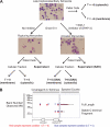
Fig. 1.
Fig. 1.
PROTOMAP analysis of parasites rupturing from host red blood cells. A, experimental setup used to generate samples for analysis by PROTOMAP. Synchronously grown parasites were isolated in the late trophozoite/early schizont stage (approximately 42 h postinfection). Samples for analysis (bold labels) were prepared as indicated. Representative stained blood smears are shown to indicate parasite morphology at each sample point. Final parasite or supernatant samples were subjected to SDS-PAGE, and each lane was excised into 26 slices followed by analysis by LC-MS/MS. B, representative peptograph generated from the data set collected from the samples in A. Peptides (colored bars) are mapped to the location on the protein sequence (top of gel image). Approximate molecular weight (MW) positions are shown at left, and total spectral counts for each peptide are shown at right. The colors are used to compare data obtained for this protein in each of two samples. In this case, peptides identified in the T = −6 are shown in red, and peptides identified in the T = 0 sample are shown in blue. A conserved domain map based on published predictions is shown at the bottom of the peptograph (21, 22). Spectral count data error bars are standard error of the mean.
Fig. 2.
Fig. 2.
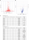
Analysis of PROTOMAP data set confirms stage-specific expression of proteins. A, plot of the total number of spectra counts observed for each protein versus the log2 -fold change between the early (T = −6) and late (T = 0) time points. This allows selection of proteins with the greatest life cycle stage-specific expression. B, plot of the log10 p values for each protein identified relative to the log2 -fold change in spectral counts from the early to late time points. A line of p = 0.05 is shown. C, list of the proteins most significantly enriched in the late time point (top) and early time point (bottom). Accession numbers, number of hours postinfection of maximum and minimum gene expression, and corresponding gene ontology functional annotations as reported in PlasmoDB (www.plasmodb.org) are shown. Pf-iRBC, P. falciparum-infected RBC; min expr, minimum expression; max expr, maximum expression; spz, sporozoite; Gam, gametocyte.
Fig. 3.
Fig. 3.
Identification of signal peptide removal and proteolytic maturation of proteins using PROTOMAP data. A, list of predicted signal peptides for which corresponding semitryptic peptides corresponding to the resultant mature N terminus were found. Predicted signal peptides were determined from neural network (NN) and hidden Markov model (HMM) predictions obtained from PlasmoDB. Protein accession numbers, gene ontology annotations, and total spectral counts for each peptide are also listed. Note that two peptides did not match the top predicted signal peptide cleavage site but matched a second, more weakly predicted site (indicated with **). B, list of identified proteolytic maturation sites of seven proteins known to undergo proteolytic maturation. The gene accession numbers and proteins names are shown along with the type of predicted processing expected and total number of spectral counts for identified semitryptic peptides. Periods in sequences represent cleavage sites. HAP, is plasmepsin III or histo-aspartic protease.
Fig. 4.
Fig. 4.
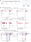
Peptographs identify time-dependent proteolytic processing events. A, peptographs for falcipain-3 (PF11_0162) at time T = −6 (red) and T = 0 (blue) for membrane (left) and cytosolic (right) samples. The sequences of the semitryptic peptide identified as the N-terminal most peptide in the mature 25-kDa fragment are shown in both membrane and cytosolic samples. The tryptic peptide corresponding to the mature N terminus plus an additional two N-terminal residues was identified in the full-length proform as shown. A Western blot of the same sample used for PROTMAP analysis using an anti-falcipain-3 antibody is shown for comparison with location of peptide identified in the peptographs. B, peptographs showing time-dependent processing of SERA-4 and SERA-7. The predicted Sub-1 cleavage site on SERA-4 that was identified as a semitryptic peptide is shown. * identifies the predicted size of full-length proteins (PF11_0162, 56.6 kDa; PFB0345c, 108.6 kDa; PFB0330c, 109.6 kDa). Periods in sequences represent cleavage sites.
Fig. 5.
Fig. 5.
Identification of proteolytic processing events that depend on DPAP3 activity. A, list and Venn diagram of the total number of proteins identified that were either dependent or independent of DPAP3 activity. Proteins listed as both showed multiple processing events with at least one event dependent on DPAP3 activity. B, examples of peptographs that show DPAP3-dependent processing events. C, example of a peptograph that shows multiple processing events with only one showing DPAP3 dependence. * identifies the predicted size of full-length proteins (PFB0330c, 109.6 kDa; PF08_0087, 61.1 kDa; PFI1475w, 195.7 kDa).
Fig. 6.
Fig. 6.
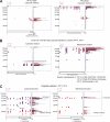
Mining PROTOMAP data set for additional biologically relevant data. A, peptographs of two proteins comparing T = 0 (blue) with T = −6 (red) samples to demonstrate constitutive processing to produce two stable fragments. B, peptographs comparing T = 0 (blue) with T = −6 (red) for both membrane and cytosolic samples, demonstrating a time-independent processing event that results in conversion of a full-length membrane-bound protein to a cytosolic peptide fragment. C, peptographs from the cytosolic fraction showing greater processing of DPAP1 at T = −6 compared with T = 0. * identifies the predicted size of full-length proteins (PF10_0320, 246.7 kDa; PFL2505c, 263.1 kDa; PF14_0541, 76.4 kDa; PF11_0174, 80.4 kDa).

Similar articles

Cited by

References

    1. Arastu-Kapur S., Ponder E. L., Fonoviæ U. P., Yeoh S., Yuan F., Fonoviæ M., Grainger M., Phillips C. I., Powers J. C., Bogyo M. (2008) Identification of proteases that regulate erythrocyte rupture by the malaria parasite Plasmodium falciparum. Nat. Chem. Biol. 4, 203–213 - PubMed
    1. Chandramohanadas R., Davis P. H., Beiting D. P., Harbut M. B., Darling C., Velmourougane G., Lee M. Y., Greer P. A., Roos D. S., Greenbaum D. C. (2009) Apicomplexan parasites co-opt host calpains to facilitate their escape from infected cells. Science 324, 794–797 - PMC - PubMed
    1. Yeoh S., O'Donnell R. A., Koussis K., Dluzewski A. R., Ansell K. H., Osborne S. A., Hackett F., Withers-Martinez C., Mitchell G. H., Bannister L. H., Bryans J. S., Kettleborough C. A., Blackman M. J. (2007) Subcellular discharge of a serine protease mediates release of invasive malaria parasites from host erythrocytes. Cell 131, 1072–1083 - PubMed
    1. Gevaert K., Goethals M., Martens L., Van Damme J., Staes A., Thomas G. R., Vandekerckhove J. (2003) Exploring proteomes and analyzing protein processing by mass spectrometric identification of sorted N-terminal peptides. Nat. Biotechnol. 21, 566–569 - PubMed
    1. Impens F., Colaert N., Helsens K., Ghesquière B., Timmerman E., De Bock P. J., Chain B. M., Vandekerckhove J., Gevaert K. (2010) A quantitative proteomics design for systematic identification of protease cleavage events. Mol. Cell. Proteomics 9, 2327–2333 - PMC - PubMed

Publication types

MeSH terms