. 2010 Dec 10;285(50):39303-13.
doi: 10.1074/jbc.M110.181610. Epub 2010 Oct 13.

Activity and cellular functions of the deubiquitinating enzyme and polyglutamine disease protein ataxin-3 are regulated by ubiquitination at lysine 117

Affiliations

Activity and cellular functions of the deubiquitinating enzyme and polyglutamine disease protein ataxin-3 are regulated by ubiquitination at lysine 117

Sokol V Todi et al. J Biol Chem. .

Abstract

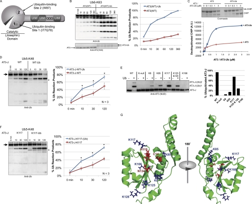
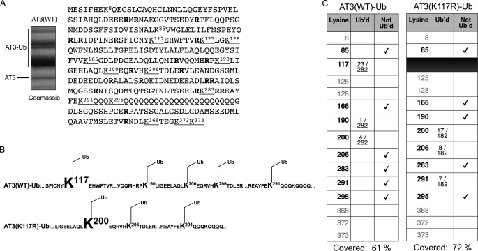
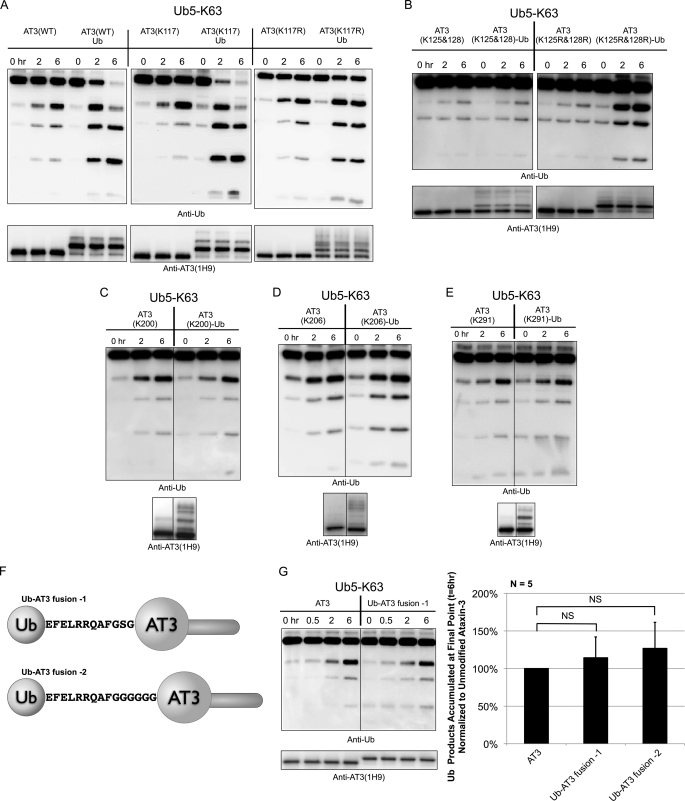
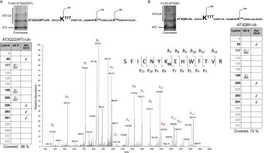
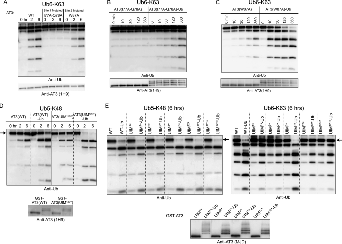
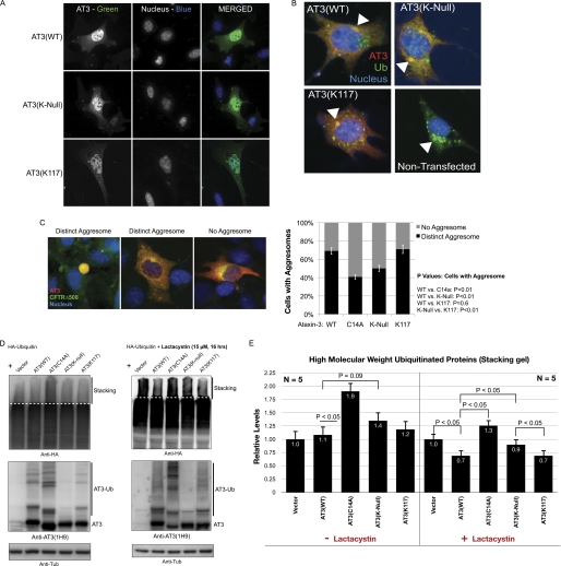
Deubiquitinating enzymes (DUbs) play important roles in many ubiquitin-dependent pathways, yet how DUbs themselves are regulated is not well understood. Here, we provide insight into the mechanism by which ubiquitination directly enhances the activity of ataxin-3, a DUb implicated in protein quality control and the disease protein in the polyglutamine neurodegenerative disorder, Spinocerebellar Ataxia Type 3. We identify Lys-117, which resides near the catalytic triad, as the primary site of ubiquitination in wild type and pathogenic ataxin-3. Further studies indicate that ubiquitin-dependent activation of ataxin-3 at Lys-117 is important for its ability to reduce high molecular weight ubiquitinated species in cells. Ubiquitination at Lys-117 also facilitates the ability of ataxin-3 to induce aggresome formation in cells. Finally, structure-function studies support a model of activation whereby ubiquitination at Lys-117 enhances ataxin-3 activity independent of the known ubiquitin-binding sites in ataxin-3, most likely through a direct conformational change in or near the catalytic domain.


Figures

FIGURE 1.
FIGURE 1.
Lysine 117 in the Josephin domain is the preferred site of ubiquitination. A, ataxin-3 domain structure. The catalytic (Josephin) domain comprises the catalytic site and two Ub-binding sites. Site 1 is close to the catalytic groove, centered on residues Ile-77 and Gln-78; site 2, distant from the catalytic triad, is centered on Trp-87. The C-terminal half of AT3 contains three UIMs flanking the poly(Q) region. B, left, Ub6-Lys-63 chains were incubated with unmodified, wild type AT3 (AT3(WT)) or ubiquitinated, wild type AT3 (AT3(WT)-Ub), and fractions were collected at the indicated time points. Right, densitometry analysis of data from the left. The results are representative of over 10 similar independent experiments. Lys-63-linked chains were used as the substrate because they are cleaved preferentially to Lys-48-linked Ub chains by full-length AT3 in vitro (18). AT3-Ub species persist over time in DUb reactions because AT3 does not deubiquitinate itself completely (28). C, top, deubiquitination of monoubiquitinated CHIP by AT3 or AT3-Ub. CHIP was monoubiquitinated in vitro, purified, and then mixed with either unmodified AT3 or ubiquitinated AT3 for 10 min. Bottom, densitometry analysis of data from the top. The example is representative of three independently conducted experiments. A.U., arbitrary units. D, left, unmodified Josephin domain (AT3-J) and ubiquitinated AT3-J (prepared as in panel E) were incubated with Ub5-Lys-48 chains, and fractions were collected at the indicated time points. Right, densitometry analyses of the experiment from the left panel and other similar experiments, n = 3. Error bars indicate S.D. Asterisks: p < 0.05. Lys-48-linked chains were used as the substrate in panels D and F because they are cleaved preferentially to Lys-63-linked chains by the Josephin domain (17). E, GST-tagged, Josephin domain constructs (AT3-J) that were wild type (WT), lacking all six lysines (K-null), or retaining only specific lysine residues as indicated were ubiquitinated in vitro by CHIP-Ubch5c, affinity-purified, cleaved from GST, and analyzed by immunoblot. Bands correspond to unmodified and mono- and diubiquitinated AT3-J. These mixtures of ubiquitinated and unmodified AT3-J species were used in DUb reactions in panels D and F. Densitometry analyses of species in the left panel are shown on the right. Results are representative of independent experiments conducted at least three times. F, left, samples prepared as in panel E were incubated with Ub5-Lys-48 chains, and fractions were collected at the indicated time points. Right, densitometry analyses of the experiment from the left and other similar experiments. n = 3. Error bars indicate S.D. Asterisks: p < 0.05. For the left panels in D and F, the arrow denotes the disappearance of the starting substrate (Ub5-K48). The asterisk near blots denotes ubiquitinated AT3-J, which is detectable by anti-Ub antibody. G, solution structure of the Josephin domain highlighting catalytic residues (red) and lysine residues (blue; based on the structure from Ref. 34). AT3-J-K117-(Ub), ubiquitinated AT3-J containing only Lys-117.
FIGURE 2.
FIGURE 2.
Lys-117 is the preferred ubiquitination site for full-length AT3. A, left, GST-tagged, wild type AT3 (AT3(WT)) was ubiquitinated in vitro by CHIP-Ubch5c, purified, and cleaved from GST. The picture shows a Coomassie Blue stain of the AT3 bands analyzed by LC-MS/MS. Right, amino acid sequence of AT3. Underlined and bolded letters indicate lysine residues and arginine residues, respectively. Trypsin digests for MS analyses occur at the Lys and Arg residues. B, top, summary of LC-MS/MS analyses of ubiquitinated, wild type AT3 (AT3(WT)-Ub), which showed that AT3 is predominantly ubiquitinated at Lys-117. Ubiquitination was also observed less frequently at Lys-190, Lys-200, Lys-206, and Lys-291 among different runs. LC-MS/MS analyses were conducted on three different samples, with Lys-117 being the predominantly ubiquitinated species in each run. Bottom, for AT3 with Lys-117 mutated to arginine and ubiquitinated in vitro (AT3(K117R)-Ub), Lys-200 became the predominant site of ubiquitination. C, sample of data collected by LC-MS/MS. Fractions indicate number of ubiquitinated (Ub'd) lysine hits per total number of peptides identified for that run.
FIGURE 3.
FIGURE 3.
Ubiquitination at Lys-117 is sufficient for activation of AT3. A, wild type AT3 (AT3(WT)), AT3 containing Lys-117 but no other lysines (AT3(K117)), or AT3 mutated only at Lys-117 (AT3(K117R)) was ubiquitinated in vitro, purified, and then incubated with Ub5-Lys-63 chains to monitor DUb activity when compared with unmodified counterparts. Fractions were collected at the indicated time points. Examples are representative of five independent experiments each. B, AT3 containing Lys-125 and Lys-128 but no other lysine residues (AT3(K125&128)) or AT3 mutated only at Lys-125 and Lys-128 (AT3(K125R&128R)) was ubiquitinated in vitro and then incubated with Ub5-Lys-63 chains to monitor DUb activity when compared with unmodified counterparts. Fractions were collected at the indicated time points. Examples are representative of five independent experiments. C–E, AT3 containing only the individual lysines indicated were ubiquitinated in vitro, and DUb activity toward Ub5-Lys-63 chains was compared with the unmodified AT3 counterparts. Examples are representative of independent experiments conducted at least three times each. AT3(K200), AT3 containing Lys-200 but no other lysine residues; AT3(K206)-Ub, ubiquitinated AT3 containing Lys-206 but no other lysine residues; AT3(K206), AT3 containing Lys-206 but no other lysine residues; AT3(K291), AT3 containing Lys-291 but no other lysine residues; AT3(K291)-Ub, ubiquitinated AT3 containing Lys-291 but no other lysine residues. F, Ub-AT3 fusion constructs used in DUb reactions. G, left, DUb activity of unmodified AT3 or an Ub-AT3 fusion toward Ub5-Lys-63 chains was assessed for the indicated time points. Right, densitometry analyses of DUb reaction products at the final time point (6 h) from the left panel and other similar experiments. Shown are means normalized to unmodified AT3, ± S.D.; n = 5. Results are not significantly different (NS) from one another (p > 0.1). AT3(K200)-Ub, ubiquitinated AT3 with only Lys-200 present.
FIGURE 4.
FIGURE 4.
Normal and poly(Q)-expanded AT3 are both preferentially ubiquitinated at Lys-117 in cells. FLAG-tagged, wild type AT3 (panel A; FLAG-AT3Q22(WT)) or expanded AT3 (panel B; FLAG-AT3Q80) was co-expressed with Ub in Cos-7 cells. Cells were harvested 24–48 h after transfection, and FLAG-AT3 was immunopurified with anti-FLAG antibody. For both panels, top, Coomassie Blue staining of the bands analyzed by LC-MS/MS and ubiquitinated species that were identified. Bottom, example of data collected by LC-MS/MS. Fractions indicate the number of ubiquitinated (Ub'd) lysine hits per total number of peptides identified for that run. The MS/MS spectrum in panel A is representative of cellular AT3 ubiquitinated on Lys-117 (see “Experimental Procedures” for details). The inset shows the ubiquitinated peptide (amino acids 111–124) encompassing the modified Lys-117. Observed b and y ion series are indicated. Diglycine signature, characteristic of the ubiquitinated tryptic peptides, is denoted by Kgg. LC-MS/MS analyses were conducted on two different samples each, and Lys-117 was consistently the predominantly ubiquitinated species for both wild type and pathogenic AT3.
FIGURE 5.
FIGURE 5.
Ubiquitin-binding domains on AT3 are not necessary for ubiquitination-dependent activation. A, Ub6-Lys-63 chains were incubated with recombinant wild type AT3 (WT), AT3 with site 1 mutated (I77A/Q78A), or AT3 with site 2 mutated (W87A). Fractions were collected at the indicated time points. B and C, unmodified AT3 or ubiquitinated AT3 with site 1 (B) or site 2 (C) mutated was incubated with Ub6-Lys-63 chains, and fractions were collected as indicated. Examples in panels A–C are representative of at least four independent experiments each. D, GST-tagged, wild type AT3 (AT3(WT)) or AT3 with all three UIMs mutated (UIM123*) was ubiquitinated in vitro, and DUb activity toward Lys-48-linked, penta-Ub (Ub5-Lys-48) chains was compared with the activity of the respective, unmodified AT3 counterparts. E, unmodified or in vitro ubiquitinated, GST-tagged wild type AT3 (WT) or AT3 with single UIMs mutated (UIM*) was incubated with Ub5-Lys-48 chains (left) or Ub6-Lys-63 chains (right). Only the final time point (6 h) is shown for each reaction (an intermediate time point is shown in supplemental Fig. 2). The arrows in panels D and E indicate loss of the main substrate band during reactions. Anti-AT3 blots in panels A–E show the enzymes used in each reaction. Examples in panels D and E are representative of experiments repeated at least three times each. AT3(I77a-Q78A), AT3 with site 1 mutated; AT3(I77A-Q78A)-Ub, ubiquitinated AT3 with site 1 mutated; AT3(W87A), AT3 with site 2 mutated; AT3(W87A)-Ub, ubiquitinated AT3 with site 2 mutated.
FIGURE 6.
FIGURE 6.
AT3 ubiquitination is important for its cellular functions. A, Atxn3 knock-out MEFs were transiently transfected with plasmids encoding wild type AT3 (AT3(WT)), AT3 with no lysines present (AT3(K-Null)), or AT3 with only Lys-117 present (AT3(K117)). Cells were fixed 48 h after transfection and immunolabeled with anti-AT3 monoclonal 1H9 antibody and polyclonal anti-Ub antibody. B, Atxn3 knock-out MEFs were transfected as in panel A. Cells were treated with the proteasome inhibitor lactacystin (15 μm) for 10 h before fixation and immunolabeling as indicated. The arrowheads indicate perinuclear, ubiquitin-positive aggresomes that are also AT3-immunoreactive in cells expressing the indicated AT3 constructs. C, Atxn3 knock-out MEFs were co-transfected with GFP-CFTRΔ508 and AT3 constructs as indicated. 48 h after transfection, cells were treated for 4 h with the proteasome inhibitor MG132 (15 μm), fixed, stained with monoclonal anti-AT3 antibody (1H9), and visualized. Left, types of GFP-CFTRΔ508 aggregates observed in MEFs. Right, quantification of data from three similar, independent experiments. Error bars indicate S.D. Only cells co-expressing GFP-CFTRΔ508 and AT3 were counted. D, Cos-7 cells were transfected as indicated, treated with or without the proteasomal inhibitor lactacystin (15 μm for 16 h) at 48 h after transfection, and analyzed by immunoblot. The area above the dotted lines is the stacking portion of the gel and the focus of our analyses. AT3(C14A), catalytically inactive AT3. E, densitometry analyses of ubiquitinated species in the stacking gel portion from panel D and other similar experiments. n = 5. Error bars indicate S.E.

Similar articles

Cited by

References

    1. Amerik A. Y., Hochstrasser M. (2004) Biochim. Biophys Acta 1695, 189–207 - PubMed
    1. Komander D., Clague M. J., Urbé S. (2009) Nat. Rev. Mol. Cell. Biol. 10, 550–563 - PubMed
    1. Nijman S. M., Luna-Vargas M. P., Velds A., Brummelkamp T. R., Dirac A. M., Sixma T. K., Bernards R. (2005) Cell 123, 773–786 - PubMed
    1. Reyes-Turcu F. E., Ventii K. H., Wilkinson K. D. (2009) Annu. Rev. Biochem. 78, 363–397 - PMC - PubMed
    1. Fischer J. A. (2003) Int. Rev. Cytol. 229, 43–72 - PubMed

Publication types