. 2010 Dec 17;285(51):40148-62.
doi: 10.1074/jbc.M110.145961. Epub 2010 Oct 13.

The dyslexia-associated KIAA0319 protein undergoes proteolytic processing with {gamma}-secretase-independent intramembrane cleavage

Affiliations

The dyslexia-associated KIAA0319 protein undergoes proteolytic processing with {gamma}-secretase-independent intramembrane cleavage

Antonio Velayos-Baeza et al. J Biol Chem. .

Abstract

The KIAA0319 gene has been associated with reading disability in several studies. It encodes a plasma membrane protein with a large, highly glycosylated, extracellular domain. This protein is proposed to function in adhesion and attachment and thought to play an important role during neuronal migration in the developing brain. We have previously proposed that endocytosis of this protein could constitute an important mechanism to regulate its function. Here we show that KIAA0319 undergoes ectodomain shedding and intramembrane cleavage. At least five different cleavage events occur, four in the extracellular domain and one within the transmembrane domain. The ectodomain shedding processing cleaves the extracellular domain, generating several small fragments, including the N-terminal region with the Cys-rich MANEC domain. It is possible that these fragments are released to the extracellular medium and trigger cellular responses. The intramembrane cleavage releases the intracellular domain from its membrane attachment. Our results suggest that this cleavage event is not carried out by γ-secretase, the enzyme complex involved in similar processing in many other type I proteins. The soluble cytoplasmic domain of KIAA0319 is able to translocate to the nucleus, accumulating in nucleoli after overexpression. This fragment has an unknown role, although it could be involved in regulation of gene expression. The absence of DNA-interacting motifs indicates that such a function would most probably be mediated through interaction with other proteins, not by direct DNA binding. These results suggest that KIAA0319 not only has a direct role in neuronal migration but may also have additional signaling functions.


Figures

FIGURE 1.
FIGURE 1.
Schematic representation of human KIAA0319 constructs. Schemes of the KIAA0319 cDNA (numbers indicate coding exons) and its encoded protein are shown (top) (12). KIAA0319 protein domains are: signal peptide (SP), MANEC domain, PKD (1–5) domains, C6 domain, and TM domain (see Introduction for more details). Vertical lines represent N-glycosylation consensus sites (glycosylation is not predicted in the first site in PKD5 (short line)). The different constructs used in this work appear numbered, grouped according to the expected localization of the encoded proteins (membrane-attached, secreted, or cytoplasmic). The name of each protein is shown on the right; other details are described in supplemental Table 1. Non-modified regions appear as a gray bar, except the SP, which is shown as a white bar. Deleted regions of the protein are represented as a line. Mutated Cys-rich domains are indicated as a black bar. In construct #11, A indicates Y995A mutation. HA, EGFP, and myc+His tags are shown as filled, semi-filled, and open areas, respectively, just below each construct. Vector backbones are pcDNA4-TO-mycHis (#1 to 18) and pEGFP-C1 (#19–23). PM, plasma membrane; Cytop., cytoplasm.
FIGURE 2.
FIGURE 2.
Detection and analysis of proteolytic processing in the KIAA0319 protein. 293T cells were transiently transfected with different KIAA0319 plasmids (see Fig. 1 and supplemental Table 1), and cell lysates or media were analyzed by Western blotting (WB) as indicated. A, several C-terminal fragments originated after ectodomain shedding were detected with specific antiserum R7 from the full-length protein (Km) but not from non-membrane proteins, whereas antiserum R2 only detects the non-cleaved proteins. B, shown is analysis of glycosylation in C-terminal fragments by treatment with peptide N-glycosidase F (PF). C, ectodomain shedding in differently tagged human and mouse proteins is shown. D, introduction of N-terminal HA tag does not affect C-terminal cleavage and allows detection of cleaved fragments in conditioned medium. E, PMA induces ectodomain shedding and allows detection of an extra KIAA0319 C-terminal fragment (KCT); the different fragments are indicated according to their apparent size. F, C-terminal cleavage is up-regulated in the deletion protein KHAmd3-14b (Δ) compared with the full-length (FL) KIAA0319, with the KCT18 band clearly detectable without PMA-induction. In lane Δ: arrowhead, non-cleaved deletion protein, similar to KCT48; asterisks, multimers of deletion protein. G, PMA induction is inhibited by the protein kinase C inhibitor BIM. H, shown is a schematic representation of the proteolytic processing of KIAA0319. The approximate location of the different cleavage events detected and the fragments originated from each of these events are shown. Protein domains, N-glycosylation sites (short horizontal lines) and epitopes recognized by the R2 and R7 specific antisera are represented (see also Fig. 1). PM, plasma membrane. KCT18, KIAA0319 C-terminal fragment of about 18 kDa. N-terminal HA tag and C-terminal myc-His tags are represented as dark rectangles and included in the size estimation of the cleavage fragments.
FIGURE 3.
FIGURE 3.
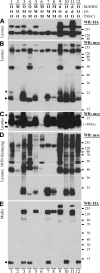
Effect of alteration of Cys-rich domains in the cleavage of KIAA0319. 293T cells were transiently transfected with constructs encoding KIAA0319 proteins with their MANEC, C6, and TM-Ct domains unmodified (O), deleted (d), or all-Cys residues mutated (M). Lysates and conditioned media were analyzed by Western blotting (WB) under reducing conditions, except in D, as indicated. A, detection of HA-tagged fragments in cell lysates is shown; only the bands shown were detected in the blot. B, detection of myc-tagged fragments in cell lysates is shown. C, procedures were the same as in B but after PMA induction; only short fragments are shown. D, procedures were the same as in B but using non-reducing conditions; the white dashed box indicates the dimeric forms of the KCT20 fragments. E, HA-tagged fragments were detected in conditioned media. Some cleavage fragments are indicated: asterisk, KCT26; arrowhead, KCT20; arrow, KCT18; circle, KNT12.
FIGURE 4.
FIGURE 4.
Effect of endocytosis impairment in the cleavage of KIAA0319. Conditioned media (A) and cell lysates (B and C) of 293T cells transiently overexpressing KIAA0319 full-length protein (WT) or the endocytosis-impaired mutant Y995A (A) (see Fig. 1) were analyzed by Western blotting (WB). A, the N-terminal cleavage fragment is detected with both wild-type and mutant constructs. B, the C-terminal fragments originated by ectodomain shedding are absent or highly reduced in lysates of cells transfected with the Y995A mutant. The second panel of myc Western blotting is a longer exposure of the blot shown on the left (diff. exp.). C, induction with PMA shows that the C-terminal fragment originated after intramembrane cleavage is detected with both constructs.
FIGURE 5.
FIGURE 5.
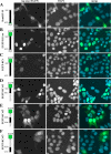
Subcellular localization of KIAA0319 cytoplasmic domain proteins. Several constructs overexpressing different fragments of the cytoplasmic domain of KIAA0319 were transiently transfected into 293T cells. Each of these constructs is represented on the left of the figure; tags and the first and last residues of the KIAA0319 protein are shown (EGFP tag not at scale). Green indicates the tag used for detection of these proteins by IF; light gray indicates regions of the cytoplasmic domain deleted in constructs shown in panels D, E, and F. The Myc tag was detected with anti-myc antibody. Nuclei are detected by DAPI staining. A 10-μm bar is shown in the top left panel; all panels are at the same scale.
FIGURE 6.
FIGURE 6.
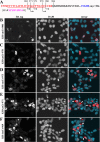
Analysis of the effect of N-terminal elongation of the cytoplasmic domain of KIAA0319 proteins in subcellular localization. Several constructs overexpressing the cytoplasmic domain of KIAA0319 with different numbers of residues from the TM domain, with N-terminal HA tag and C-terminal myc+His tags (KHAmCt; constructs #24–28 in supplemental Table 1), were transiently transfected into 293T cells, and the subcellular localization of the encoded proteins were analyzed by IF with rabbit anti-HA antibody. A, shown is a representation of the residues encoded by KIAA0319 exons 19–21 (19 in red, 20 in black, 21 in blue; dots represent residues not shown). Predicted TM domain residues are underlined. N-terminal tagging, including the HA tag in purple, is shown in square brackets. The positions of the first residues from the KIAA0319 protein in these constructs are indicated. B–F, detection by IF of the protein encoded by the construct indicated on the left is shown. Cytoplasmic aggregation of these proteins is indicated in the merge panels by white arrows. Nuclei are detected by DAPI staining. A 10-μm bar is shown in the top left panel; all panels are at the same scale.
FIGURE 7.
FIGURE 7.
Co-localization of KIAA0319 C-terminal fragment protein KHAmCt-980 with subnuclear markers. KHAmCt-980 protein was overexpressed in 293T cells as described above, and its subnuclear localization was analyzed by IF with chicken anti-HA antibody (red in merge panels) and compared with that obtained with specific markers (green in merge panels) for nucleoli (B23; A), Cajal bodies (coilin; B), and speckles (sc-35; C). Nuclei are detected by DAPI staining. A 10-μm bar is shown in the top left panel; all panels are at the same scale.
FIGURE 8.
FIGURE 8.
Analysis of intramembrane cleavage of KIAA0319 by luciferase assay. A, shown is a schematic representation of the proteins encoded by the constructs used in these assays (see supplemental Table 1 and “Experimental Procedures”). B, shown is a graphic representation of the luciferase assay results on 293T cells transfected with the indicated constructs plus pFR-Luc under control conditions (DMSO) or treated with the γ-secretase inhibitor L-685458 (Inh.). C, procedures were as in B, but using WT and γ-secretase-deficient PS1−/−, PS2−/− double KO MEF cells. Values are normalized for the luciferase activities obtained in the same conditions with the soluble positive control pSG5-Gal4-VP16 (GV) and presented as means of three assays ± S.E.

Similar articles

Cited by

References

    1. Paracchini S., Scerri T., Monaco A. P. (2007) Annu. Rev. Genomics Hum. Genet. 8, 57–79 - PubMed
    1. Fisher S. E., Francks C. (2006) Trends Cogn. Sci. 10, 250–257 - PubMed
    1. McGrath L. M., Smith S. D., Pennington B. F. (2006) Trends Mol. Med. 12, 333–341 - PubMed
    1. Galaburda A. M., LoTurco J., Ramus F., Fitch R. H., Rosen G. D. (2006) Nat. Neurosci. 9, 1213–1217 - PubMed
    1. Gabel L. A., Gibson C. J., Gruen J. R., LoTurco J. J. (2010) Neurobiol. Dis. 38, 173–180 - PMC - PubMed

Publication types

MeSH terms

Substances

LinkOut - more resources