. 2010 Dec 17;285(51):40050-9.
doi: 10.1074/jbc.M110.154955. Epub 2010 Oct 13.

Calcineurin signaling regulates human islet {beta}-cell survival

Affiliations

Calcineurin signaling regulates human islet {beta}-cell survival

Scott A Soleimanpour et al. J Biol Chem. .

Abstract

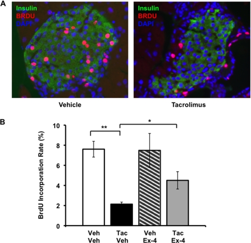
The calcium-regulated phosphatase calcineurin intersects with both calcium and cAMP-mediated signaling pathways in the pancreatic β-cell. Pharmacologic calcineurin inhibition, necessary to prevent rejection in the setting of organ transplantation, is associated with post-transplant β-cell failure. We sought to determine the effect of calcineurin inhibition on β-cell replication and survival in rodents and in isolated human islets. Further, we assessed whether the GLP-1 receptor agonist and cAMP stimulus, exendin-4 (Ex-4), could rescue β-cell replication and survival following calcineurin inhibition. Following treatment with the calcineurin inhibitor tacrolimus, human β-cell apoptosis was significantly increased. Although we detected no human β-cell replication, tacrolimus significantly decreased rodent β-cell replication. Ex-4 nearly normalized both human β-cell survival and rodent β-cell replication when co-administered with tacrolimus. We found that tacrolimus decreased Akt phosphorylation, suggesting that calcineurin could regulate replication and survival via the PI3K/Akt pathway. We identify insulin receptor substrate-2 (Irs2), a known cAMP-responsive element-binding protein target and upstream regulator of the PI3K/Akt pathway, as a novel calcineurin target in β-cells. Irs2 mRNA and protein are decreased by calcineurin inhibition in both rodent and human islets. The effect of calcineurin on Irs2 expression is mediated at least in part through the nuclear factor of activated T-cells (NFAT), as NFAT occupied the Irs2 promoter in a calcineurin-sensitive manner. Ex-4 restored Irs2 expression in tacrolimus-treated rodent and human islets nearly to baseline. These findings reveal calcineurin as a regulator of human β-cell survival in part through regulation of Irs2, with implications for the pathogenesis and treatment of diabetes following organ transplantation.


Figures

FIGURE 1.
FIGURE 1.
Calcineurin inhibition induces human β-cell apoptosis. A, representative image (magnification, ×40) of vehicle- (upper) and tacrolimus- (lower) treated human islets. Lower right, increased magnification of focal area of β-cell apoptosis in a tacrolimus-treated human islet. Green, insulin; red, TUNEL; blue, DAPI. B, quantitation of human β-cell TUNEL rates after 48-h treatment with vehicle, 10 ng/ml tacrolimus, 10 nm Ex-4, or a combination of tacrolimus and Ex-4 at the same doses. Data are presented as mean ± S.E. (error bars) of three independent human islet preparations and are expressed as fold compared with the vehicle-treated group. Absolute rates of apoptosis are presented in supplemental Fig. 1. 5,000–30,000 human β-cells were counted/treatment group for each preparation. *, p < 0.05.
FIGURE 2.
FIGURE 2.
Ex-4 attenuates the tacrolimus-induced decrease in murine β-cell proliferation. A, representative image of islets from mice treated with vehicle or tacrolimus. Insulin is seen in green, BrdU in red, DAPI in blue. B, quantitation of β-cell BrdU incorporation rates over 14 days of continuous administration in drinking water. All data points are mean ± S.E. (error bars). Bar graphs, open bar, vehicle/vehicle; black bar, 1.0 mg/kg per day tacrolimus/vehicle; hatched bar, vehicle/20 nmol per kg per day Ex-4; gray bar, 1.0 mg/kg per day tacrolimus/20 nmol/kg per day Ex-4. n = 7–8 mice/group except for vehicle/Ex-4 (n = 5). *, p < 0.05; **, p < 0.01.
FIGURE 3.
FIGURE 3.
Calcineurin inhibition reduces phosphorylation of Akt and Akt substrates. A, Western blot analysis of INS-1 cells following treatment with vehicle, 30 ng/ml tacrolimus, and/or 10 μm forskolin for 6 h, probed with anti-phospho-Akt (Thr308) (representative of four independent experiments). B, band intensity quantitation of p-Akt/Akt protein ratio in INS-1 cells by Western blotting. Data are presented as fold change from vehicle controls and as the mean ± S.E. (error bars) of four experiments performed in duplicate. C and D, representative Western blot of phospho-Akt substrate protein expression in INS-1 cells (C) and human islets (D) following treatment with vehicle or tacrolimus for 6 h. For human islets, duplicate cultures of islets derived from a single donor are shown (data are representative of experiments performed on islets from three independent donors). E and F, Western blot analysis of phospho- and total GSK3 (E) and phospho- and total S6 (F) expression in human islets following treatment with vehicle or10 ng/ml tacrolimus for 6 h. G, quantitative PCR analysis of Akt isoform mRNA expression in cultured human and rat islets. Isoform expression is presented relative to Akt1 mRNA levels. Data are presented as mean ± S.E. (error bars) of three independent rat islet preparations and four independent human islet preparations (each analyzed in triplicate). *, p < 0.05; **, p < 0.01.
FIGURE 4.
FIGURE 4.
Tacrolimus decreases Irs2 expression, which is restored by cAMP stimuli. A and B, Irs2 mRNA (A) and protein (B) levels were assessed in INS-1 cells after a 6-h incubation with tacrolimus (30 ng/ml), forskolin (10 μm), or their vehicles as indicated. Each Western blot lane represents a separate culture dish. C and D, quantitative PCR analysis of Irs2 mRNA expression in rat islets following vehicle, tacrolimus, and/or Ex-4 treatment for 3 h (C) or 6 h (D). Data are expressed as mean ± S.E. (error bars) of three experiments performed in triplicate. E, quantitative PCR analysis of Irs2 mRNA expression following treatment with vehicle, 10 ng/ml tacrolimus, and/or 10 nm Ex-4 for 6 h in three independent human islet preparations (studied in triplicate). Data are presented as the mean ± S.E. (error bars) fold mRNA level versus vehicle. F, Irs2 protein expression in representative Western blot of human islets following treatment with vehicle, 10 ng/ml tacrolimus, and/or 10 nm Ex-4 for 6 h. Each Western blot lane represents a separate culture dish from one donor. The experiment was performed with islets from three separate donors in duplicate. *, p < 0.05; **, p < 0.01; †, p < 0.001.
FIGURE 5.
FIGURE 5.
NFATc1 occupies two regions of the Irs2 promoter in a calcineurin-sensitive manner. Quantitative ChIP analysis in Min6 cells following treatment with vehicle or 30 ng/ml tacrolimus, is expressed as fold enrichment (αNFAT versus normal mouse IgG). Data are displayed as mean ± S.D. (error bars) of a single representative ChIP experiment. Experiment was performed three times. Primers designed to amplify a known NFATc1 binding region of the cyclin D1 and insulin promoter and a cis-control, located in an intergenic region nearly 70 kb upstream of the Irs2 transcriptional start site, were used as positive and negative controls, respectively.
FIGURE 6.
FIGURE 6.
Tacrolimus reduces D-cyclin expression in rodent islets. A–C, quantitative PCR analysis of cyclin D1 (A), cyclin D2 (B), and Cdk4 (C) expression in cultured rat islets following vehicle, tacrolimus, and/or Ex-4 treatment for 3 or 6 h. Data are expressed as mean ± S.E. (error bars) of three experiments performed in triplicate. *, p < 0.05; **, p < 0.01; †, p < 0.001.

References

    1. Cosio F. G., Kudva Y., van der Velde M., Larson T. S., Textor S. C., Griffin M. D., Stegall M. D. (2005) Kidney Int. 67, 2415–2421 - PubMed
    1. Kasiske B. L., Snyder J. J., Gilbertson D., Matas A. J. (2003) Am. J. Transplant. 3, 178–185 - PubMed
    1. Baid S., Cosimi A. B., Farrell M. L., Schoenfeld D. A., Feng S., Chung R. T., Tolkoff-Rubin N., Pascual M. (2001) Transplantation 72, 1066–1072 - PubMed
    1. Bonner-Weir S. (2000) Endocrinology 141, 1926–1929 - PubMed
    1. Davidson J., Wilkinson A., Dantal J., Dotta F., Haller H., Hernández D., Kasiske B. L., Kiberd B., Krentz A., Legendre C., Marchetti P., Markell M., van der Woude F. J., Wheeler D. C. (2003) Transplantation 75, SS3–S24 - PubMed

Publication types

MeSH terms

LinkOut - more resources