. 2010 Dec 24;285(52):41019-33.
doi: 10.1074/jbc.M110.185850. Epub 2010 Oct 12.

Crystal structure of hexokinase KlHxk1 of Kluyveromyces lactis: a molecular basis for understanding the control of yeast hexokinase functions via covalent modification and oligomerization

Affiliations

Crystal structure of hexokinase KlHxk1 of Kluyveromyces lactis: a molecular basis for understanding the control of yeast hexokinase functions via covalent modification and oligomerization

E Bartholomeus Kuettner et al. J Biol Chem. .

Abstract

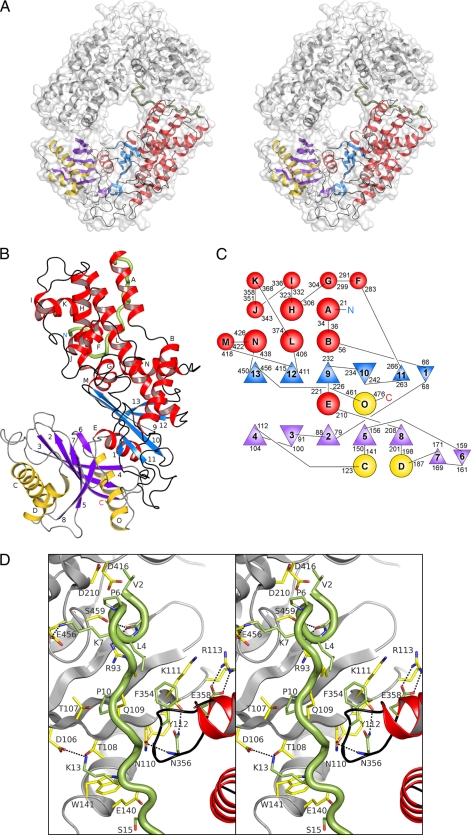
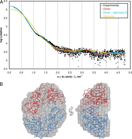
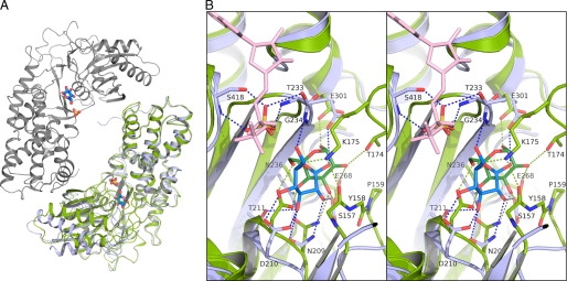
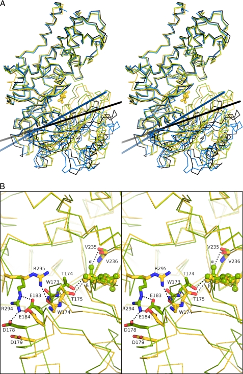
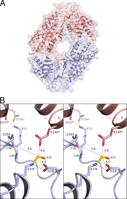
Crystal structures of the unique hexokinase KlHxk1 of the yeast Kluyveromyces lactis were determined using eight independent crystal forms. In five crystal forms, a symmetrical ring-shaped homodimer was observed, corresponding to the physiological dimer existing in solution as shown by small-angle x-ray scattering. The dimer has a head-to-tail arrangement such that the small domain of one subunit interacts with the large domain of the other subunit. Dimer formation requires favorable interactions of the 15 N-terminal amino acids that are part of the large domain with amino acids of the small domain of the opposite subunit, respectively. The head-to-tail arrangement involving both domains of the two KlHxk1 subunits is appropriate to explain the reduced activity of the homodimer as compared with the monomeric enzyme and the influence of substrates and products on dimer formation and dissociation. In particular, the structure of the symmetrical KlHxk1 dimer serves to explain why phosphorylation of conserved residue Ser-15 may cause electrostatic repulsions with nearby negatively charged residues of the adjacent subunit, thereby inducing a dissociation of the homologous dimeric hexokinases KlHxk1 and ScHxk2. Two complex structures of KlHxk1 with bound glucose provide a molecular model of substrate binding to the open conformation and the subsequent classical domain closure motion of yeast hexokinases. The entirety of the novel data extends the current concept of glucose signaling in yeast and complements the induced-fit model by integrating the events of N-terminal phosphorylation and dissociation of homodimeric yeast hexokinases.


Figures

FIGURE 1.
FIGURE 1.
Crystal structure of dimeric KlHxk1. A, stereoview of homodimeric KlHxk1. Monomer A is shown with blue β-strands and red helices for the large domain and violet β-strands and yellow helices for the small domain. The N terminus (residues 2–20) is shown in green. B, monomer structure with labeled α-helices (A–O) and β-strands (1–13). C, topology diagram of KlHxk1 with residue numbers. The circles and triangles correspond to α-helices and β-strands, respectively. D, interactions of the N terminus of chain A (green) with amino acids of the adjacent monomer (gray) within a 4 Å distance. Side chain carbon atoms are colored green for chain A and yellow for chain B. Hydrogen bonds (≤3.5Å) are depicted as dotted lines.
FIGURE 2.
FIGURE 2.
Small-angle x-ray scattering analysis of KlHxk1. A, merged KlHxk1 solution-scattering data (black) obtained at protein concentrations of 6.9 and 3.5 mg/ml. Scattering curves calculated for the ring-shaped dimer (crystal form IV, both chains comprising amino acids 2–485), for its rigid body-fitted form and for a monomer (crystal form III, chain A, comprising amino acids 2–485) are shown in red, blue, and yellow, respectively. B, ab initio shape model of KlHxk1 in solution. The transparent gray spheres represent the SAXS envelope model obtained without employing symmetry constraints. The crystallographic ring-shaped dimer (crystal form III, both chains, comprising amino acids 2–485) has been manually superimposed (monomer A in blue and monomer B in red).
FIGURE 3.
FIGURE 3.
Glucose binding by KlHxk1. A, dimer structure of KlHxk1 crystal form VII (blue and gray polypeptide chains, bound glucose and sulfate molecules shown as sticks) with superimposed monomer structure of crystal form XI (green polypeptide chain). B, stereoview of the glucose binding sites of KlHxk1 crystal form VII, chain A (light blue), and crystal form XI (green). For crystal form VII, a sulfate ion is shown with a yellow sulfur atom. Polar interactions are shown as dotted lines. An ADP molecule (light red atoms) is superimposed from human brain hexokinase structure (PDB entry 1DGK (55)).
FIGURE 4.
FIGURE 4.
Comparison of the domain flexibility of yeast hexokinases KlHxk1, ScHxk1 and ScHxk2 (stereoview). The structures have been superimposed based on the Cα atoms of the large domains (shown in the upper part of the images). A, superposition of KlHxk1 crystal forms XI (green) and IX (blue) and ScHxk1 (yellow) (29) and ScHxk2 (black) (30). The interdomain rotation axes of KlHxk1 crystal form IX and of ScHxk2 are calculated relative to KlHxk1 crystal form XI or ScHxk1, respectively, using the same color code. Glucose molecules from the structures of ScHxk1 (29) and of KlHxk1 crystal form XI are shown in yellow and green, respectively. B, domain interfaces of KlHxk1 crystal form XI (green) and ScHxk1 (yellow) (29) with selected residues shown as sticks. The configurations of the glucose molecules are labeled at their 1-OH positions. Polar interactions are shown as dotted lines.
FIGURE 5.
FIGURE 5.
Determination of the dynamic domains of KlHxk1 by comparison of the most open (crystal form IX) and the most closed (crystal form XI) conformers. A, description of domain rotation by three dynamic domains (stereoview). The small (red) and large (blue) domains move relative to the central domain (yellow). The rotation axes are indicated in red. In all panels, the bending residues are shown in green. B, cluster analysis of the rotation vectors for the three-domain description. C, alternative description of KlHxk1 domain rotation based on two dynamic domains (stereoview). The view is along the rotation axes, indicated in red. D, cluster analysis of the rotation vectors for the two-domain description.
FIGURE 6.
FIGURE 6.
Crystal structures of the homodimeric SmHxk1 and KlHxk1 hexokinases. The crystals of SmHxk1 (59) contain a dimer (A) that closely resembles the symmetric KlHxk1 dimer (B). The large domains of both enzymes are shown in red and the small domains in yellow.
FIGURE 7.
FIGURE 7.
Environment of KlHxk1 residue Ser-15 at the dimer interface. A, dimer structure of KlHxk1 with Ser-15 residues shown as sticks (yellow, carbon atoms). The residues of chain A of the symmetric dimer are depicted in light blue and of chain B of the opposite monomer in light red. *, marks residues of the adjacent monomer. B, stereoview of the dimer interface around Ser-15 of chain A. Only charged amino acids and their binding partners are shown. Selected hydrogen bonds are plotted as black dotted lines, and distances from the Cβ atom of residue Ser-15 are presented as red dotted lines.

Similar articles

Cited by

References

    1. Moreno F., Ahuatzi D., Riera A., Palomino C. A., Herrero P. (2005) Biochem. Soc. Trans. 33, 265–268 - PubMed
    1. Rolland F., Sheen J. (2005) Biochem. Soc. Trans. 33, 269–271 - PubMed
    1. Matschinsky F. M., Magnuson M. A., Zelent D., Jetton T. L., Doliba N., Han Y., Taub R., Grimsby J. (2006) Diabetes 55, 1–12 - PubMed
    1. Rolland F., Baena-Gonzalez E., Sheen J. (2006) Annu. Rev. Plant Biol. 57, 675–709 - PubMed
    1. Rolland F., Winderickx J., Thevelein J. M. (2001) Trends Biochem. Sci. 26, 310–317 - PubMed

LinkOut - more resources