. 2010 Dec 24;285(52):40635-44.
doi: 10.1074/jbc.M110.168187. Epub 2010 Oct 12.

Crystal structure of cardiac-specific histone methyltransferase SmyD1 reveals unusual active site architecture

Affiliations

Crystal structure of cardiac-specific histone methyltransferase SmyD1 reveals unusual active site architecture

Nualpun Sirinupong et al. J Biol Chem. .

Abstract

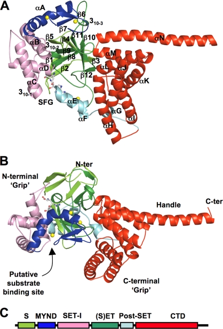
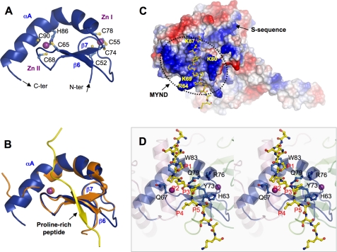
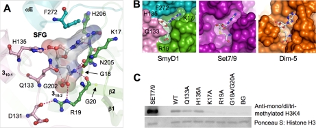
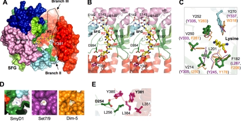
SmyD1 is a cardiac- and muscle-specific histone methyltransferase that methylates histone H3 at lysine 4 and regulates gene transcription in early heart development. The unique domain structure characterized by a "split" SET domain, a conserved MYND zinc finger, and a novel C-terminal domain (CTD) distinguishes SmyD1 from other SET domain containing methyltransferases. Here we report the crystal structure of full-length SmyD1 in complex with the cofactor analog sinefungin at 2.3 Å. The structure reveals that SmyD1 folds into a wrench-shaped structure with two thick "grips" separated by a large, deep concave opening. Importantly, our structural and functional analysis suggests that SmyD1 appears to be regulated by an autoinhibition mechanism, and that unusually spacious target lysine-access channel and the presence of the CTD domain both negatively contribute to the regulation of this cardiovascularly relevant methyltransferase. Furthermore, our structure also provides a structural basis for the interaction between SmyD1 and cardiac transcription factor skNAC, and suggests that the MYND domain may primarily serve as a protein interaction module and cooperate SmyD1 with skNAC to regulate cardiomyocyte growth and maturation. Overall, our data provide novel insights into the mechanism of SmyD1 regulation, which would be helpful in further understanding the role of this protein in heart development and cardiovascular diseases.


Figures

FIGURE 1.
FIGURE 1.
Ribbon diagram of the overall structure of SmyD1 in complex with sinefungin. A, top, and B, side views. Secondary structures of SmyD1, α-helices, 310-helices, and β-strands are labeled and numbered according to their position in the primary sequence. The S-sequence, MYND, SET-I, core SET, post-SET, and CTD domains are depicted in light green, blue, pink, green, cyan, and red, respectively, whereas sinefungin (SFG) is represented by ball-and-stick and zinc ions by yellow spheres. Sinefungin is a structural analog of AdoMet and a potent inhibitor of AdoMet-dependent methyltransferases. Using sinefungin instead of AdoHcy or AdoMet in co-crystallization was necessary to obtain diffraction quality crystals. C, schematic diagram of SmyD1 domain structures.
FIGURE 2.
FIGURE 2.
Sequence alignment of SmyD family proteins. The alignment, which includes mouse SmyD1 and human SmyD1, -2, and -3, was performed by ClustalW (31). Identical residues are shown as white on black, and similar residues appear shaded in cyan. Secondary structure elements of SmyD1 are displayed above the sequences, colored and labeled according to Fig. 1. Sequence numbering is displayed to the left of the sequences, with every 10th residue marked by a dot above the alignment.
FIGURE 3.
FIGURE 3.
Zinc finger MYND domain. A, ribbon diagram of the MYND domain structure. Zinc ligands are labeled and represented by ball-and-stick, and zinc ions by purple spheres. The N- and C termini are indicated. B, superposition of the MYND domains of SmyD1 (blue) and AML1/ETO (PDB code 2ODD) (orange). The proline-rich peptide from AML1/ETO containing the PPPLI motif is displayed as ribbon and colored in yellow. C, surface representation of SmyD1 with coloring according to the electrostatic potential: red, white, and blue correspond to negative, neutral, and positive potentials, respectively. The surface of MYND, outlined by a dotted line, exhibits numerous positive charges due to the clustering of basic amino acids. The conserved arginine and lysine residues are labeled. The modeled proline-rich peptide, represented by ball-and-stick with carbon atoms colored in yellow, is seen to bind on the surface of SmyD1. The N terminus of the peptide projects toward the S-sequence, which appears to extend the positively charged MYND surface. D, stereo view ribbon diagram illustrates the interaction between the MYND domain and the modeled proline-rich peptide. Residues in SmyD1 that are potentially important for peptide interaction are represented by ball-and-stick with carbon atoms colored in blue. Residues in the modeled peptide are denoted according to their position in the PXLXP motif, with the 1st proline as P1 and last as P5.
FIGURE 4.
FIGURE 4.
Cofactor binding pocket. A, detailed interactions between SmyD1 and sinefungin. SmyD1 residues are represented by ball-and-stick with their carbon atoms colored as described in the legend to Fig. 1. Sinefungin are also depicted by ball-and-stick overlaid by its transparent molecular surface. Hydrogen bonds are illustrated as dashed lines. B, surface representation of cofactor binding sites illustrates that the cofactor is more buried in SmyD1 than Set7/9 or Dim-5. C, effects of the mutations on the enzymatic activity of SmyD1. Methylation of H3K4 by SmyD1 was detected by Western blotting with mono-, di-, and tri-methylated H3K4-specific antibody. The reaction without cofactor AdoMet was used as a background control, Set7/9 as a positive control, and Ponceau S stained histone H3 as a loading control.
FIGURE 5.
FIGURE 5.
Putative substrate binding site. A, surface representation of SmyD1 reveals a Y-shaped cleft that runs across the entire molecule. Black dotted line outlines the cleft with 3 branches. The surface is colored according to domain with the same scheme as described in the legend to Fig. 1. The modeled H3 peptide (1–10) based on structure comparison with Set7/9 (PDB code 1O9S) is displayed as ball-and-stick with carbon atoms colored in yellow, which is seen to bind in branch I. Sinefungin, located on the opposite face of SmyD1, is indicated. Some conserved residues in branch II are labeled and colored white on the surface. B, stereo view ribbon diagram of the putative substrate binding site (branch I), illustrating the interaction between SmyD1 and the modeled H3 peptide. The view is rotated ∼90° clockwise about the perpendicular axis relative to A. Residues in SmyD1 that are potentially important for H3 recognition are represented by ball-and-stick, whereas residues in the peptide are labeled and numbered according to the H3 sequence. C, superposition of the target lysine-access channels of SmyD1, Set7/9, and Dim-5. Residues in SmyD1 are represented by ball-and-stick, whereas residues in Set7/9 and Dim-5 are displayed as stick in purple and orange, respectively. The target lysine is colored yellow. D, surface representation of lysine-access channels illustrates that the channel in SmyD1 is more spacious than Set7/9 or Dim-5. Sinefungin and AdoHcy are shown with C-NH3 amine colored in blue and sulfur atom in yellow, respectively. E, hydrophobic cluster and hydrogen bonding interaction between the CTD and SET domains in branch II. Residues are colored according to the domain in which they reside, and hydrogen bond is indicated as dashed line.
FIGURE 6.
FIGURE 6.
Mutation analysis of the substrate binding site. A, biotin pulldown assay using biotinylated H3 peptide (1–21) reveals that SmyD1 has a significantly lower binding affinity to histone H3 than Set7/9. The binding experiments were analyzed by SDS-PAGE and visualized by Coomassie Blue staining. Streptavidin (SA) beads alone were used as a control for nonspecific binding. B, biotin pulldown assay examines the effect of mutations on SmyD1 binding to H3. The binding experiments were analyzed by Western blotting using anti-SmyD1 antibody. The amount of SmyD1 proteins in each reaction is shown in the lower panel. C, histone methyltransferase activity of SmyD1 and its mutants. The experimental procedure is essentially the same as that described in the legend to Fig. 4C.

Similar articles

Cited by

References

    1. Kwon C., Qian L., Cheng P., Nigam V., Arnold J., Srivastava D. (2009) Nat. Cell Biol. 11, 951–957 - PMC - PubMed
    1. Gottlieb P. D., Pierce S. A., Sims R. J., Yamagishi H., Weihe E. K., Harriss J. V., Maika S. D., Kuziel W. A., King H. L., Olson E. N., Nakagawa O., Srivastava D. (2002) Nat. Genet. 31, 25–32 - PubMed
    1. Tan X., Rotllant J., Li H., De Deyne P., Du S. J. (2006) Proc. Natl. Acad. Sci. U.S.A. 103, 2713–2718 - PMC - PubMed
    1. Li D., Niu Z., Yu W., Qian Y., Wang Q., Li Q., Yi Z., Luo J., Wu X., Wang Y., Schwartz R. J., Liu M. (2009) Nucleic Acids Res. 37, 7059–7071 - PMC - PubMed
    1. Borlak J., Thum T. (2003) FASEB J. 17, 1592–1608 - PubMed

Publication types

MeSH terms

Associated data

LinkOut - more resources