. 2010 Oct 13;30(41):13644-55.
doi: 10.1523/JNEUROSCI.2487-10.2010.

Interdependent roles for accessory KChIP2, KChIP3, and KChIP4 subunits in the generation of Kv4-encoded IA channels in cortical pyramidal neurons

Affiliations

Interdependent roles for accessory KChIP2, KChIP3, and KChIP4 subunits in the generation of Kv4-encoded IA channels in cortical pyramidal neurons

Aaron J Norris et al. J Neurosci. .

Abstract

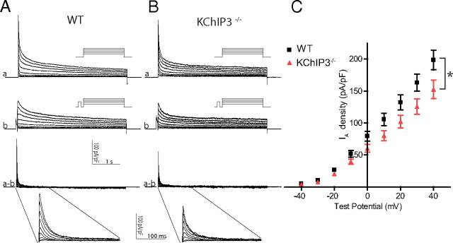
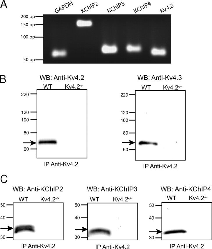
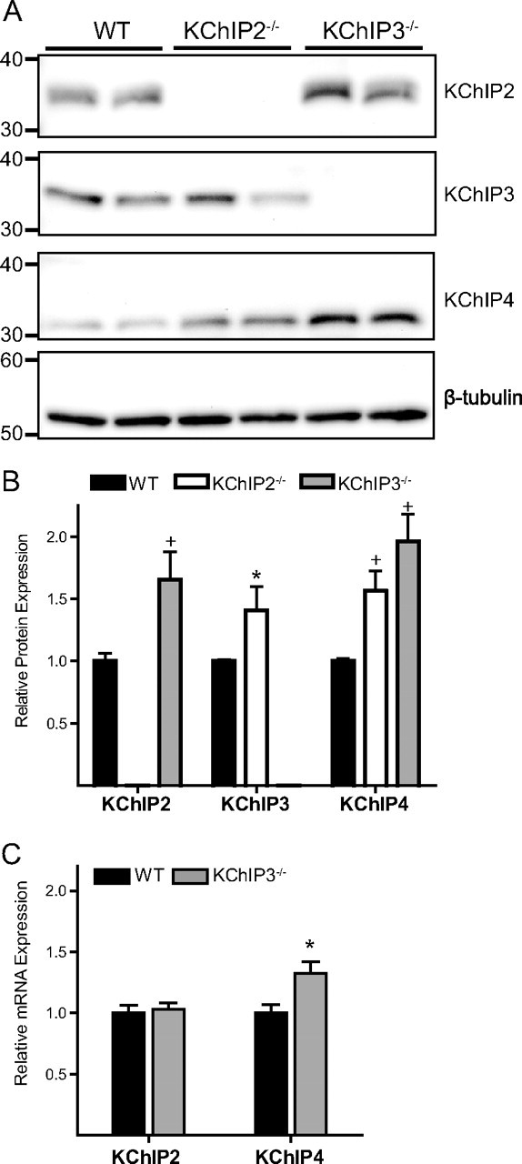
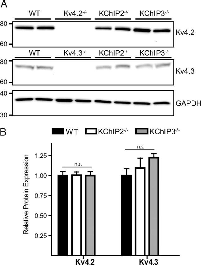
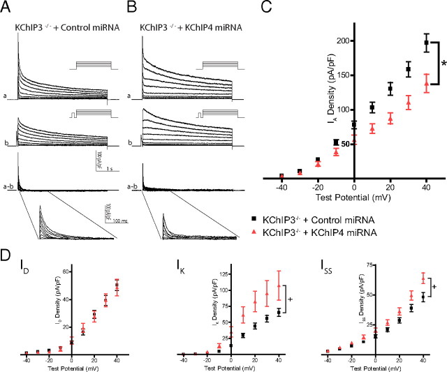
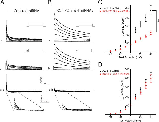
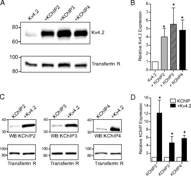
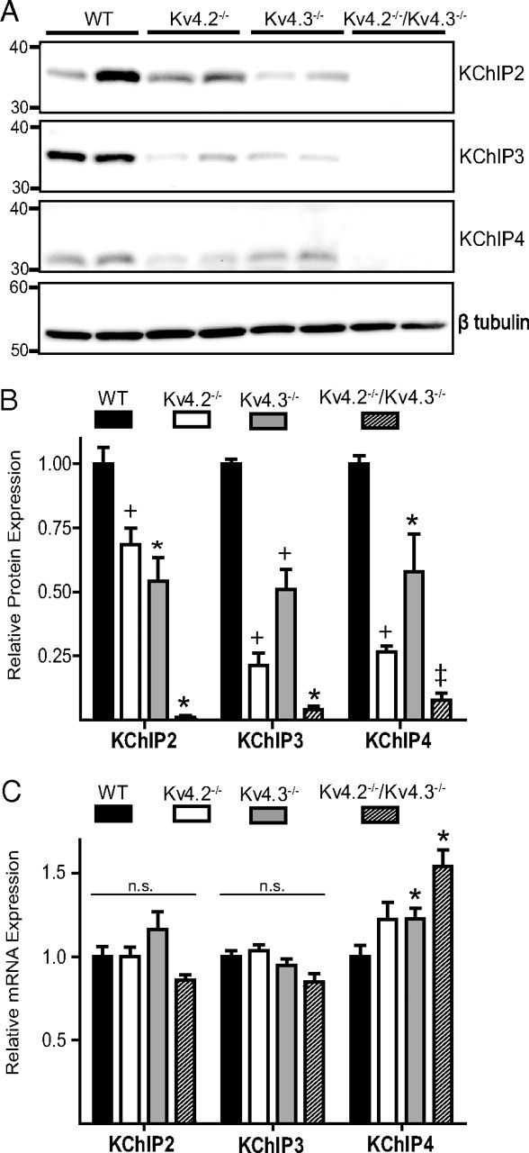
The rapidly activating and inactivating voltage-dependent outward K(+) (Kv) current, I(A), is widely expressed in central and peripheral neurons. I(A) has long been recognized to play important roles in determining neuronal firing properties and regulating neuronal excitability. Previous work demonstrated that Kv4.2 and Kv4.3 α-subunits are the primary determinants of I(A) in mouse cortical pyramidal neurons. Accumulating evidence indicates that native neuronal Kv4 channels function in macromolecular protein complexes that contain accessory subunits and other regulatory molecules. The K(+) channel interacting proteins (KChIPs) are among the identified Kv4 channel accessory subunits and are thought to be important for the formation and functioning of neuronal Kv4 channel complexes. Molecular genetic, biochemical, and electrophysiological approaches were exploited in the experiments described here to examine directly the roles of KChIPs in the generation of functional Kv4-encoded I(A) channels. These combined experiments revealed that KChIP2, KChIP3, and KChIP4 are robustly expressed in adult mouse posterior (visual) cortex and that all three proteins coimmunoprecipitate with Kv4.2. In addition, in cortical pyramidal neurons from mice lacking KChIP3 (KChIP3(-/-)), mean I(A) densities were reduced modestly, whereas in mean I(A) densities in KChIP2(-/-) and WT neurons were not significantly different. Interestingly, in both KChIP3(-/-) and KChIP2(-/-) cortices, the expression levels of the other KChIPs (KChIP2 and 4 or KChIP3 and 4, respectively) were increased. In neurons expressing constructs to mediate simultaneous RNA interference-induced reductions in the expression of KChIP2, 3, and 4, I(A) densities were markedly reduced and Kv current remodeling was evident.


Figures

Figure 1.
Figure 1.
IA density is reduced in KChIP3−/− cortical pyramidal neurons. A, B, Whole-cell Kv current recordings were obtained from cortical pyramidal neurons isolated from WT and KChIP3 −/− mice. Representative recordings from WT (A) and KChIP3−/− (B) neurons are illustrated. In each cell, Kv currents were elicited by depolarizing voltage steps ranging from −40 to +40 mV in 10 mV increments from a holding potential of −70 mV (a). Recordings were then obtained from the same cell using a prepulse paradigm in which the same depolarizing steps, preceded by a prepulse of 60 ms to −10 mV to selectively inactive IA, were presented (b). The paradigms are illustrated in the insets. For each cell, currents recorded with the prepulse (b) were subtracted off-line from the control records (a) to isolated IA (a − b). Similar recordings were obtained from WT (n = 22) and KChIP3−/− (n = 24) neurons. C, IA densities were calculated from the subtracted records, and mean ± SEM IA densities are plotted. Mean ± SEM IA densities were modestly, but significantly (*p < 0.05), lower in KChIP3−/−, compared with WT, neurons.
Figure 2.
Figure 2.
Expression of KChIP2, KChIP3, and KChIP4 in cortex and coimmunoprecipitation with Kv4.2. To examine the expression of KChIP transcripts, RNA was extracted from samples prepared from the posterior (∼1 mm) cortex (containing visual cortex) of adult WT mice. A, Using sequence-specific primer pairs and RT-PCR, transcripts coding for KChIP2, KChIP3, and KChIP4 were readily detected. Experiments were also performed to examine KChIP protein expression and association with Kv4.2. Immunoprecipitations using a rabbit anti-Kv4.2 antibody were conducted on lysates prepared from the posterior cortices of WT or Kv4.2−/− mice, and blots of fractionated immunoprecipitated samples were probed using subunit-specific antibodies. B, In immunoprecipitated samples from WT cortices, but not in those from Kv4.2−/− cortices, both Kv4.2 and Kv4.3 were readily detected. C, In immunoprecipitated samples from WT, but not Kv4.2−/−, cortices, KChIP2, KChIP3, and KChIP4 were also detected indicating that all three KChIP proteins are expressed and assemble with Kv4.2 in cortex. Molecular masses are indicated on the blots in kilodaltons.
Figure 3.
Figure 3.
KChIPs are upregulated in KChIP2−/− and KChIP3−/− cortices. A, Lysates prepared from the posterior (∼1 mm) cortices of WT, KChIP2−/−, and KChIP3−/− mice (n = 6 animals for each genotype) were fractionated, transferred to PVDF membranes, and probed with a specific anti-KChIP2, anti-KChIP3, or anti-KChIP4 antibody. All three KChIPs were detected in samples from WT mice. Confirming the specificities of the anti-KChIP2 and anti-KChIP3 antibodies, no signal was detected with the anti-KChIP2 or the anti-KChIP3 antibody in samples from KChIP2−/− or KChIP3−/− cortices, respectively. Blots were also probed with antibodies against β-tubulin to confirm equal loading of proteins. In each lane, anti-KChIP antibody signals were quantified and normalized to the anti-β-tubulin antibody signals. B, In KChIP2−/− cortices, the mean ± SEM expression levels of KChIP3 and KChIP4 proteins were significantly (+p < 0.01) higher than in WT cortices. Similarly, the mean ± SEM expression levels of the KChIP2 and KChIP4 proteins were significantly (*p < 0.05 and +p < 0.01, respectively) higher in KChIP3−/− cortices. C, QRT-PCR analysis revealed that the mean ± SEM expression level of KChIP2 transcript was not significantly different in WT (n = 6) and KChIP3−/− (n = 6) cortices, whereas the mean ± SEM expression level of KChIP4 transcript was slightly, but significantly (*p > 0.05), higher in cortices from KChIP3−/−, compared with WT, mice. Molecular masses are indicated on the blots in kilodaltons.
Figure 4.
Figure 4.
Maintained expression of Kv4.2 and Kv4.3 proteins in KChIP2−/− and KChIP3−/− cortices. A, Lysates were prepared from the posterior (∼1 mm) cortices of WT, KChIP2−/−, and KChIP3−/− mice (n = 6 of each genotype) and fractionated by SDS-PAGE. After transfer, membranes were probed with a monoclonal anti-Kv4.2 or anit-Kv4.3 antibody and, subsequently, with an anti-GAPDH antibody, to verify equal loading of proteins in each lane. Signals from the anti-Kv4.2 and anti-Kv4.3 antibodies in each lane were quantified and normalized to signals from the anti-GAPDH antibody in the same lane. Molecular masses are indicated on the blots in kilodaltons. B, Mean ± SEM levels of Kv4.2 and Kv4.3 proteins are not significantly different in either KChIP2−/− or KChIP3−/−, compared with WT, cortices.
Figure 5.
Figure 5.
Validation of miRNA constructs to mediate RNAi based knockdown of KChIP2, KChIP3 or KChIP4 in neurons. As described in Materials and Methods, plasmids encoding human miR30, with substituted targeting sequences and a fluorescent protein (YFP, CFP, or tdTomato) on a single transcript, were generated. A, The miR30 sequence was placed on an intron downstream of the CMV promoter and upstream of the sequence coding for the fluorescent protein (CFP, YFP, or tdTomato). B, Transfections of these plasmids into neurons allowed for visual identification of neurons expressing one or all three of the plasmids for subsequent electrophysiological recording. C, Specific sequences targeting KChIP2, KChIP3, and KChIP4 were screened in HEK-293 cells. The targeted KChIP (KChIP2, 3, or 4) was coexpressed with either a control (nontargeting) miRNA construct or with a miRNA construct containing sequence complementary to the sequence of the targeted KChIP. Lysates were prepared from transfected HEK-293 cells, fractionated by SDS-PAGE, transferred to membranes, and probed for KChIP2, KChIP3, or KChIP4. Blots were also probed with an anti-transferrin receptor (Transferrin R) antibody to verify equal loading of proteins. Targeting sequences found to reduce the expression of each of the targeted KChIPs are illustrated and were used in subsequent experiments, on cortical neurons.
Figure 6.
Figure 6.
Knockdown of KChIP4 in KChIP3−/− cortical pyramidal neurons results in decreased IA density and upregulation of IK and ISS. A, B, Whole-cell Kv currents, elicited in response to depolarizing voltage steps, were recorded from transfected cortical pyramidal neurons isolated from KChIP3−/− mice. Neurons were transfected by electroporation using the Amaxa Nucleofector system at the time of isolation with a miRNA construct containing either sequence targeting KChIP4 or a control nontargeting sequence; whole-cell recordings were obtained on the second and third days after transfections. IA was isolated and quantified using the prepulse paradigm and off-line subtraction method described in the legend to Figure 1. C, Analysis of the subtracted records (a − b) revealed that mean ± SEM IA densities were significantly (*p < 0.05) lower in KChIP3−/− neurons expressing miRNA targeting KChIP4 (n = 16) compared with KChIP3−/− neurons expressing control miRNA (n = 37). D, Analysis of the inactivation phases of the Kv currents also revealed that, in neurons expressing KChIP4 targeting miRNA, the mean ± SEM densities of IK and ISS were significantly (+p < 0.01) higher than in control miRNA-expressing neurons.
Figure 7.
Figure 7.
Concurrent knockdown of KChIP2, KChIP3, and KChIP4 in Kv1.4−/− cortical pyramidal neurons results in marked reductions in Kv4-encoded IA densities and KV current remodeling. To examine the combined role(s) of the KChIPs in the generation of Kv4-encoded IA channels, cortical pyramidal neurons were isolated from Kv1.4−/− mice and transfected with the validated miRNA constructs targeting KChIP2, KChIP3, and KChIP4 or with plasmids containing control (nontargeting) sequences. Because each KChIP miRNA construct also encoded for a distinct fluorescent protein (CFP, YFP, or tdTomato), cells expressing all three KChIP targeting miRNA constructs could be identified. A, B, Recordings were obtained from neurons expressing control plasmids (A) or all three targeting plasmids (B). Surprisingly, no prominent rapidly inactivating component was observed in approximately one-half (11 of 20) of the neurons expressing the KChIP targeting miRNA constructs and delayed rectifier currents were increased. In all cells, IA was isolated and quantified using the prepulse paradigm described in the legend to Figure 1. Analyses of subtracted records (a − b) revealed residual Kv4-encoded IA in all neurons expressing the three KChIP targeting miRNAs simultaneously. The mean ± SEM IA density was significantly (p < 0.001) lower (C) in neurons expressing the three KChIP targeting miRNA constructs (n = 20) than in neurons expressing control constructs (n = 21). D, Consistent with the upregulation of delayed rectifier currents, analysis of the peak current (IPeak) revealed no significant reduction in mean ± SEM IPeak density in neurons expressing KChIP targeting miRNA compared with those expressing control constructs, despite the marked reduction in IA densities (c).
Figure 8.
Figure 8.
Coexpression of Kv4.2 with KChIP2, KChIP3, or KChIP4 results in the costabilization of both the Kv4.2 and KChIP proteins. HEK-293 cells were transfected with DNA constructs encoding Kv4.2 alone (n = 9), one of the KChIPs (KChIP2, n = 6; KChIP3, n = 9; KChIP4, n = 6) alone, or Kv4.2 in combination with KChIP2 (n = 6), KChIP3 (n = 9), or KChIP4 (n = 6). A, Western blots on lysates prepared from transfected HEK-293 cells were probed with the monoclonal anti-Kv4.2 antibody. Blots were also probed with anti-transferrin receptor antibody (Transferrin R) to verify equal loading of proteins in each lane. The anti-Kv4.2 antibody signals were measured and normalized to the signals from the anti-transferrin receptor in the same lane. B, Quantitative analyses revealed a significant (*p < 0.05) increase in Kv4.2 protein in cells expressing Kv4.2 plus one of the three KChIPs, compared with cells expressing Kv4.2 alone. C, Western blots conducted on HEK-293 cell lysates using the anti-KChIP2, anti-KChIP3, or anti-KChIP4 antibody also revealed that KChIP protein expression was increased in cells coexpressing Kv4.2, compared with cells expressing KChIP2, 3, or 4 alone. D, Mean ± SEM levels of KChIP2, 3, and 4 protein expression were significantly (*p < 0.05; +p < 0.01) higher in cells coexpressing Kv4.2 compared with cells expressing either of the KChIP proteins alone.
Figure 9.
Figure 9.
Endogenous KChIP2, KChIP3, and KChIP4 protein expression is dependent on the expression of Kv4 α-subunits. A, Representative Western blots of fractionated lysates prepared from posterior (∼1 mm) cortices of WT (n = 6), Kv4.2−/− (n = 6), Kv4.3−/− (n = 6), and Kv4.2−/−/Kv4.3−/− (n = 3) mice were probed with the anti-KChIP antibodies. KChIP protein levels were differentially affected by the loss of Kv4.2 or Kv4.3, although drastic reductions in all three proteins were evident with the loss of both Kv4.2 and Kv4.3. For quantification, blots were also probed with an anti-β-tubulin antibody to confirm equal protein loading, in each lane, and signals from the anti-KChIP2, 3, or 4 antibodies were normalized against the signals from the anti-β-tubulin antibody in the same lane. B, Analysis of mean (±SEM) normalized data revealed that the expression levels of KChIP2, KChIP3, and KChIP4 proteins in Kv4.2−/− and Kv4.3−/− cortices were significantly (*p < 0.05, +p < 0.01, or p < 0.001) lower than in WT cortices. In Kv4.2−/−Kv4.3−/− cortices, KChIP2, KChIP3, and KChIP4 protein expression levels were extremely low. C, QRT-PCR of analysis of RNA isolated from the posterior cortices of WT (n = 6), Kv4.2−/− (n = 6), Kv4.3−/− (n = 6), and Kv4.2−/−/Kv4.3−/− (n = 3) mice revealed no reductions in KChIP transcripts. The mean ± SEM transcript expression level of KChIP4 was, however, significantly (p < 0.05) higher in Kv4.3−/− and in Kv4.2−/−/Kv4.3−/−, compared with WT, cortices.

Similar articles

Cited by

References

    1. Aghajanian GK. Modulation of a transient outward current in serotonergic neurones by alpha 1-adrenoceptors. Nature. 1985;315:501–503. - PubMed
    1. Aimond F, Kwak SP, Rhodes KJ, Nerbonne JM. Accessory Kvbeta1 subunits differentially modulate the functional expression of voltage-gated K+ channels in mouse ventricular myocytes. Circ Res. 2005;96:451–458. - PubMed
    1. Alexander JC, McDermott CM, Tunur T, Rands V, Stelly C, Karhson D, Bowlby MR, An WF, Sweatt JD, Schrader LA. The role of calsenilin/DREAM/KChIP3 in contextual fear conditioning. Learn Mem. 2009;16:167–177. - PMC - PubMed
    1. An WF, Bowlby MR, Betty M, Cao J, Ling HP, Mendoza G, Hinson JW, Mattsson KI, Strassle BW, Trimmer JS, Rhodes KJ. Modulation of A-type potassium channels by a family of calcium sensors. Nature. 2000;403:553–556. - PubMed
    1. Anderson D, Rehak R, Hameed SW, Hamish M, Zamponi GW, Turner RW. Regulation of the KV4.2 complex by CaV3.1 calcium channels. Channels (Austin) 2010a;4:163–167. - PubMed

Publication types