. 2010 Oct 13;30(41):13656-69.
doi: 10.1523/JNEUROSCI.2200-10.2010.

Haploinsufficiency in peptidylglycine alpha-amidating monooxygenase leads to altered synaptic transmission in the amygdala and impaired emotional responses

Affiliations

Haploinsufficiency in peptidylglycine alpha-amidating monooxygenase leads to altered synaptic transmission in the amygdala and impaired emotional responses

Eric D Gaier et al. J Neurosci. .

Abstract

The mammalian amygdala expresses various neuropeptides whose signaling has been implicated in emotionality. Many neuropeptides require amidation for full activation by peptidylglycine α-amidating monooxygenase (PAM), a transmembrane vesicular cuproenzyme and regulator of the secretory pathway. Mice heterozygous for the Pam gene (PAM(+/-)) exhibit physiological and behavioral abnormalities related to specific peptidergic pathways. In the present study, we evaluated emotionality and examined molecular and cellular responses that characterize neurophysiological differences in the PAM(+/-) amygdala. PAM(+/-) mice presented with anxiety-like behaviors in the zero maze that were alleviated by diazepam. PAM(+/-) animals were deficient in short- and long-term contextual and cued fear conditioning and required higher shock intensities to establish fear-potentiated startle than their wild-type littermates. Immunohistochemical analysis of the amygdala revealed PAM expression in pyramidal neurons and local interneurons that synthesize GABA. We performed whole-cell recordings of pyramidal neurons in the PAM(+/-) amygdala to elucidate neurophysiological correlates of the fear behavioral phenotypes. Consistent with these observations, thalamic afferent synapses in the PAM(+/-) lateral nucleus were deficient in long-term potentiation. This deficit was apparent in the absence and presence of the GABA(A) receptor antagonist picrotoxin and was abolished when both GABA(A) and GABA(B) receptors were blocked. Both evoked and spontaneous excitatory signals were enhanced in the PAM(+/-) lateral nucleus. Phasic GABAergic signaling was also augmented in the PAM(+/-) amygdala, and this difference comprised activity-independent and -dependent components. These physiological findings represent perturbations in the PAM(+/-) amygdala that may underlie the aberrant emotional responses in the intact animal.


Figures

Figure 1.
Figure 1.
PAM+/− mice display anxiety-like behaviors that are alleviated with diazepam. Wild-type (Wt) and PAM+/− littermates received vehicle, 0.5 or 1 mg/kg diazepam (intraperitoneally) and were tested in the zero maze 30 min later. A, Effects of diazepam on percentage of time spent in the open areas by Wt and PAM+/− mice. B, Diazepam effects on the latency to first enter the open areas. *p ≤ 0.05, compared with Wt mice; + p < 0.05, compared with vehicle treatment within genotype; ∧p < 0.05, compared with vehicle-treated Wt mice between genotypes (ANOVA, Bonferroni-corrected pairwise comparisons). n = 10–13 mice per genotype per treatment.
Figure 2.
Figure 2.
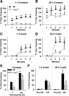
PAM+/− mice are deficient in fear conditioning. Wild-type (Wt) and PAM+/− littermates were conditioned and tested 1 or 24 h later in context- and cue-dependent fear conditioning. A, B, Percentage of time freezing during context testing 1 h (A) or 24 h (B) after conditioning. Animals were tested in the same chamber in which they had been conditioned, but in the absence of the CS and UCS. C, D, Percentage of time freezing during cued testing 1 h (C) or 24 h (D) after conditioning. In cued testing, animals were tested in a novel chamber, and after 2 min, the CS was presented in the absence of the UCS. E, Mean percentage of time freezing by Wt and PAM+/− mice over the 5 min context test, conducted 1 and 24 h after conditioning. F, Mean percentage of time freezing by PAM+/− mice during cued testing at 1 h (left) and 24 h (right) after conditioning; the pre-CS results are averaged over the first 2 min interval, and the CS results are over the final 3 min of testing during CS presentation. *p < 0.05, compared with Wt mice; + p < 0.05, compared with freezing responses at 1 h; ∧p < 0.05, compared with the pre-CS interval in the cued test (ANOVA, RMANOVA, Bonferroni-corrected pairwise comparisons). n = 10 mice pre genotype per test condition and time.
Figure 3.
Figure 3.
PAM+/− mice show abnormal fear-potentiated startle. Wild-type (Wt) and PAM+/− littermates were conditioned with two different intensities of shock (0.4 and 0.6 mA). A, Percentage of potentiation by Wt and PAM+/− mice to a 12 kHz, 70 dB tone preceded by a 100, 105, or 110 dB white-noise startle stimulus; animals were conditioned with 0.4 mA (left) or 0.6 mA (right) shock. B, Sensitivity of Wt and PAM+/− mice to varying intensities of scrambled footshock; behavioral responses are represented by composite scores during the application of the shock. *p < 0.05, compared with Wt mice; + p < 0.05, compared with responses to the 100 dB startle stimulus; # p < 0.05, compared with responses to the 105 dB startle stimulus; ∧p < 0.05, compared with responses after 0.4 mA shock (ANOVA, RMANOVA, Bonferroni-corrected pairwise comparisons). n = 9–10 mice per genotype per shock intensity for fear-potentiated startle; n = 10 mice per genotype for shock-threshold testing.
Figure 4.
Figure 4.
PAM is expressed in pyramidal neurons and interneurons in the basolateral complex. Coronal sections (15 μm) through the amygdala of wild-type (Wt) and PAM+/− mice were immunostained simultaneously with antisera to PAM (green) and GAD67 (red). A, B, Low-power merged images of Wt and PAM+/− amygdalae. LA, Lateral amygdala; BLA, basolateral amygdala; CeA, central amygdala; LPCS, lateral paracapsular cells; MPCS, medial paracapsular cells. Scale bars, 100 μm. C, D, High-power images of the area in LA of Wt and PAM+/− amygdalae outlined in A and B. Arrows indicate GAD67-positive interneurons. Asterisks indicate pyramidal neurons surrounded by GAD67-immunoreactive puncta representing axo-somatic GABAergic terminals. Staining for PAM was apparent in the soma of both interneurons and pyramidal neurons. Scale bars, 20 μm.
Figure 5.
Figure 5.
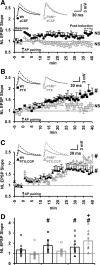
PAM+/− mice exhibit a deficiency in thalamic afferent LTP that relies on GABAergic transmission. Thalamic afferents were stimulated, and pyramidal neuron membrane responses were recorded in whole-cell mode in the lateral nuclei of wild-type (Wt) and PAM+/− amygdala slices. Insets above each plot depict representative averaged traces for each genotype before (solid) and after (dashed) LTP induction as indicated (baseline and after induction). A–C, Time course (1 min bins) of averaged LTP experiments using an action potential (AP) pairing induction paradigm. Synaptic efficacy was assessed using the rise slope of EPSPs that were normalized (NL) for genotypic comparisons. Experiments were conducted in aCSF control solution (A; n = 10 Wt, 11 PAM+/−), aCSF with 100 μm PTX (B; n = 9 Wt, 7 PAM+/−), or aCSF with PTX and CGP35348 (1 μm) (C; n = 12 Wt, 10 PAM+/−). D, Values of LTP assessed at 40 min after induction for individual pyramidal neurons are plotted by experimental condition (data from A–C). Bars depict mean values, and error bars depict the SEM. # p < 0.05, compared with baseline; + p < 0.05, compared with aCSF within genotype; NS, not significant compared to baseline; (Wilcoxon signed rank test).
Figure 6.
Figure 6.
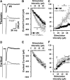
Baseline synaptic efficacy is enhanced at thalamic but not at cortical inputs to the PAM+/− lateral nucleus. Recordings of current responses (V holding = −55 mV) to graded intensities of single stimuli applied to thalamic and cortical afferent pathways of wild-type (Wt) and PAM+/− lateral amygdala slices. Responses consisted of a monosynaptic glutamatergic inward current followed by a disynaptic GABAergic outward current, representing feedforward inhibition. A, D, Representative traces depict pyramidal neuron responses to thalamic (A) and cortical (D) stimulation. B, E, Amplitude of evoked monosynaptic inward/excitatory current plotted as a function of stimulus intensity for thalamic (B) and cortical (E) inputs. C, F, Amplitude of evoked disynaptic outward/inhibitory current plotted as a function of stimulus intensity for thalamic (C) and cortical (F) inputs. Thalamic: n = 9 Wt, 11 PAM+/−; cortical: n = 8 Wt, 10 PAM+/−. *p < 0.05, compared with Wt (unpaired t test at individual stimulation intensities).
Figure 7.
Figure 7.
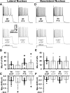
Active membrane properties of PAM+/− amygdalar pyramidal neurons are mostly intact but show subtle signs of hyperexcitability. A series of depolarizing 500 ms current steps were applied to wild-type (Wt) and PAM+/− pyramidal neurons in the presence and absence of PTX (100 μm). A, D, Representative active membrane voltage responses recorded in control solution (aCSF; left) and in the presence of PTX (right). Insets below indicate current step amplitude corresponding to displayed voltage traces. B, E, Action potentials (APs) elicited by current steps were counted, and the maximum numbers of APs are plotted by genotype and treatment condition for lateral (B; n = 17 Wt, 22 PAM+/−) and basolateral (E; n = 24 Wt, 26 PAM+/−) nuclei pyramidal neurons. Scatter points represent values for individual neurons, and bars represent group means with SEs. C, F, Peak amplitudes of the AHPs that occurred at the offset of each step were measured, and the maximum value for each neuron is plotted by genotype and treatment condition for lateral (C) and basolateral (F) nucleus pyramidal neurons. *p < 0.05, compared with Wt within treatment; + p < 0.05, compared with aCSF within genotype (two-way ANOVA, Bonferroni-corrected pairwise comparisons).
Figure 8.
Figure 8.
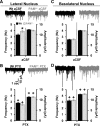
Enhanced spontaneous glutamatergic activity in the PAM+/− lateral nucleus is dependent on GABAA receptors. sEPSC from wild-type (Wt) and PAM+/− lateral and basolateral nucleus pyramidal neurons were recorded at V holding = −70 mV in the presence and absence of PTX (100 μm). Traces are representative examples of sEPSC activity for each genotype and treatment condition. A, C, sEPSC activity from Wt and PAM+/− pyramidal neurons in the lateral (A; n = 25 Wt, 25 PAM+/−; 38 events per neuron) and basolateral (C; n = 20 Wt, 24 PAM+/−; 63 events per neuron) nuclei recorded in control solution (aCSF). Frequency and amplitude of sEPSC events are plotted for each genotype (right). B, D, sEPSC activity recorded in the presence of PTX. Data are from Wt and PAM+/− pyramidal neurons in the lateral (B; n = 24 Wt, 21 PAM+/−) and basolateral (D; n = 19 Wt, 23 PAM+/−) nuclei. *p < 0.05, compared with Wt; + p < 0.05, compared with aCSF within genotype (two-way ANOVA, Bonferroni-corrected pairwise comparisons).
Figure 9.
Figure 9.
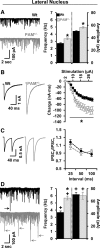
GABAergic signaling is enhanced in the PAM+/− lateral nucleus. Wild-type (Wt) and PAM+/− pyramidal neurons in the lateral nucleus were dialyzed with a high [Cl]-containing pipette solution, and IPSCs were recorded under glutamatergic blockade. A, mIPSCs were recorded in the presence of 1 μm TTX. Representative current traces depicting mIPSCs recorded from Wt and PAM+/− pyramidal neurons (left) are shown. Mean mIPSC frequency and amplitude are plotted for both genotypes (right; n = 28 Wt, 25 PAM+/−; 37 events per neuron). B, Local stimulation elicited “evoked” IPSCs in Wt and PAM+/− neurons only in the absence of TTX. Two superimposed representative current traces from each genotype in response to a 20 μA single stimulus are shown (left). Charge (the integral of current) was used as a measure of synaptic strength and is plotted as a function of stimulation intensity (right; n = 21 Wt, 16 PAM+/−). *p < 0.05, compared with Wt (unpaired t tests at individual stimulus intensities). C, Paired pulses were applied under the same conditions as in B. Averaged traces for each genotype depict representative current responses to paired pulses evoked at an intensity that elicited half-maximal responses for individual neurons (left). Ratios for both genotypes are plotted for 25, 50, and 100 ms intervals (right; n = 16 Wt, 13 PAM+/−). D, sIPSCs were recorded from Wt and PAM+/− neurons in the absence of TTX and local stimulation. Representative current traces depicting sIPSCs recorded from Wt and PAM+/− pyramidal neurons (left) are shown. Arrows indicate putative action potential-dependent GABAergic events. sIPSC frequency and amplitude are plotted for both genotypes (right; n = 25 Wt, 21 PAM+/−; 36 events per neuron). *p < 0.05, compared with Wt; + p < 0.05, compared with mIPSC value within genotype (ANOVA, Bonferroni-corrected pairwise comparisons for mIPSC and sIPSC data).
Figure 10.
Figure 10.
Roles of various amygdalar neuropeptides in neurophysiological alterations in the PAM+/− amygdala. PAM and neuropeptides are expressed in glutamatergic pyramidal neurons (PN) and GABAergic interneurons (IN) of the basolateral complex of the amygdala. GRP is expressed and secreted by PNs of the basolateral complex. GRP binds to the GRP receptor, which is expressed on INs and signals to increase GABA release (Shumyatsky et al., 2002). TRH also signals to increase GABA release (Gaier et al., unpublished data). The major neuropeptides expressed by local interneurons include CCK and NPY. CCK depolarizes interneurons to increase GABA release (Chung and Moore, 2007, 2009). NPY acts directly on PNs, hyperpolarizing them and reducing membrane resistance (Sosulina et al., 2008). In the PAM+/− amygdala (right), we found signs of enhanced GABA release, increased GABAergic synapse number, increased baseline glutamatergic signaling, reduced LTP at thalamic afferent synapses, and impairment of PN AHP.

Similar articles

Cited by

References

    1. Alam MR, Steveson TC, Johnson RC, Back N, Abraham B, Mains RE, Eipper BA. Signaling mediated by the cytosolic domain of peptidylglycine alpha-amidating monooxygenase. Mol Biol Cell. 2001;12:629–644. - PMC - PubMed
    1. Anagnostaras SG, Josselyn SA, Frankland PW, Silva AJ. Computer-assisted behavioral assessment of Pavlovian fear conditioning in mice. Learn Mem. 2000;7:58–72. - PMC - PubMed
    1. Baldwin GS, Patel O, Shulkes A. Phylogenetic analysis of the sequences of gastrin-releasing peptide and its receptors: biological implications. Regul Pept. 2007;143:1–14. - PubMed
    1. Bale TL, Vale WW. CRF and CRF receptors: role in stress responsivity and other behaviors. Annu Rev Pharmacol Toxicol. 2004;44:525–557. - PubMed
    1. Bauer EP, Ledoux JE. Heterosynaptic long-term potentiation of inhibitory interneurons in the lateral amygdala. J Neurosci. 2004;24:9507–9512. - PMC - PubMed

Publication types

MeSH terms

LinkOut - more resources