. 2010 Oct 13;30(41):13679-89.
doi: 10.1523/JNEUROSCI.1523-10.2010.

Prototypic seizure activity driven by mature hippocampal fast-spiking interneurons

Affiliations

Prototypic seizure activity driven by mature hippocampal fast-spiking interneurons

Yoko Fujiwara-Tsukamoto et al. J Neurosci. .

Abstract

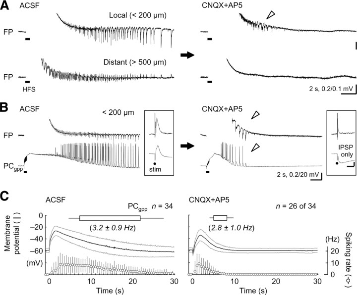
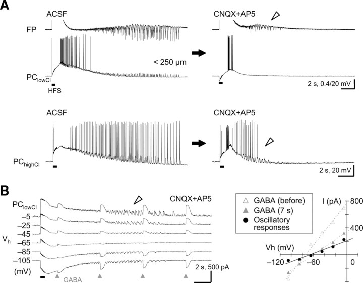
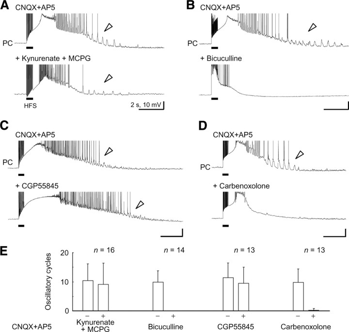
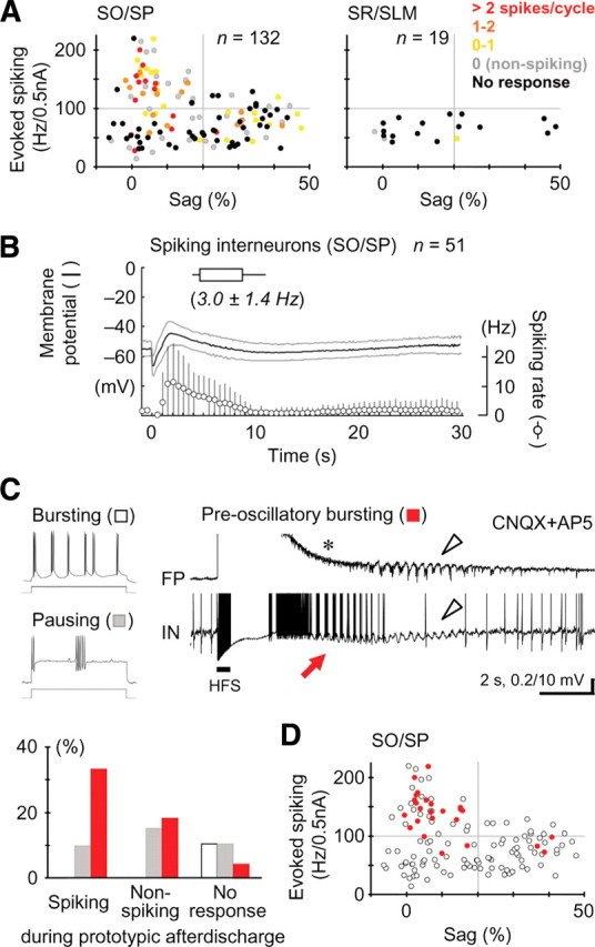
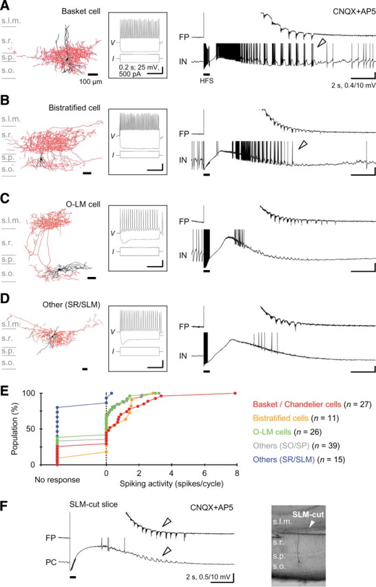
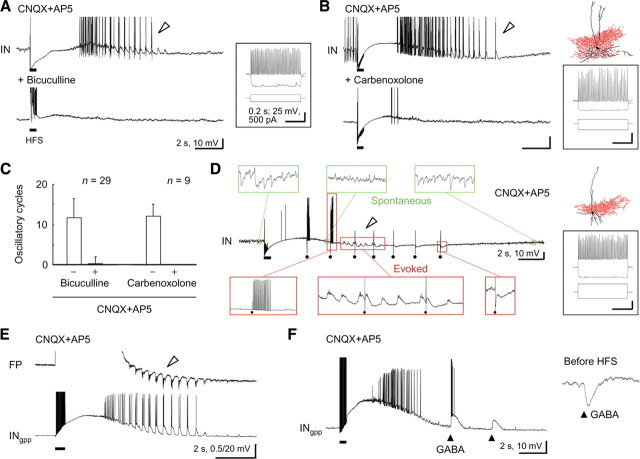
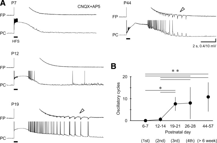
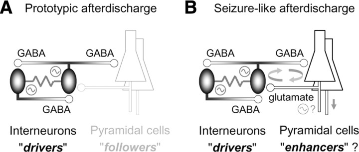
A variety of epileptic seizure models have shown that activation of glutamatergic pyramidal cells is usually required for rhythm generation and/or synchronization in hippocampal seizure-like oscillations in vitro. However, it still remains unclear whether GABAergic interneurons may be able to drive the seizure-like oscillations without glutamatergic transmission. Here, we found that electrical stimulation in rat hippocampal CA1 slices induced a putative prototype of seizure-like oscillations ("prototypic afterdischarge," 1.8-3.8 Hz) in mature pyramidal cells and interneurons in the presence of ionotropic glutamate receptor antagonists. The prototypic afterdischarge was abolished by GABA(A) receptor antagonists or gap junction blockers, but not by a metabotropic glutamate receptor antagonist or a GABA(B) receptor antagonist. Gramicidin-perforated patch-clamp and voltage-clamp recordings revealed that pyramidal cells were depolarized and frequently excited directly through excitatory GABAergic transmissions in each cycle of the prototypic afterdischarge. Interneurons that were actively spiking during the prototypic afterdischarge were mostly fast-spiking (FS) interneurons located in the strata oriens and pyramidale. Morphologically, these interneurons that might be "potential seizure drivers" included basket, chandelier, and bistratified cells. Furthermore, they received direct excitatory GABAergic input during the prototypic afterdischarge. The O-LM cells and most of the interneurons in the strata radiatum and lacunosum moleculare were not essential for the generation of prototypic afterdischarge. The GABA-mediated prototypic afterdischarge was observed later than the third postnatal week in the rat hippocampus. Our results suggest that an FS interneuron network alone can drive the prototypic form of electrically induced seizure-like oscillations through their excitatory GABAergic transmissions and presumably through gap junction-mediated communications.


Figures

Figure 1.
Figure 1.
Induction of the prototypic afterdischarge under a glutamate-blocking condition in hippocampal pyramidal cells. A, Local field potentials (FP) showing HFS-induced seizure-like afterdischarge in normal ACSF (left) and prototypic afterdischarge in the presence of the ionotropic glutamate receptor antagonists CNQX (10 μm) and AP5 (50 μm; right). Whereas the seizure-like afterdischarge was observed at both local (<200 μm from the site of stimulation) and distant (>500 μm) recording sites, the prototypic afterdischarge (arrowhead) was detected only at the local recording site in the same hippocampal CA1 slice. B, Oscillatory depolarizing responses during the seizure-like afterdischarge (left) and the prototypic afterdischarge (right) in a pyramidal cell recorded in the gramicidin-perforated patch-clamp method (PCgpp). Note that these responses were synchronous with the population spikes in the field potential (top, arrowheads) and that the generation of spikes indicates not only depolarizing but also excitatory responses in the pyramidal cell. Insets, Synaptic responses evoked by a single shock of electrical stimulation (stim). Calibration: 25 ms, 0.2 and 2.5 mV. C, Summary of the seizure-like afterdischarge (left) and the prototypic afterdischarge (right) in the pyramidal cells. Top, Time course of the seizure-like and prototypic afterdischarges in the pyramidal cells (boxes and bars indicate the mean and SD of start and end time, respectively). Middle, Membrane potential changes after the electrical induction (spikes were clipped in advance; black and gray lines indicate mean and SD, respectively). Bottom, Spiking rate changes after the electrical induction (circles and bars indicate mean and SD, respectively).
Figure 2.
Figure 2.
Oscillatory responses mediated by direct GABAergic input in pyramidal cells. A, Top, No clear oscillatory depolarizing responses were detected in pyramidal cells recorded in the low intracellular Cl condition (PClowCl) during the prototypic afterdischarge in the field potential (arrowhead). Bottom, The oscillatory responses in the prototypic afterdischarge (arrowhead) observed in pyramidal cells recorded in the high intracellular Cl condition (PChighCl). The oscillatory responses appeared to depend on Cl conductance. B, Left, Voltage-clamp recordings of the oscillatory responses in the prototypic afterdischarge (arrowhead) and exogenous GABA responses (triangles, 2, 7, 12, and 17 s after HFS) in a pyramidal cell. Right, Similar reversal potentials of oscillatory responses (filled circles) and exogenous GABA responses (gray triangles) in the same pyramidal cell. These reversal potentials may be artificially low because of the low-Cl-based electrode solution.
Figure 3.
Figure 3.
Pharmacological characterization of the prototypic afterdischarge in pyramidal cells. A, No effect of additional application of the nonspecific ionotropic glutamate receptor antagonist kynurenate (2 mm) and the metabotropic glutamate receptor antagonist MCPG (0.5 mm) on the expression of the prototypic afterdischarge in pyramidal cells (arrowheads). Action potentials are truncated for display. B, Complete abolishment of the expression of the prototypic afterdischarge by additional application of the GABAA receptor antagonist bicuculline (25 μm). C, No effect of the GABAB receptor antagonist CGP55845 (1 μm) on the prototypic afterdischarge expression (arrowheads). D, Abolishment of the prototypic afterdischarge expression by the gap junction blocker carbenoxolone (100 μm). E, Summary of the effects of these antagonists or the blocker on the expression of the prototypic afterdischarge in pyramidal cells. All of the pyramidal cells were recorded through the high Cl electrodes for reliable detection of the prototypic afterdischarge (see Fig. 2).
Figure 4.
Figure 4.
Electrophysiological properties of hippocampal interneurons showing the prototypic afterdischarge. A, Spiking activity in the prototypic afterdischarge cycles in individual interneurons with different electrophysiological properties (spiking ability and sag amplitude) in the strata oriens and pyramidale (left) and the strata radiatum and lacunosum moleculare (right). Each dot represents a single interneuron, and the color indicates the spiking ability [spikes/cycle; 0 (nonspiking), oscillatory responses with no spiking; No response, no oscillatory responses]. We defined an FS interneuron as >100 Hz/0.5 nA spiking and <20% sag amplitude (left, top left quadrant) (see Materials and Methods). B, Time course of the prototypic afterdischarge (top), membrane potential changes (middle), and spiking rate changes (bottom) in the interneurons with spiking oscillatory responses (compare Fig. 1C). C, Top, Representative traces illustrating an intrinsically bursting property (left, top) and a spike-pausing property (left, bottom) in response to depolarizing current injection and a preoscillatory bursting (right). Note that the preoscillatory bursting (red arrow) appeared in the interneuron, but not in the field potential (asterisk), earlier than the prototypic afterdischarge (arrowheads). Bottom, Population ratios of interneurons showing the bursting property (open), pausing property (gray), and preoscillatory bursting (red) in the interneurons with spiking and nonspiking oscillatory responses and with no oscillatory responses. D, Electrophysiological properties of interneurons showing the preoscillatory bursting activity (red) in the strata oriens and pyramidale (A, left). Most of these interneurons were categorized as FS interneurons.
Figure 5.
Figure 5.
Morphological identification of interneurons showing the prototypic afterdischarge. A, A basket cell showing oscillatory depolarizing responses with robust spiking (arrowhead) during the prototypic afterdischarge in the field potential. Left, Morphology of the recorded and visualized neuron (black, soma and dendrites; red, axons). Middle, Membrane potential responses to depolarizing and hyperpolarizing current injections. Right, Field potential and membrane potential traces during the prototypic afterdischarge. Calibrations are the same in AD. B, A bistratified cell showing oscillatory depolarizing responses with spiking during the prototypic afterdischarge (arrowhead). These two interneurons were categorized as FS interneurons. C, An O-LM cell showing oscillatory depolarizing responses without spiking during the prototypic afterdischarge. D, Another type of interneuron located in the s. radiatum close to the s. lacunosum moleculare showing no oscillatory responses during the prototypic afterdischarge. E, Cumulative distribution of spiking activity in the prototypic afterdischarge cycles (zero means nonspiking oscillatory responses) in different groups of morphologically visualized interneurons. F, Preservation of the prototypic afterdischarge generation (left, arrowheads) without the s. lacunosum moleculare (right). s.l.m., s. lacunosum moleculare; s.r., s. radiatum; s.p., s. pyramidale; s.o., s. oriens.
Figure 6.
Figure 6.
The prototypic afterdischarge mediated through GABAA receptors and gap junctions among interneurons. A, Abolishment of the prototypic afterdischarge in an FS interneuron by the application of 25 μm bicuculline. Calibrations are the same in A, B, and D. B, Abolishment of the prototypic afterdischarge in a basket cell by 100 μm carbenoxolone. C, Summary of the effects of bicuculline and carbenoxolone on the prototypic afterdischarge in interneurons. D, Spontaneous (green boxes) and evoked (red boxes) postsynaptic potentials around the prototypic afterdischarge expression (arrowhead) in another basket cell. Note that spontaneous hyperpolarizing postsynaptic potentials appeared only in the resting state (top, left and right) and that single shocks of electrical stimulation (dots) evoked excitatory (bottom left), depolarizing (bottom middle), and hyperpolarizing (bottom right) responses. E, Excitatory (spike-evoking) oscillatory responses during the prototypic afterdischarge in an interneuron recorded with the gramicidin-perforated patch-clamp method (INgpp). F, Excitatory/depolarizing responses to external GABA application (triangles) after HFS in the gramicidin-perforated patch-clamp condition.
Figure 7.
Figure 7.
Lack of the prototypic afterdischarge in an early developmental stage. A, Representative trials of electrical induction of the prototypic afterdischarge on postnatal days 7, 12, 19, and 44. Note that the prototypic afterdischarge appeared in field potential and pyramidal cells after the third postnatal week (P19 and P44; arrowheads). B, Postnatal development of the prototypic afterdischarge in pyramidal cells. *p < 0.05; **p < 0.01. All of the pyramidal cells were recorded through the high-Cl electrodes for reliable detection of the prototypic afterdischarge (see Fig. 2).
Figure 8.
Figure 8.
Schematic diagrams on network mechanisms underlying prototypic afterdischarge and seizure-like afterdischarge. A, Rhythm generation among FS interneurons through excitatory GABAergic transmissions and gap junctions during prototypic afterdischarge. B, Enhancement of afterdischarge activity by glutamatergic pyramidal cells during seizure-like afterdischarge. See Discussion for details.

Similar articles

Cited by

References

    1. Anderson WW, Lewis DV, Swartzwelder HS, Wilson WA. Magnesium-free medium activates seizure-like events in the rat hippocampal slice. Brain Res. 1986;398:215–219. - PubMed
    1. Avoli M, Psarropoulou C, Tancredi V, Fueta Y. On the synchronous activity induced by 4-aminopyridine in the CA3 subfield of juvenile rat hippocampus. J Neurophysiol. 1993;70:1018–1029. - PubMed
    1. Avoli M, Barbarosie M, Lücke A, Nagao T, Lopantsev V, Köhling R. Synchronous GABA-mediated potentials and epileptiform discharges in the rat limbic system in vitro. J Neurosci. 1996;16:3912–3924. - PMC - PubMed
    1. Babb TL, Pretorius JK, Kupfer WR, Crandall PH. Glutamate decarboxylase-immunoreactive neurons are preserved in human epileptic hippocampus. J Neurosci. 1989;9:2562–2574. - PMC - PubMed
    1. Ben-Ari Y. Excitatory actions of GABA during development: the nature of the nurture. Nat Rev Neurosci. 2002;3:728–739. - PubMed

Publication types

MeSH terms

Substances

LinkOut - more resources