. 2010 Oct 13;30(41):13690-8.
doi: 10.1523/JNEUROSCI.0303-09.2010.

Glucocorticoids exacerbate lipopolysaccharide-induced signaling in the frontal cortex and hippocampus in a dose-dependent manner

Affiliations

Glucocorticoids exacerbate lipopolysaccharide-induced signaling in the frontal cortex and hippocampus in a dose-dependent manner

Carolina Demarchi Munhoz et al. J Neurosci. .

Abstract

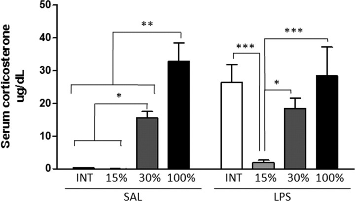
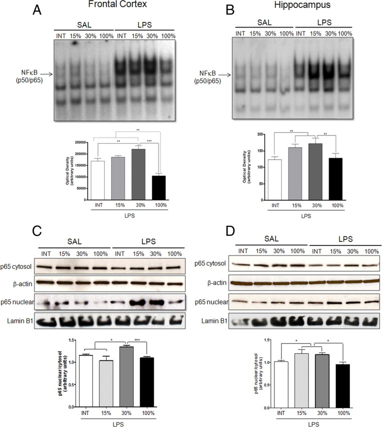
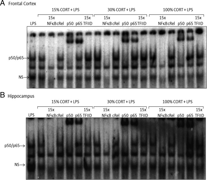
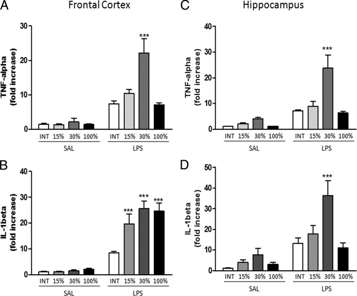
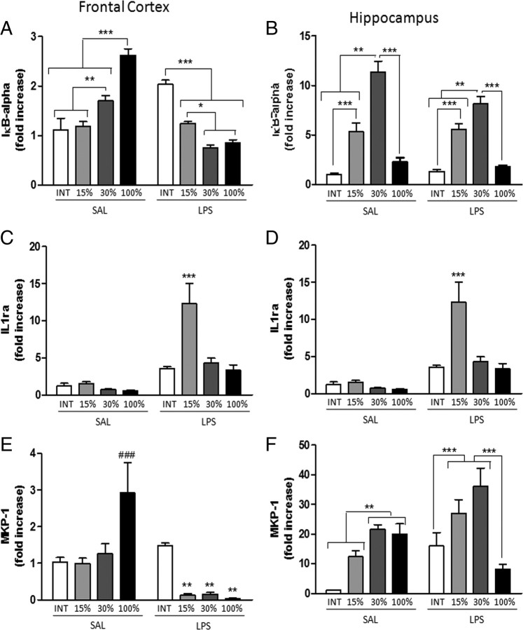
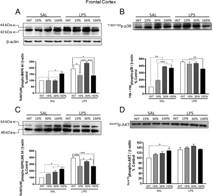
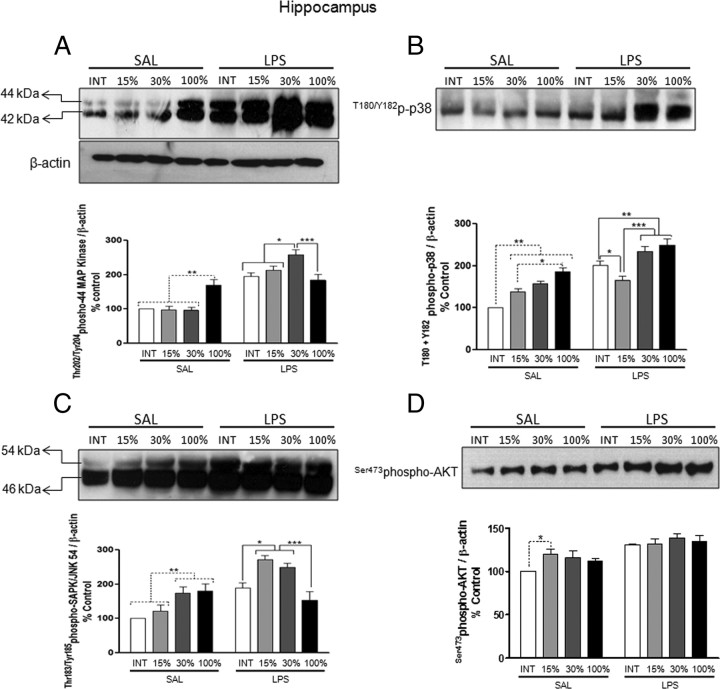
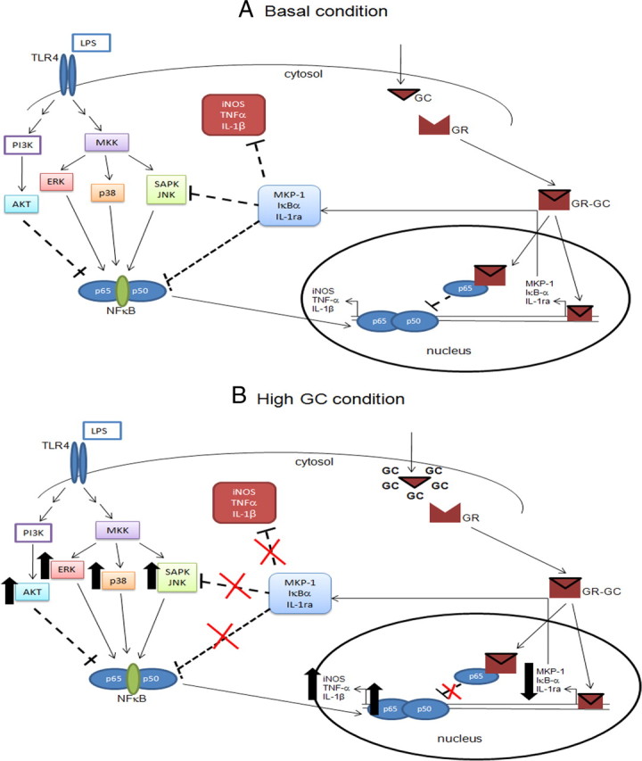
Although the anti-inflammatory actions of glucocorticoids (GCs) are well established, evidence has accumulated showing that proinflammatory GC effects can occur in the brain, in a poorly understood manner. Using electrophoretic mobility shift assay, real-time PCR, and immunoblotting, we investigated the ability of varying concentrations of corticosterone (CORT, the GC of rats) to modulate lipopolysaccharide (LPS)-induced activation of NF-κB (nuclear factor κB), expression of anti- and proinflammatory factors and of the MAP (mitogen-activated protein) kinase family [ERK (extracellular signal-regulated kinase), p38, and JNK/SAPK (c-Jun N-terminal protein kinase/stress-activated protein kinase)], and AKT. In the frontal cortex, elevated CORT levels were proinflammatory, exacerbating LPS effects on NF-κB, MAP kinases, and proinflammatory gene expression. Milder proinflammatory GCs effects occurred in the hippocampus. In the absence of LPS, elevated CORT levels increased basal activation of ERK1/2, p38, SAPK/JNK, and AKT in both regions. These findings suggest that GCs do not uniformly suppress neuroinflammation and can even enhance it at multiple levels in the pathway linking LPS exposure to inflammation.


Conflict of interest statement

The authors report no biomedical financial interests or potential conflicts of interest.

Figures

Figure 1.
Figure 1.
Serum CORT levels produced by CORT/cholesterol pellets in rats treated with saline (SAL) or LPS. Results are presented as mean ± SEM (n = 6 animals per group). Bonferroni test: *p < 0.05; **p < 0.01; ***p < 0.001.
Figure 2.
Figure 2.
Influence of CORT levels on NF-κB activation induced by LPS or saline (SAL) in the frontal cortex and hippocampus. Top, Representative EMSA autoradiography of frontal cortex (A) and hippocampus (B). NF-κB-specific band is indicated by arrow. Representative autoradiography from Western blot assay of the translocation of p65-NF-κB subunit in frontal cortex (C) and hippocampus (D). Bottom, Densitometric analysis of the NF-κB-specific band (representing p65/p50 heterodimers) or cytosolic and nuclear ratio presented in the top (mean ± SEM; n = 5 animals per group). Bonferroni test: **p < 0.01; ***p < 0.001.
Figure 3.
Figure 3.
Competition studies and supershift assays were performed on nuclear extract from frontal cortex (A) and hippocampus (B) of rats implanted with CORT pellets and treated with saline or LPS (1 mg/kg, i.v., 2 h) in the absence or presence of unlabeled specific (NF-κB consensus sequence, 15-fold molar excess) or nonspecific (TFIID consensus sequence, 15-fold molar excess) oligonucleotide, as indicated. Supershift assays were performed with the same nuclear extract (10 μg) incubated in the absence and presence of antibodies against subunits p50 (1:20 dilution), p65 (1:20 dilution), and cRel (1:20 dilution), as indicated. The position of specific NF-κB/DNA binding complex p50/p65 is indicated. NS represents nonspecific binding. The localization of the probe is also indicated. Results are representative of three independent experiments.
Figure 4.
Figure 4.
Influence of CORT levels on proinflammatory genes (IL-1β and TNF-α) in the absence (SAL) or presence of LPS in the frontal cortex (A, C) and hippocampus (B, D). Real-time PCRs were performed in duplicate and results are represented as mean ± SEM; n = 6 animals per group. Bonferroni test: *p < 0.05; **p < 0.01; ***p < 0.001 vs INT/LPS.
Figure 5.
Figure 5.
Influence of CORT levels on anti-inflammatory genes (IκB-α, IL-1ra, and MKP-1) in the absence (SAL) or presence of LPS in the frontal cortex (A, C, E) and hippocampus (B, D, F). Real-time PCRs were performed in duplicate and results are represented as mean + SEM; n = 6 animals per group. Bonferroni test: *p < 0.05; **p < 0.01; ***p < 0.001. For MKP-1: ###p < 0.001 vs INT/SAL; **p < 0.05 vs INT/LPS.
Figure 6.
Figure 6.
Influence of CORT levels on phosphorylated forms of MAP kinases 42/44 MAP kinase (A), p38 MAP kinase (B), SAPK/JNK (C), and AKT (D) in the absence (SAL) or presence of LPS in the frontal cortex. Top, Representatives of Western blot autoradiographies. Bottom, Densitometric analysis of the specific bands of SAL- or LPS-treated groups presented in the top panels (mean ± SEM; n = 5 animals per group). Protein levels are presented as ratios of specific kinases to β-actin levels. Bonferroni test: *p < 0.05; **p < 0.01; ***p < 0.001.
Figure 7.
Figure 7.
Influence of CORT levels on phosphorylated forms of MAP kinases 42/44 MAP kinase (A), p38 MAP kinase (B), SAPK/JNK (C), and AKT (D) in the absence (SAL) or presence of LPS in the hippocampus. Top, Representatives of Western blot photographs. Bottom, Densitometric analysis of the specific bands of SAL- or LPS-treated groups presented in the top (mean ± SEM; n = 5 animals per group). Protein levels are presented as ratios of specific kinases to β-actin levels. Bonferroni test: *p < 0.05; **p < 0.01; ***p < 0.001.
Figure 8.
Figure 8.
Schematic of the proposed role of high CORT levels on the proinflammatory response triggered by LPS and related to MAP kinase–NF-κB pathway in rat brain. Normally, LPS binds to TLR4 and activates the MAP kinase pathway, thus activating NF-κB and increasing expression of proinflammatory cytokines. Basal levels of GCs inhibit this pathway through a number of steps, including (1) GR interacting with the p65 subunit of NF-κB, (2) increasing the expression of the inhibitory protein IκB-α, and (3) increasing expression of anti-inflammatory genes such as IL-1ra and MKP-1 (A). However, when GC exposure is elevated and prolonged, the hormone increases and potentiates the proinflammatory response related to MAP kinase–NF-κB pathway (B). Straight lines indicate activation and dotted lines indicate inhibition.

Similar articles

Cited by

References

    1. Aljada A, Ghanim H, Assian E, Mohanty P, Hamouda W, Garg R, Dandona P. Increased IkappaB expression and diminished nuclear NF-kappaB in human mononuclear cells following hydrocortisone injection. J Clin Endocrinol Metab. 1999;84:3386–3389. - PubMed
    1. Almawi WY, Melemedjian OK. Negative regulation of nuclear factor-kappaB activation and function by glucocorticoids. J Mol Endocrinol. 2002;28:69–78. - PubMed
    1. Annane D, Sébille V, Bellissant E, Ger-Inf-05 Study Group Effect of low doses of corticosteroids in septic shock patients with or without early acute respiratory distress syndrome. Crit Care Med. 2006;34:22–30. - PubMed
    1. Beishuizen A, Thijs LG. Endotoxin and the hypothalamo-pituitary-adrenal (HPA) axis. J Endotoxin Res. 2003;9:3–24. - PubMed
    1. Bhattacharyya S, Brown DE, Brewer JA, Vogt SK, Muglia LJ. Macrophage glucocorticoid receptors regulate Toll-like receptor 4-mediated inflammatory responses by selective inhibition of p38 MAP kinase. Blood. 2007;109:4313–4319. - PMC - PubMed

Publication types

MeSH terms