

Mitogen-activated protein kinase-activated protein kinase 2 (MK2) contributes to secondary damage after spinal cord injury
- PMID: 20943915
- PMCID: PMC6633726
- DOI: 10.1523/JNEUROSCI.2998-10.2010
Mitogen-activated protein kinase-activated protein kinase 2 (MK2) contributes to secondary damage after spinal cord injury
Abstract
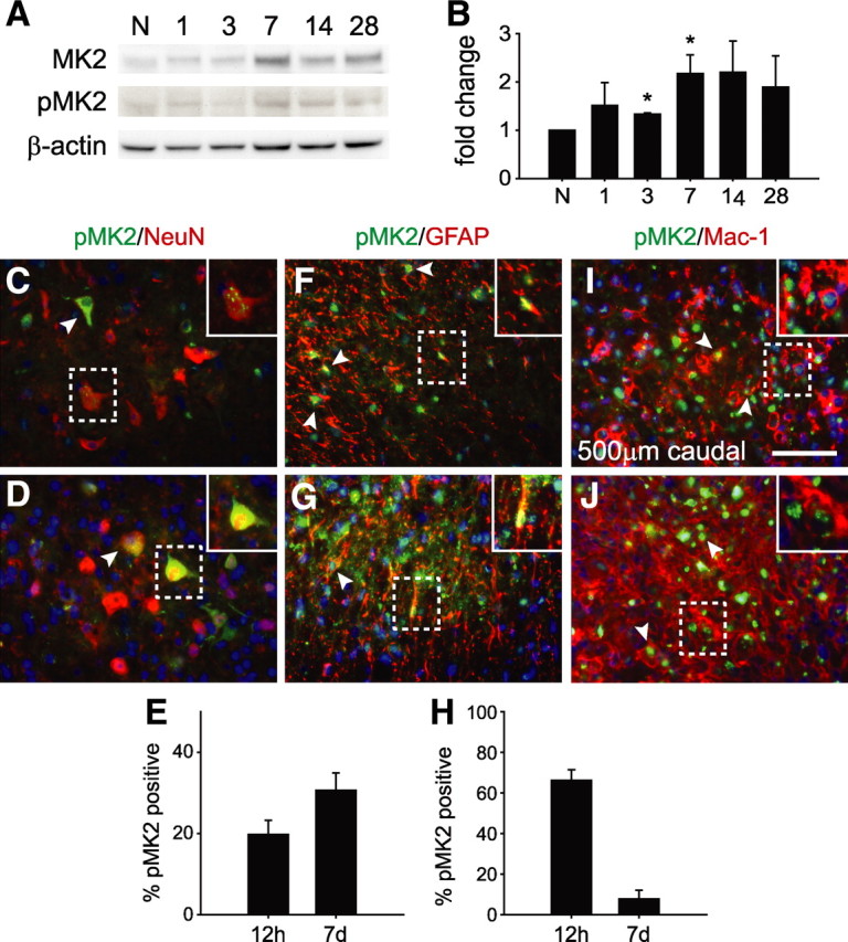
The inflammatory response contributes importantly to secondary tissue damage and functional deficits after spinal cord injury (SCI). In this work, we identified mitogen-activated protein kinase (MAPK)-activated protein kinase 2 (MAPKAPK2 or MK2), a downstream substrate of p38 MAPK, as a potential target using microarray analysis of contused spinal cord tissue taken at the peak of the inflammatory response. There was increased expression and phosphorylation of MK2 after SCI, with phospho-MK2 expressed in microglia/macrophages, neurons and astrocytes. We examined the role of MK2 in spinal cord contusion injury using MK2(-/-) mice. These results show that locomotor recovery was significantly improved in MK2(-/-) mice, compared with wild-type controls. MK2(-/-) mice showed reduced neuron and myelin loss, and increased sparing of serotonergic fibers in the ventral horn caudal to the injury site. We also found differential expression of matrix metalloproteinase-2 and 9 in MK2(-/-) and wild-type mice after SCI. Significant reduction was also seen in the expression of proinflammatory cytokines and protein nitrosylation in the injured spinal cord of MK2(-/-) mice. Our previous work has shown that macrophages lacking MK2 have an anti-inflammatory phenotype. We now show that there is no difference in the number of macrophages in the injured spinal cord between the two mouse strains and little if any difference in their phagocytic capacity, suggesting that macrophages lacking MK2 have a beneficial phenotype. These findings suggest that a lack of MK2 can reduce tissue damage after SCI and improve locomotor recovery. MK2 may therefore be a useful target to treat acute SCI.
Figures


Similar articles
-
MicroRNA-24-3p Inhibits Microglia Inflammation by Regulating MK2 Following Spinal Cord Injury.Neurochem Res. 2021 Apr;46(4):843-852. doi: 10.1007/s11064-020-03211-y. Epub 2021 Jan 13. Neurochem Res. 2021. PMID: 33439430
-
Ultrashort Wave Combined with Human Umbilical Cord Mesenchymal Stem Cell (HUC-MSC) Transplantation Inhibits NLRP3 Inflammasome and Improves Spinal Cord Injury via MK2/TTP Signalling Pathway.Biomed Res Int. 2020 Dec 5;2020:3021750. doi: 10.1155/2020/3021750. eCollection 2020. Biomed Res Int. 2020. PMID: 33376718 Free PMC article.
-
Expression and detrimental role of hematopoietic prostaglandin D synthase in spinal cord contusion injury.Glia. 2011 Apr;59(4):603-14. doi: 10.1002/glia.21128. Epub 2011 Feb 3. Glia. 2011. PMID: 21294159
-
Stress-Activated Protein Kinases in Spinal Cord Injury: Focus on Roles of p38.Int J Mol Sci. 2018 Mar 15;19(3):867. doi: 10.3390/ijms19030867. Int J Mol Sci. 2018. PMID: 29543752 Free PMC article. Review.
-
Novel Therapeutic Potential of Mitogen-Activated Protein Kinase Activated Protein Kinase 2 (MK2) in Chronic Airway Inflammatory Disorders.Curr Drug Targets. 2019;20(4):367-379. doi: 10.2174/1389450119666180816121323. Curr Drug Targets. 2019. PMID: 30112991 Review.
Cited by
-
PTEN/PI3K and MAPK signaling in protection and pathology following CNS injuries.Front Biol (Beijing). 2013 Aug 1;8(4):10.1007/s11515-013-1255-1. doi: 10.1007/s11515-013-1255-1. Front Biol (Beijing). 2013. PMID: 24348522 Free PMC article.
-
MK2 and Fas receptor contribute to the severity of CNS demyelination.PLoS One. 2014 Jun 25;9(6):e100363. doi: 10.1371/journal.pone.0100363. eCollection 2014. PLoS One. 2014. PMID: 24964076 Free PMC article.
-
Macrophage polarization: a key event in the secondary phase of acute spinal cord injury.J Cell Mol Med. 2017 May;21(5):941-954. doi: 10.1111/jcmm.13034. Epub 2016 Dec 13. J Cell Mol Med. 2017. PMID: 27957787 Free PMC article. Review.
-
Harnessing the Secretome of Mesenchymal Stromal Cells for Traumatic Spinal Cord Injury: Multicell Comparison and Assessment of In Vivo Efficacy.Stem Cells Dev. 2020 Nov 15;29(22):1429-1443. doi: 10.1089/scd.2020.0079. Epub 2020 Oct 21. Stem Cells Dev. 2020. PMID: 32962528 Free PMC article.
-
Molecular and cellular changes in the lumbar spinal cord following thoracic injury: regulation by treadmill locomotor training.PLoS One. 2014 Feb 10;9(2):e88215. doi: 10.1371/journal.pone.0088215. eCollection 2014. PLoS One. 2014. PMID: 24520355 Free PMC article.
References
-
- Alford KA, Glennie S, Turrell BR, Rawlinson L, Saklatvala J, Dean JL. Heat shock protein 27 functions in inflammatory gene expression and transforming growth factor-beta-activated kinase-1 (TAK1)-mediated signaling. J Biol Chem. 2007;282:6232–6241. - PubMed
-
- Antri M, Orsal D, Barthe JY. Locomotor recovery in the chronic spinal rat: effects of long-term treatment with a 5-HT2 agonist. Eur J Neurosci. 2002;16:467–476. - PubMed
-
- Bao F, Chen Y, Dekaban GA, Weaver LC. Early anti-inflammatory treatment reduces lipid peroxidation and protein nitration after spinal cord injury in rats. J Neurochem. 2004;88:1335–1344. - PubMed
-
- Bartholdi D, Schwab ME. Expression of pro-inflammatory cytokine and chemokine mRNA upon experimental spinal cord injury in mouse: an in situ hybridization study. Eur J Neurosci. 1997;9:1422–1438. - PubMed
Publication types
MeSH terms
Substances
Grants and funding
LinkOut - more resources
Full Text Sources
Medical
Molecular Biology Databases