. 2010 Oct 13;30(41):13794-807.
doi: 10.1523/JNEUROSCI.1567-10.2010.

RBPJkappa-dependent signaling is essential for long-term maintenance of neural stem cells in the adult hippocampus

Affiliations

RBPJkappa-dependent signaling is essential for long-term maintenance of neural stem cells in the adult hippocampus

Oliver Ehm et al. J Neurosci. .

Abstract

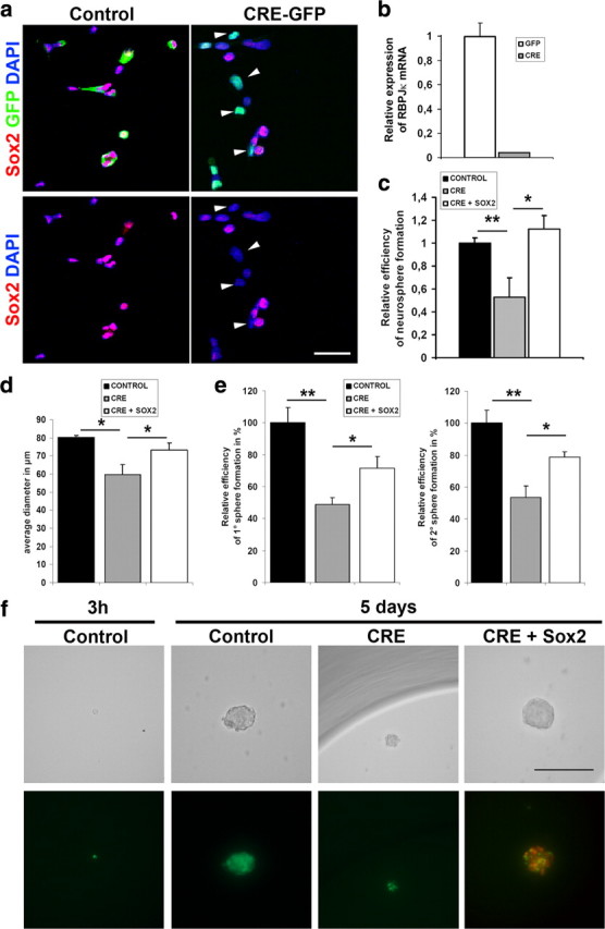
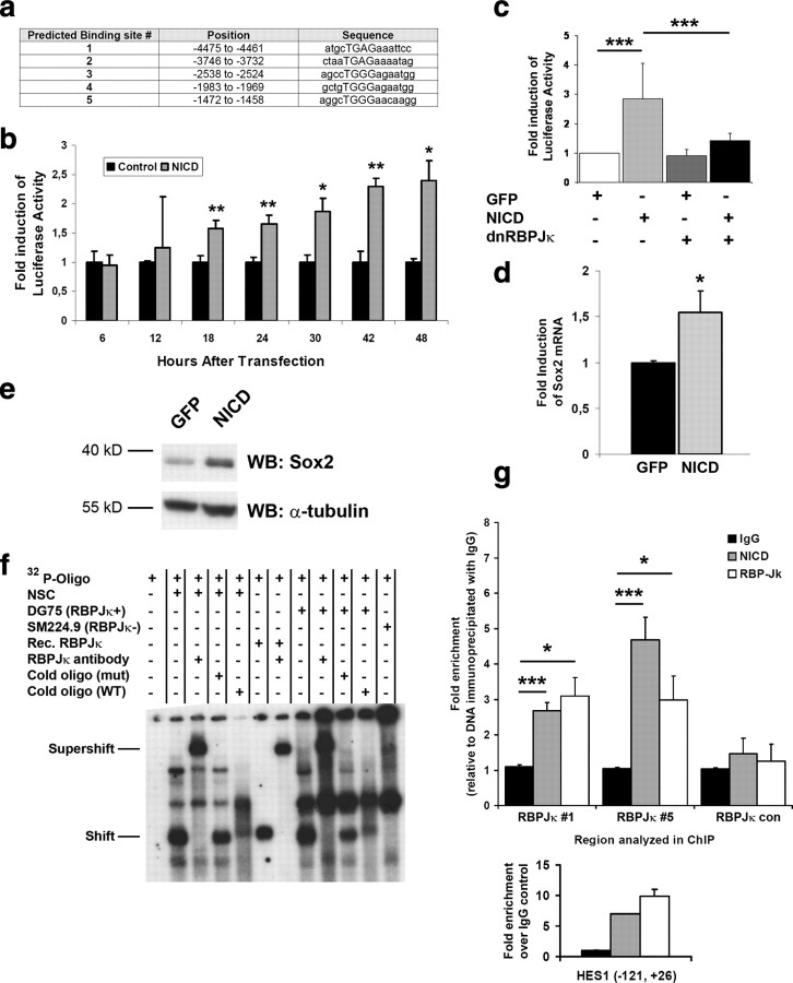
The generation of new neurons from neural stem cells in the adult hippocampal dentate gyrus contributes to learning and mood regulation. To sustain hippocampal neurogenesis throughout life, maintenance of the neural stem cell pool has to be tightly controlled. We found that the Notch/RBPJκ-signaling pathway is highly active in neural stem cells of the adult mouse hippocampus. Conditional inactivation of RBPJκ in neural stem cells in vivo resulted in increased neuronal differentiation of neural stem cells in the adult hippocampus at an early time point and depletion of the Sox2-positive neural stem cell pool and suppression of hippocampal neurogenesis at a later time point. Moreover, RBPJκ-deficient neural stem cells displayed impaired self-renewal in vitro and loss of expression of the transcription factor Sox2. Interestingly, we found that Notch signaling increases Sox2 promoter activity and Sox2 expression in adult neural stem cells. In addition, activated Notch and RBPJκ were highly enriched on the Sox2 promoter in adult hippocampal neural stem cells, thus identifying Sox2 as a direct target of Notch/RBPJκ signaling. Finally, we found that overexpression of Sox2 can rescue the self-renewal defect in RBPJκ-deficient neural stem cells. These results identify RBPJκ-dependent pathways as essential regulators of adult neural stem cell maintenance and suggest that the actions of RBPJκ are, at least in part, mediated by control of Sox2 expression.


Figures

Figure 1.
Figure 1.
a, Analysis of adult Tg(Cp-EGFP)25Gaia mice shows that Sox2-expressing cells (blue) in the SGZ of the dentate gyrus are positive for the GFP reporter (green, arrowheads). In contrast, adjacent NeuroD-expressing cells (red) are GFP reporter negative. GFP expression is also present in scattered cells of the dentate granule cell layer. Scale bar, 10 μm. b, Analysis of adult Hes5-GFP mice reveals activity of the GFP reporter (green) in Sox2-positive (red) radial glia-like and nonradial stem cells. GFAP is shown in blue. Scale bar, 20 μm. c, Hes5-GFP reporter is active in Sox2-positive cells (red, arrowheads) but not in NeuroD-expressing cells (blue).
Figure 2.
Figure 2.
a, Experimental strategy to study the role of RBPJκ signaling in adult hippocampal stem cells and neurogenesis. GLAST::CreERT2; RBPJκloxp/loxp; R26::EYFP (RBPJκ-cKO) and GLAST::CreERT2; RBPJκloxp/+; R26::EYFP (control) were treated for 5 d with TAM to induce recombination in the RBPJκ and the R26::EYFP locus. Animals were analyzed 21 or 60 d after the final TAM injection. b–d, Loss of RBPJκ in radial glia-like stem cells decreases stem cell numbers 3 weeks after TAM-induced recombination. b, Representative confocal images of RBPJκ-cKO and control mice. A large proportion of YFP-positive recombined cells (green) in RBPJκ-cKO does not express Sox2 (red) and GFAP (blue) and does not display a radial glia-like morphology. Staining for Sox2 shows reduced density of Sox2 cells in the SGZ of RBPJκ-cKO. Staining for GFAP demonstrates an overall reduction in radial glia-like stem cells in the dentate gyrus. In addition, the overall number of YFP-positive cells is increased in RBPJκ-cKO. DAPI is shown in gray. Scale bar, 20 μm. c, The fraction of all Sox2-expressing cells, radial glia-like stem cells (type 1 cells, identified by Sox2/GFAP expression and radial morphology), and nonradial stem cells (type 2 cells, identified by Sox2 expression and localization in the SGZ) among the recombined cells is significantly decreased in RBPJκ-cKO mice. **p < 0.01; ***p < 0.001. d, The density of Sox2-expressing cells, radial glia-like stem cells (type 1 cells), and nonradial stem cells (type 2 cells) in the SGZ is significantly decreased in RBPJκ-cKO mice. *p < 0.05; **p < 0.01).
Figure 3.
Figure 3.
Loss of RBPJκ in radial glia-like stem cells increases neurogenesis 3 weeks after recombination. a, Representative confocal images of RBPJκ-cKO and control mice. The overall number of proliferating cells identified by expression of PCNA (red) as well as the fraction of PCNA-positive cells among the YFP-positive recombined cells (green, arrowheads) is increased in RBPJκ-cKO mice. Scale bar, 20 μm. b, Quantification of the density of YFP-positive cells. *p < 0.01. c, e, Quantification of the density of PCNA positive cells (c) (**p < 0.01) and the fraction of PCNA positive cells (e) among the recombined cells (***p < 0.001). d, The overall number of NeuroD-expressing newly generated neurons (red) as well as the fraction of NeuroD-positive immature neurons among the YFP-positive recombined cells (green, arrowheads) is strongly increased in RBPJκ-cKO mice. DCX is shown in blue. Scale bar, 20 μm. f, Quantification of the density of NeuroD-positive cells (***p < 0.001). g, The overall number of newly generated neurons identified by expression of DCX (red) as well as the fraction of DCX-positive immature neurons among the YFP-positive recombined cells (green) is strongly increased in RBPJκ-cKO mice. Note the pronounced increase in the overall number of YFP-expressing cells. Scale bar, 20 μm. h, Quantification of the density of DCX-positive immature neurons and the fraction of DCX-expressing cells among the recombined cells (***p < 0.001).
Figure 4.
Figure 4.
Loss of RBPJκ in radial glia-like stem cells increases proliferation in YFP-positive and YFP-negative cells. Proliferating Sox2-positive stem cells (blue) are present in the YFP-positive (green, red arrowhead) and YFP-negative (white arrowhead) compartment (left). Similarly, proliferating DCX-positive immature neurons are found among YFP-positive (red arrowhead) and YFP-negative cells (right). PCNA is shown in red, and DAPI is shown in gray. Scale bar, 20 μm.
Figure 5.
Figure 5.
Loss of RBPJκ in radial glia-like stem cells decreases hippocampal neurogenesis and leads to persistent loss of Sox2-expressing stem cells 2 months after recombination. a, Representative confocal images of RBPJκ-cKO (right) and control mice (left). In RBPJκ-cKO mice, YFP-positive recombined cells (green) do not express Sox2 (red) or GFAP (blue). Staining for GFAP demonstrates a strong reduction in the number of radial glia-like stem cells in the dentate gyrus. The vast majority of YFP-positive cells in RBPJκ-cKO is located in the granule cell layer. DAPI is shown in gray. Scale bar, 20 μm. b, Sox2-expressing cells, radial glia-like stem cells (type 1 cells), and nonradial stem cells (type 2 cells) are depleted from the SGZ of the dentate gyrus in RBPJκ-cKO mice (**p < 0.01). c, Phenotyping of YFP-positive cells demonstrates that the vast majority recombined radial glial like stem cells have left the stem cell compartment. Almost no YFP-positive cells express DCX, indicating that recombined cells do not contribute to the generation of new neurons 2 months after induction of recombination. Cells were phenotyped according to the following criteria: type 1, Sox2+ GFAP+ and radial morphology; type 2, Sox2+ GFAP−; immature neurons, DCX+; mature neurons, NeuN+. d, Representative confocal images of RBPJκ-cKO and control mice. In RBPJκ-cKO mice, DCX-expressing immature neurons (red) are virtually absent. YFP is shown in green, and DAPI is shown in blue. Scale bar, 20 μm. e, The density of DCX-expressing immature neurons in the dentate gyrus is severely reduced (*p < 0.05). f, Representative confocal images of RBPJκ-cKO and control mice. In RBPJκ-cKO mice, proliferating cells identified by the expression of PCNA (red) are virtually absent. DAPI is shown in blue. Scale bar, 20 μm.
Figure 6.
Figure 6.
a, Immunocytochemical analysis of neurospheres derived from adult RBPJκloxp/loxp mice 48 h after transduction with the CAG-GFP (control) or the CAG-GFPnlsCRE (CRE-GFP) retrovirus (green). Cells were dissociated and fixed onto slides. Note that a number of CRE-GFP transduced cells are negative for Sox2 (red, arrowheads). b, Quantitative PCR reveals significantly decreased (p < 0.001) expression of RBPJκ mRNA in CAG-GFPnlsCRE (CRE) transduced RBPJκloxp/loxp neurospheres. c–f, Neurosphere assays of RBPJκloxp/loxp NSCs after transduction with CAG-GFP (control), CAG-GFPnlsCRE (CRE), or CAG-GFPnlsCRE and CAG-Sox2 IRES dsRED (CRE + SOX2). c, Analysis of neurosphere-forming efficiency of RBPJκloxp/loxp NSCs in single-cell assays (*p < 0.05; **p < 0.01). d, Analysis of neurosphere diameter of RBPJκloxp/loxp NSCs 7 d after transduction (*p < 0.05). e, Analysis of primary and secondary neurosphere-forming efficiency of RBPJκloxp/loxp NSCs in low-density cell assays (*p < 0.05; **p < 0.01). f, Left, Representative image of a CAG-GFP transduced single cell 3 h after seeding into a miniwell. Right, Representative images of neurospheres formed by RBPJκloxp/loxp NSCs 5 d after plating. Top, Bright field; bottom, fluorescent analysis. Scale bar, 100 μm.
Figure 7.
Figure 7.
a, Summary of in silico analysis of the 5.5 kb mouse Sox2 promoter. The positions of the predicted RBPJκ-binding sites are presented in relation to the transcriptional start site. b, Reporter assays in HEK293 cells using the Sox2-luciferase show significant activation 18 h after transfection with NICD (*p < 0.05; **p < 0.01). c, Reporter assays in adult hippocampal NSCs using a 5.5 kb Sox2-luciferase demonstrate that the Sox2 promoter is activated by Notch signaling. This activation was inhibited by expression of a dominant-negative form of RBPJκ (***p < 0.001). d, Quantitative RT-PCR analysis of Sox2 mRNA expression in NSCs 6 h after transfection with an expression vector for NICD or a control expression vector encoding for GFP (*p < 0.05). e, Western blot analysis of adult hippocampal NSCs after overexpression of NICD or GFP as control. Enhanced activation of Notch signaling by overexpression of NICD increases expression of Sox2. The loading control is α-tubulin. f, EMSA shows binding of adult hippocampal NSC-derived RBPJκ to sequences (predicted binding site #5) in the 5.5 kb Sox2 promoter. g, ChIP analysis demonstrates that Sox2 is a direct target of Notch/RBPJκ signaling in adult NSCs. PCR primers were designed to surround the predicted RBPJκ-binding sites 1 (RBPJκ #1) and 5 (RBPJκ #5) and a control region (RBPJκ con) on the Sox2 promoter (*p < 0.05; ***p < 0.001). Bottom, ChIP analysis using PCR primers, which were designed to surround RBPJκ binding sites on the Hes1 promoter, shows enrichment of Notch1 and RBPJκ on the Hes1 promoter in adult hippocampal NSCs.

Similar articles

Cited by

References

    1. Ables JL, Decarolis NA, Johnson MA, Rivera PD, Gao Z, Cooper DC, Radtke F, Hsieh J, Eisch AJ. Notch1 is required for maintenance of the reservoir of adult hippocampal stem cells. J Neurosci. 2010;30:10484–10492. - PMC - PubMed
    1. Adachi K, Mirzadeh Z, Sakaguchi M, Yamashita T, Nikolcheva T, Gotoh Y, Peltz G, Gong L, Kawase T, Alvarez-Buylla A, Okano H, Sawamoto K. Beta-catenin signaling promotes proliferation of progenitor cells in the adult mouse subventricular zone. Stem Cells. 2007;25:2827–2836. - PubMed
    1. Aizawa K, Ageyama N, Terao K, Hisatsune T. Primate-specific alterations in neural stem/progenitor cells in the aged hippocampus. Neurobiol Aging. 2010 in press. - PubMed
    1. Andreu-Agullo C, Morante-Redolat JM, Delgado AC, Farinas I. Vascular niche factor PEDF modulates Notch-dependent stemness in the adult subependymal zone. Nat Neurosci. 2009;12:1514–1523. - PubMed
    1. Androutsellis-Theotokis A, Leker RR, Soldner F, Hoeppner DJ, Ravin R, Poser SW, Rueger MA, Bae SK, Kittappa R, McKay RD. Notch signalling regulates stem cell numbers in vitro and in vivo. Nature. 2006;442:823–826. - PubMed

Publication types

MeSH terms

Substances

LinkOut - more resources