. 2010 Oct 13;30(41):13814-25.
doi: 10.1523/JNEUROSCI.1943-10.2010.

Mechanisms involved in systemic nicotine-induced glutamatergic synaptic plasticity on dopamine neurons in the ventral tegmental area

Affiliations

Mechanisms involved in systemic nicotine-induced glutamatergic synaptic plasticity on dopamine neurons in the ventral tegmental area

Ming Gao et al. J Neurosci. .

Abstract

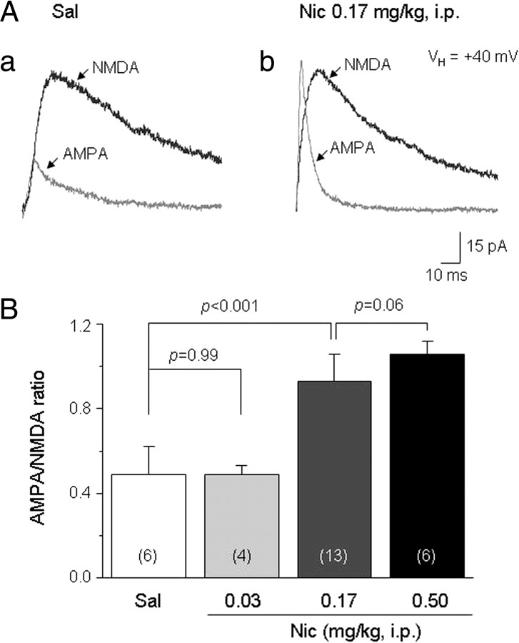
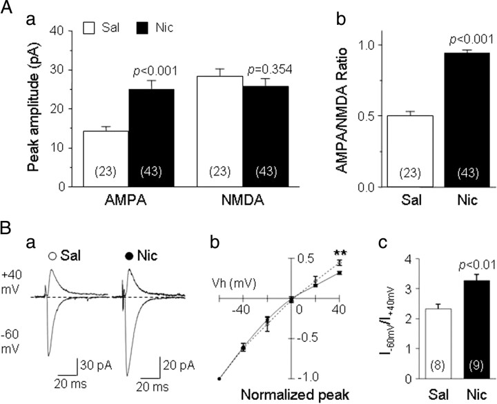
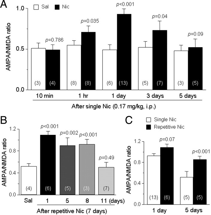
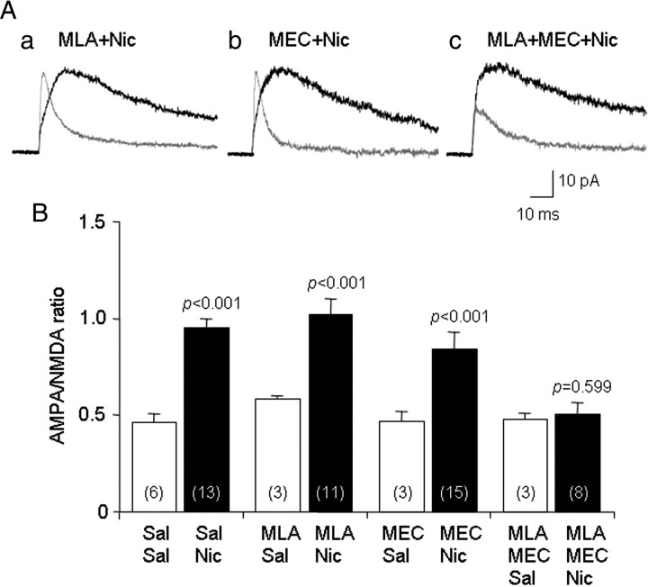
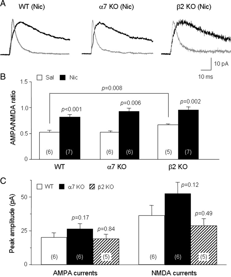
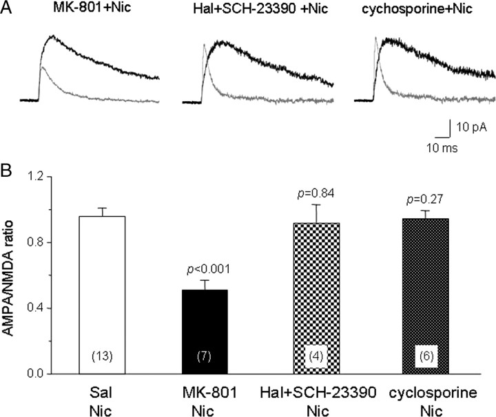
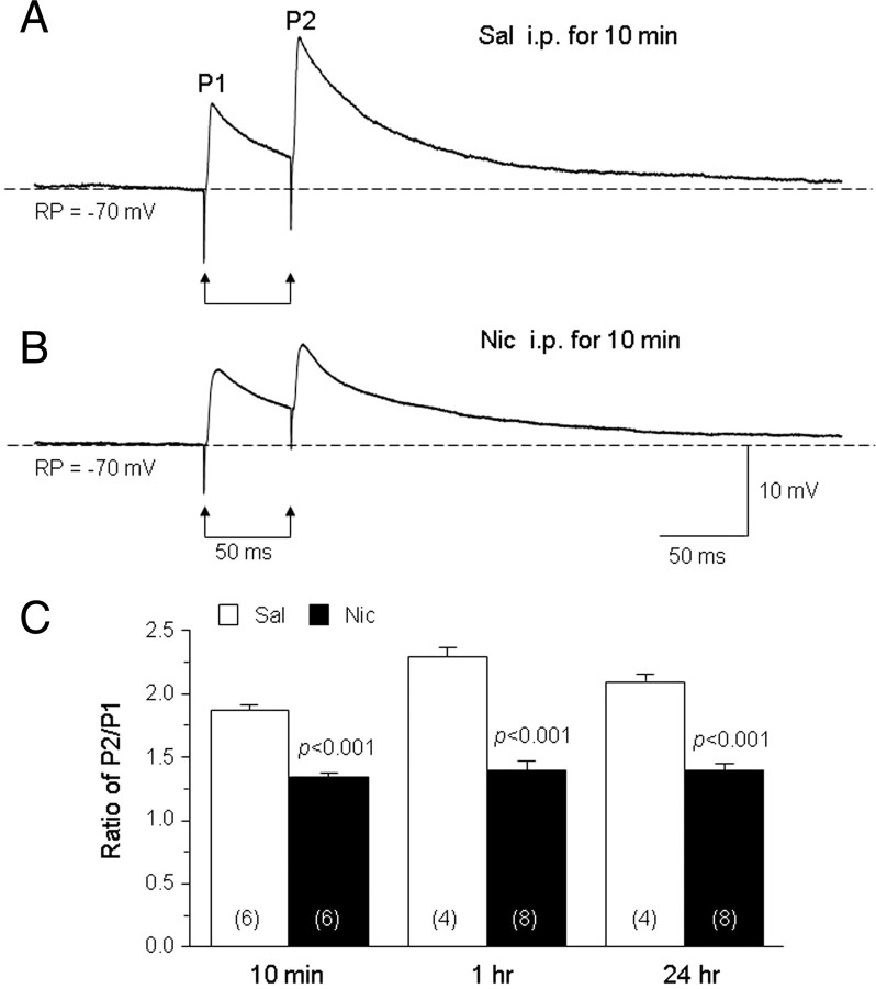
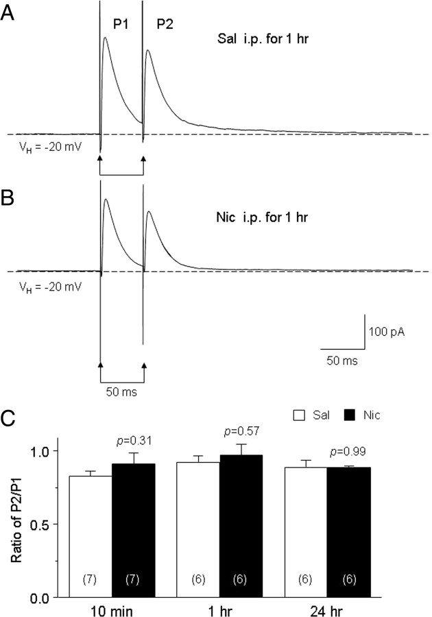
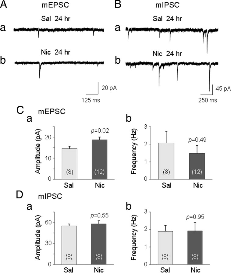
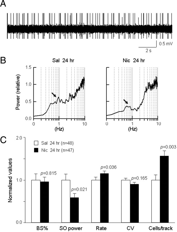
Systemic exposure to nicotine induces glutamatergic synaptic plasticity on dopamine (DA) neurons in the ventral tegmental area (VTA), but mechanisms are largely unknown. Here, we report that single, systemic exposure in rats to nicotine (0.17 mg/kg free base) increases the ratio of DA neuronal currents mediated by AMPA relative to NMDA receptors (AMPA/NMDA ratio) assessed 24 h later, based on slice-patch recording. The AMPA/NMDA ratio increase is evident within 1 h and lasts for at least 72 h after nicotine exposure (and up to 8 d after repeated nicotine administration). This effect cannot be prevented by systemic injection of either α7-nAChR (nicotinic ACh receptor)-selective [methyllycaconitine (MLA)] or β2*-nAChR-selective [mecamylamine (MEC)] antagonists but is prevented by coinjection of MLA and MEC. In either nAChR α7 or β2 subunit knock-out mice, systemic exposure to nicotine still increases the AMPA/NMDA ratio. Preinjection in rats of a NMDA receptor antagonist MK-801((+)-5-methyl-10,11-dihydro-5H-dibenzo[a,d]cyclohepten-5,10-imine maleate), but neither DA receptor antagonists [SCH-23390 (R-(+)-7-chloro-8-hydroxy-3-methyl-1-phenyl-2,3,4,5-tetrahydro-1H-3-benzazepine) plus haloperidol] nor a calcineurin inhibitor (cyclosporine), prevents the nicotine-induced increase in AMPA/NMDA ratio. After systemic exposure to nicotine, glutamatergic (but not GABAergic) transmission onto rat VTA DA neuronal inputs is enhanced. Correspondingly, DA neuronal firing measured 24 h after nicotine exposure using extracellular single-unit recording in vivo is significantly faster, and there is conversion of silent to active DA neurons. Collectively, these findings demonstrate that systemic nicotine acting via either α7- or β2*-nAChRs increases presynaptic and postsynaptic glutamatergic function, and consequently initiates glutamatergic synaptic plasticity, which may be an important, early neuronal adaptation in nicotine reward and reinforcement.


Figures

Figure 1.
Figure 1.
Single, systemic nicotine administration increases the AMPA/NMDA ratio. A, Representative traces are shown for AMPA receptor- and NMDA receptor-mediated currents (indicated by arrows) in saline- or nicotine-injected rats. Patch-clamp recording was performed from DA neurons in VTA slices 24 h after nicotine bitartrate (Nic) (equivalent free base dose is shown) or saline (Sal)injection i.p. B, A summary is shown of effects of nicotine free base delivered to achieve the indicated doses on AMPA/NMDA ratios (ordinate; mean ± SEM; n = number of cases; t test results are indicated).
Figure 2.
Figure 2.
Systemic nicotine upregulates postsynaptic AMPA receptor function. A, AMPA receptor- and NMDA receptor-mediated currents [ordinate, absolute peak amplitude (pA)] were recorded in DA neurons from VTA slices obtained from nicotine-treated (Nic) (0.17 mg/kg free base; solid bars) or saline-treated (Sal) (open bars) rats. Results show that nicotine significantly increases AMPA receptor-mediated currents (Aa), which causes an increase in the AMPA/NMDA ratio (Ab). B, Current–voltage (I–V) relationships were derived for AMPA receptor-mediated currents from DA neurons in slices from rats treated with normal saline (○) or nicotine (•) (0.17 mg/kg free base). Recordings were done using pipette electrodes containing 100 μm spermine and in the presence of bath-applied 20 μm bicuculline and 100 μm APV. Ba, Representative traces of AMPA currents at −60 and +40 mV holding potential (V h). Bb, Plot of peak AMPA current amplitudes (ordinate; normalized responses obtained at −60 mV V h) as a function of holding potential (mV) shows inward rectification at positive holding potentials. Bc, Comparison of peak AMPA current amplitudes at −60 and +40 mV V h (I −60 mV/I +40 mV; ordinate) shows enhanced inward rectification after nicotine (solid bar) relative to saline (open bar) exposure. **p < 0.01.
Figure 3.
Figure 3.
Time course for nicotine-induced increases in AMPA/NMDA ratios. A, AMPA/NMDA ratios were measured on DA neurons in rat VTA slices at the times indicated after a single nicotine injection (Nic) (0.17 mg/kg free base, i.p.; solid bars) or saline injection (Sal) (open bars). Nicotine treatment increases AMPA/NMDA ratios measured 1, 24, or 72 h but not 10 min or 5 d after injection. B, AMPA/NMDA ratios were assessed at the indicated times after repeated, once-daily injections with nicotine (0.17 mg/kg free base, i.p.) for 7 d. There was an increase in AMPA/NMDA ratios at 1, 5, or 8 d but not 11 d after the last nicotine injection. C, Comparisons of nicotine treatment effects on AMPA/NMDA ratios measured 1 or 5 d after a single injection of nicotine (open bars) or 1 or 5 d after the last of seven, once-daily injections of nicotine. Results show that repeated administration is required to maintain the change in glutamatergic response for 5 d.
Figure 4.
Figure 4.
nAChR subtypes that mediate the systemic nicotine-induced increase in the AMPA/NMDA ratio. A, Representative traces are shown for AMPA (thin) and NMDA (thick) receptor-mediated currents measured on DA neurons from rat VTA slices prepared 24 h after animals were injected to achieve doses of 5.0 mg/kg MLA, 3.0 mg/kg MEC, or both 10 min before treatment via a single injection with nicotine (Nic) (0.17 mg/kg free base, i.p.). B, Summary of effects of pretreatment with α7-nAChR (MLA) and/or β2*-nAChR (MEC) antagonists on systemic nicotine (solid bars)-induced increases in AMPA/NMDA ratios. Results also are shown for effects or antagonist pretreatment on AMPA/NMDA ratios after negative control saline (Sal) (open bars) injection. Neither MLA nor MEC alone inhibits the nicotine-induced increase in the AMPA/NMDA ratio. However, coadministration of MLA and MEC completely abolishes the nicotine-induced increase in the AMPA/NMDA ratio. Antagonists have no significant effects alone or when combined in saline-treated animals. Statistical comparisons between saline and nicotine conditions are presented above each solid bar, and statistical comparisons across saline or nicotine groups are presented in the narrative.
Figure 5.
Figure 5.
Systemic nicotine-induced increases in AMPA/NMDA ratios in nAChR α7 and β2 subunit KO mice. A, Representative traces are shown for AMPA (thin) and NMDA (thick) receptor-mediated currents measured on DA neurons from nAChR α7 subunit KO (α7 KO) or β2 subunit (β2 KO) mouse VTA slices prepared 24 h after animals were given a single injection of nicotine (Nic; 0.17 mg/kg free base, i.p). B, Summary of effects of saline (Sal) (open bars) or nicotine (solid bars) treatment on AMPA/NMDA ratios in WT, α7 KO, or β2 KO mice, showing a nicotine-induced increase in all three sets of animals. The p values indicated above solid bars make reference to the statistical difference in the increase in the AMPA/NMDA ratio in nicotine- relative to saline-treated animals. C, Summary of effects of nicotine treatment on absolute peak current amplitudes (ordinate, pA) mediated by AMPA or NMDA receptors in VTA DA neurons from WT (open bars), α7 KO (solid bars) or β2 KO (cross-hatched bars) mice. The p values indicated above the relevant KO animal bars make reference to the statistical difference in current amplitude relative to that in WT mice. Results indicate that systemic exposure to nicotine is still able to increase the AMPA/NMDA ratio in the absence of either nAChR α7 or β2 subunits.
Figure 6.
Figure 6.
Pharmacological dissection of events involved in the nicotine-induced increase in the AMPA/NMDA ratio. A, Representative traces are shown for AMPA (thin) and NMDA (thick) receptor-mediated currents measured on DA neurons from rat VTA slices prepared 24 h after animals were given a single injection of nicotine (Nic) (0.17 mg/kg free base, i.p.). B, Summary of effects of treatment before nicotine injection with MK-801 (1.0 mg/kg; 10 min pretreatment), 2 mg/kg haloperidol (Hal) plus 1 mg/kg SCH-23390 (10 min pretreatment), or 15 mg/kg cyclosporine (90 min pretreatment) on the AMPA/NMDA ratio. The p values compare the relevant results to those after positive control, saline-nicotine treatment.
Figure 7.
Figure 7.
Effects of systemic nicotine exposure on EPSPs. A, B, Representative traces are shown for paired-pulse stimulation at an interpulse interval of 50 ms of EPSPs recorded from DA neurons in rat VTA slices. Slices were prepared 10 min after animals were given a single injection of saline (Sal) (A) or nicotine (Nic) (0.17 mg/kg free base, i.p.) (B), and current-clamp recording was done at a resting potential of −70 mV in the presence of bath-applied picrotoxin to block GABAA receptors. C, Summary of results demonstrates that the EPSP paired-pulse ratio (P2/P1; ordinate) is significantly decreased 10 min or 1 or 24 h after systemic injection of nicotine (solid bars) relative to saline-treated controls (open bars; p values are shown for each pair). Absolute P1 or P2 amplitudes were measured from the same baseline, which is indicated by the horizontal dashed line in A or B, but the nicotine treatment-induced reduction in PPR is evident even if calculating P2 amplitudes from the tail of the P1 response at the time of the second pulse. These results suggest that nicotine exposure increases the probability of presynaptic glutamate release.
Figure 8.
Figure 8.
Effects of systemic nicotine exposure on IPSCs. A, B, Representative traces are shown for paired-pulse stimulation at an interpulse interval of 50 ms of IPSCs on DA neurons in rat VTA slices. Slices were prepared 1 h after animals were given a single injection of saline (Sal) (A) or nicotine (Nic) (0.17 mg/kg free base, i.p.) (B), and voltage-clamp recoding was done at a holding potential of −20 mV in the presence of bath-applied NBQX (10 μm) and d-APV (50 μm) to block glutamatergic responses. C, Summary of results demonstrates that the IPSC paired pulse ratio (P2/P1) is not significantly altered (p values are shown for each pair) 10 min or 1 or 24 h after systemic injection of nicotine (solid bars) relative to saline-treated controls (open bars). Absolute P1 or P2 amplitudes were measured from the same baseline, which is indicated by the horizontal dashed line in A or B, but equivalent results are obtained even if calculating P2 amplitudes from the tail of the P1 response These results suggest that nicotine exposure has no effect on the probability of presynaptic GABA release.
Figure 9.
Figure 9.
A single injection of nicotine significantly increases mEPSC amplitude. A, B, Typical traces of mEPSCs (A) or mIPSCs (B) recorded 24 h after a single injection of saline (Sal) (Aa) or nicotine (Nic) 0.17 mg/kg, i.p.) (Bb). C, D, Summary of results shows that, relative to normal saline controls (lightly shaded bars), systemic injection of nicotine significantly increases mEPSC amplitude (Ca, darkly shaded bar) but not mEPSC frequency (Cb), mIPSC amplitude (Da), or mIPSC frequency (Db).
Figure 10.
Figure 10.
Effects of systemic nicotine exposure on firing activity in VTA DA neurons. A, A typical trace for an extracellular, single unit recording from a VTA DA neuron in an anesthetized rat. B, Comparison of slow oscillations (0.5–1.5 Hz) in rat DA neuronal firing measured 24 h after injections with nicotine (Nic) (0.17 mg/kg free base, i.p.) or saline (Sal). C, A summary of findings showing features of VTA DA neuronal firing properties 24 h after animals were injected with saline (open bars) or nicotine (solid bars). The p values are for differences relative to saline-treated controls. Results show that a single nicotine exposure significantly decreases the power of slow oscillations (P 0.5–1.5 Hz) and increases the firing rate (Rate), but has no effect on either the number of spikes in bursts (BS%) or the coefficient of variability of interspike intervals (CV). Nicotine exposure also shows a marked increase in the number of firing DA cells in each recording track (cells/track). These findings suggest that a single nicotine injection maintains a long-lasting excitation of VTA DA neurons and switches silent to firing DA neurons.

Similar articles

Cited by

References

    1. Addy NA, Fornasiero EF, Stevens TR, Taylor JR, Picciotto MR. Role of calcineurin in nicotine-mediated locomotor sensitization. J Neurosci. 2007;27:8571–8580. - PMC - PubMed
    1. Argilli E, Sibley DR, Malenka RC, England PM, Bonci A. Mechanism and time course of cocaine-induced long-term potentiation in the ventral tegmental area. J Neurosci. 2008;28:9092–9100. - PMC - PubMed
    1. Benwell ME, Balfour DJ. The effects of acute and repeated nicotine treatment on nucleus accumbens dopamine and locomotor activity. Br J Pharmacol. 1992;105:849–856. - PMC - PubMed
    1. Bissière S, Humeau Y, Lüthi A. Dopamine gates LTP induction in lateral amygdala by suppressing feedforward inhibition. Nat Neurosci. 2003;6:587–592. - PubMed
    1. Bunney BS, Grace AA. Acute and chronic haloperidol treatment: comparison of effects on nigral dopaminergic cell activity. Life Sci. 1978;23:1715–1727. - PubMed

Publication types

MeSH terms

LinkOut - more resources