. 2010 Oct 13;30(41):13838-49.
doi: 10.1523/JNEUROSCI.3846-10.2010.

Analysis of the astray/robo2 zebrafish mutant reveals that degenerating tracts do not provide strong guidance cues for regenerating optic axons

Affiliations

Analysis of the astray/robo2 zebrafish mutant reveals that degenerating tracts do not provide strong guidance cues for regenerating optic axons

Cameron Wyatt et al. J Neurosci. .

Abstract

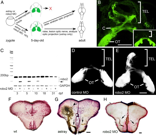
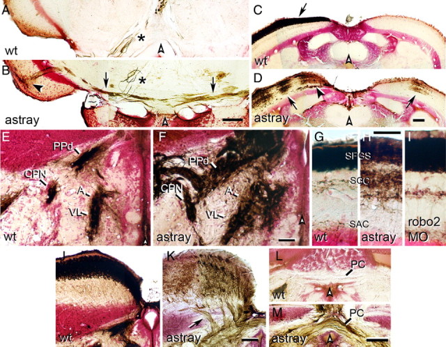
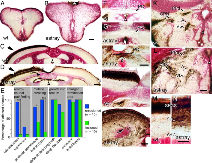
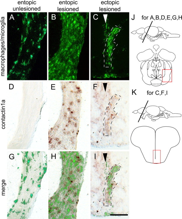
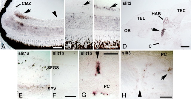
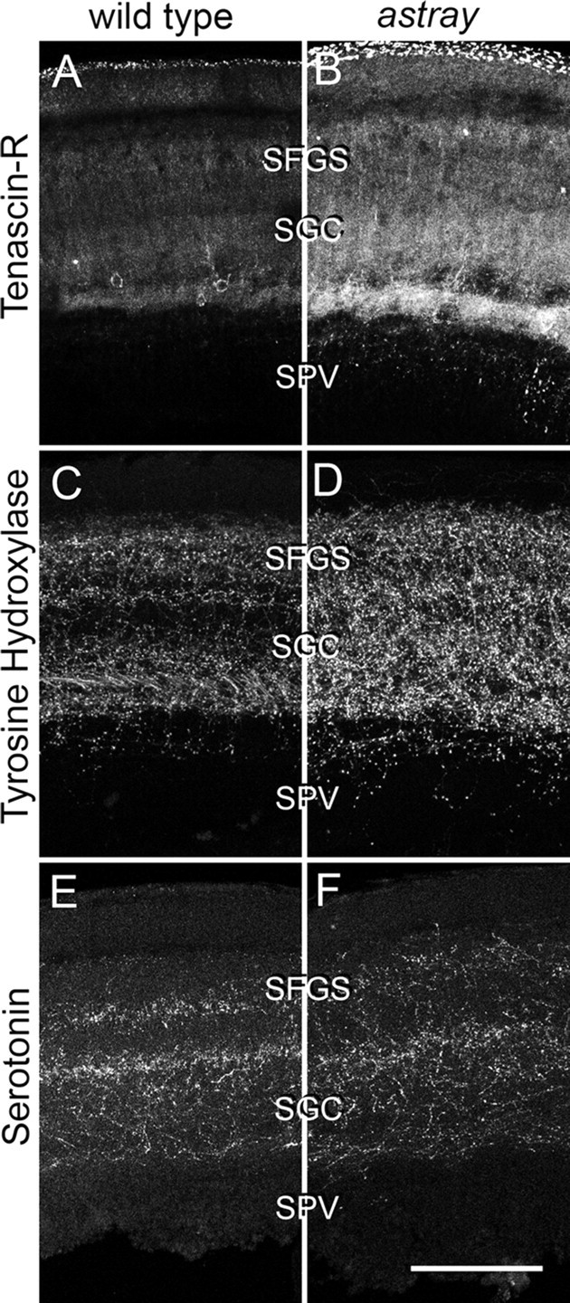
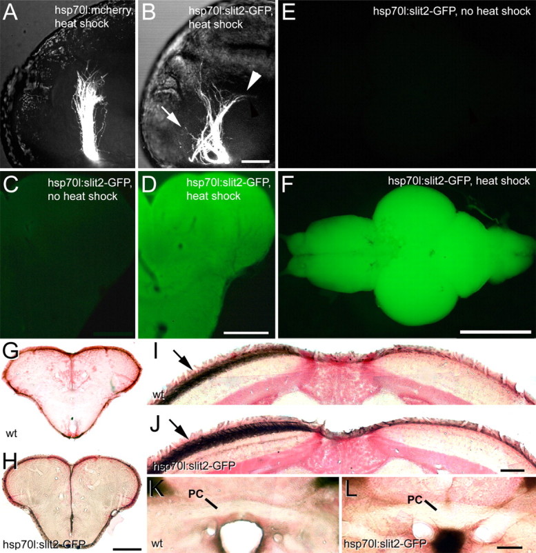
During formation of the optic projection in astray/robo2 mutant zebrafish, optic axons exhibit rostrocaudal pathfinding errors, ectopic midline crossing and increased terminal arbor size. Here we show that these errors persist into adulthood, even when robo2 function is conditionally reduced only during initial formation of the optic projection. Adult errors include massive ectopic optic tracts in the telencephalon. During optic nerve regeneration in astray/robo2 animals, these tracts are not repopulated and ectopic midline crossing is reduced compared with unlesioned mutants. This is despite a comparable macrophage/microglial response and upregulation of contactin1a in oligodendrocytes of entopic and ectopic tracts. However, other errors, such as expanded termination areas and ectopic growth into the tectum, were frequently recommitted by regenerating optic axons. Retinal ganglion cells with regenerating axons reexpress robo2 and expression of slit ligands is maintained in some areas of the adult optic pathway. However, slit expression is reduced rostral and caudal to the chiasm, compared with development and ubiquitous overexpression of Slit2 did not elicit major pathfinding phenotypes. This shows that (1) there is not an efficient correction mechanism for large-scale pathfinding errors of optic axons during development; (2) degenerating tracts do not provide a strong guidance cue for regenerating optic axons in the adult CNS, unlike the PNS; and (3) robo2 is less important for pathfinding of optic axons during regeneration than during development.


Figures

Figure 1.
Figure 1.
Pathfinding errors in the optic projection are retained in adults in astray mutants and in robo2 morphants. A, Experimental paradigm. Living 5-d-old larvae were preselected for the presence of aberrant telencephalic optic tracts and raised for adult experiments as indicated. B, Dorsal views are shown (rostral is up). Living astray larvae were selected according to the presence of GFP-positive optic axons in the telencephalon (TEL). Inset shows a wild-type projection without telencephalic tracts. (C, chiasm; OT, optic tectum). Brightly labeled neuromasts have been removed from the projection for clarity. C, PCR analysis of robo2 mRNA expression with and without robo2 splice-blocking morpholino (MO). The morpholino reduces the abundance of the wild-type transcript and an erroneous transcript (arrow) becomes detectable through at least 10 dpf. Glyceraldehyde-3-phosphate dehydrogenase is used as an internal standard. D, E, Dorsal views of DiI-traced optic projections (rostral is up) indicate astray-like pathfinding errors in robo2 morpholino-injected (E), but not in control morpholino-injected (D) 5-d-old larvae. The ectopic projection to the telencephalon is mainly unilateral. F–H, Photomicrographs show optic axons (brown) in cross sections of the adult telencephalon (counterstained in red); dorsal is up. Ectopic tracts of optic axons (arrows in G, H) are present in the telencephalon of astray (G) and robo2 morphant (MO) animals (H), but not in wild type (F). The arrowhead in G indicates a dense termination area of ectopic optic axons in the dorsal telencephalon. Scale bars: B, 100 μm (250 μm for inset); D, E, 100 μm; F, G, 200 μm; H, 100 μm.
Figure 2.
Figure 2.
Aberrations of the optic projection in adult astray mutants and robo2 morphants. Photomicrographs show optic axons (brown) in cross sections of the adult brain (counterstained in red); dorsal is up. White arrowheads indicate brain midline. A, B, Ectopic optic tracts (B, arrows) in the tegmentum of astray mutants cross the midline and terminate in the ipsilateral tectum (black arrowhead). No ectopic tracts are present in wild-type animals (A). Asterisks (A, B) indicate large diameter axons of the oculomotor nucleus that are always inadvertently retrogradely traced from the eye muscles. C, D, In wild-type fish (C), optic axons cover the entire contralateral tectum only (arrow in C). In astray mutants (D) the contralateral and ipsilateral tectal halves are innervated in ocular dominance column-like patches (D, arrows). Note deep axons growing into the tectum in astray mutants (black arrowhead in D). E, F, Innervation of the CPN, the anterior (A) and ventrolateral thalamus (VL), as well as the dorsal part of the periventricular pretectal nucleus (PPd) is expanded in astray (F) compared with wild-type (E). G–I, Innervation of tectal layers is expanded in astray mutants (H), but not in robo2 morpholino-treated animals (I) compared with wild-type (G). SFGS, Stratum fibrosum et griseum superficiale; SGC, stratum griseum centrale; SAC, stratum album centrale. J, K, In astray mutants (K), but not in wild-type animals (J), optic axons enter the tectum in several individual deep fascicles (arrow in K). L, M, Optic axons cross in the posterior commissure (PC) in astray (M), but not in wild-type fish (L). Scale bars: A, B, E–I, 50 μm; C, D, J–M, 100 μm.
Figure 3.
Figure 3.
Correction and recurrence of errors by regenerating optic axons in adult astray mutants. Optic axons are stained brown in cross sections of the adult brain (counterstained in red); dorsal is up. White arrowheads indicate the brain midline. Asterisks indicate nonspecific labeling of the meninges. A, B, No regenerated optic axons are present in wild-type (A) or astray (B) telencephalon. C, D, The regenerated optic projection in the tectum is exclusively contralateral (arrow) in wild type (C) and astray (D), but erroneous growth of deep fascicles (black arrowhead in D) recurs in astray. E, Frequencies of different astray phenotypes before and after regeneration of the optic projection in animals preselected for the presence of telencephalic tracts in larvae. *p < 0.05, ***p < 0.0001. F–H, Regenerating optic axons do not cross the posterior commissure (PC) in wild-type fish (F). Regenerated optic axons show ectopic crossing in the posterior commissure in some astray animals (H), but not in others (G). I, J, Regenerated axons enter the tectum in separate fascicles in astray (arrow in J), but not in wild type (I). K, L, Termination areas of regenerated optic axons in the pretectum are expanded in astray animals (L), compared with wild-type animals (K). M, N, In astray (N), reinnervation of tectal layers is expanded compared with wild type (M) after regeneration. For abbreviations see Figure 2. Scale bars: A, B, 200 μm; C, D, F–L, 100 μm; M, N, 50 μm.
Figure 4.
Figure 4.
Deafferented ectopic optic tracts in the telencephalon of astray mutants display macrophage/microglial cell activation and increased contactin1a mRNA expression comparable to entopic tracts. Cross sections through the adult brain are shown as indicated in J and K. Macrophage/microglial cell immunolabeling (A–C) and contactin1a mRNA labeling (D–F) is comparable between deafferented entopic (B, E, H) and ectopic astray optic tracts (C, F, I). Both signals are increased compared with unlesioned entopic tracts (A, D, G). Arrowheads in C, F, and I indicate telencephalic midline. G–I show superimposition of macrophage/microglial cell and contactin1a mRNA labeling. Scale bar, 200 μm.
Figure 5.
Figure 5.
Robo2 and slits are expressed during regeneration of the adult optic projection. Cross sections are shown, except for D. A, In the retina of unlesioned juvenile, 4-week-old animals, robo2 mRNA is expressed in recently differentiated retinal ganglion cells in the peripheral growth zone of the retina (arrow) next to the ciliary margin zone (CMZ). Older, more central retinal ganglion cells (arrowhead) do not express detectable levels of robo2 mRNA. B, C, In the adult (>3 months of age) central retina, robo2 mRNA is reexpressed in the retinal ganglion cell layer at 2 weeks postlesion (arrow in C) compared with the retinal ganglion cell layer in unlesioned controls (arrow in B). D, A sagittal section of the brain is shown (rostral left, dorsal up). Conspicuous expression of slit2 mRNA is found in the habenula (HAB) and in the ventral diencephalon (arrow) at the level of the optic chiasm (C). OB, Olfactory bulb; TEL, telencephalon; TEC, tectum mesencephali. E, F, Slit1a (E), but not slit1b (F), is expressed in the deafferented tectum at 1 week postlesion. SPV, stratum periventriculare; SFGS, stratum fibrosum et griseum superficiale. G, Strong local expression of slit1b mRNA is found at the level of the posterior commissure (PC) in cross sections of the brain. H, Low levels of slit3 mRNA expression are found in the pretectum, including the PPd area (arrow). Arrowheads in G and H indicate the brain midline. Scale bars: A, 50 μm; B, C, 50 μm; D, 200 μm; E, F, 100 μm; G, 50 μm; H, 100 μm.
Figure 6.
Figure 6.
Comparison of laminar distribution of different markers in the denervated tectum at 1 week postlesion. Cross sections through the dorsal tectum are shown (dorsal is up). Tenascin-R (A, B), tyrosine hydroxylase (C, D), and serotonin (E, F) immunoreactivities show comparable distribution in wild-type and astray animals. However, labeling intensity of Tenascin-R and tyrosine hydroxylase was increased in astray mutants relative to wild-type animals. For anatomical abbreviations see previous figures. Scale bar, 100 μm.
Figure 7.
Figure 7.
Ubiquitous overexpression of Slit2-GFP causes astray-like phenotypes in the developing, but not the regenerating optic projection. A, B, Lateral views (maximal-intensity projections) of optic axons in heat-shocked embryos at 48 hpf show an essentially wild-type projection in hsp70l:mcherry control embryos, but in hsp70l:slit2-GFP embryos the tract is severely disorganized with anterior (arrow) and posterior (arrowhead) misprojections. C–F, After repeated heat-shocks the entire brain of hsp70l:slit2-GFP animals (F) shows intense GFP fluorescence, compared with non-heat-shocked controls (E). Similarly, immunodetection of GFP in sections of the telencephalon shows homogeneous immunoreactivity after heat shock (D), but not in non-heat-shocked controls (C). G–L, Regenerating optic axons in heat-shocked wild-type (wt) and hsp70l:slit2-GFP transgenic fish do not grow into the telencephalon (G, H) or the posterior commissure (PC in K, L) and exclusively populate the contralateral tectum (arrows in I, J). Scale bars: A, B, 50 μm; C, D, 200 μm; E, F, 1 mm; G, H, 200 μm; I, J, 100 μm; K, L, 50 μm.

Similar articles

Cited by

References

    1. Ankerhold R, Leppert CA, Bastmeyer M, Stuermer CA. E587 antigen is upregulated by goldfish oligodendrocytes after optic nerve lesion and supports retinal axon regeneration. Glia. 1998;23:257–270. - PubMed
    1. Barnett SC, Riddell JS. Olfactory ensheathing cell transplantation as a strategy for spinal cord repair—what can it achieve? Nat Clin Pract Neurol. 2007;3:152–161. - PubMed
    1. Becker CG, Becker T. Repellent guidance of regenerating optic axons by chondroitin sulfate glycosaminoglycans in zebrafish. J Neurosci. 2002;22:842–853. - PMC - PubMed
    1. Becker CG, Becker T. Growth and pathfinding of regenerating axons in the optic projection of adult fish. J Neurosci Res. 2007;85:2793–2799. - PubMed
    1. Becker CG, Meyer RL, Becker T. Gradients of ephrin-A2 and ephrin-A5b mRNA during retinotopic regeneration of the optic projection in adult zebrafish. J Comp Neurol. 2000;427:469–483. - PubMed

Publication types

LinkOut - more resources