

Epothilone D improves microtubule density, axonal integrity, and cognition in a transgenic mouse model of tauopathy
- PMID: 20943926
- PMCID: PMC2958430
- DOI: 10.1523/JNEUROSCI.3059-10.2010
Epothilone D improves microtubule density, axonal integrity, and cognition in a transgenic mouse model of tauopathy
Abstract
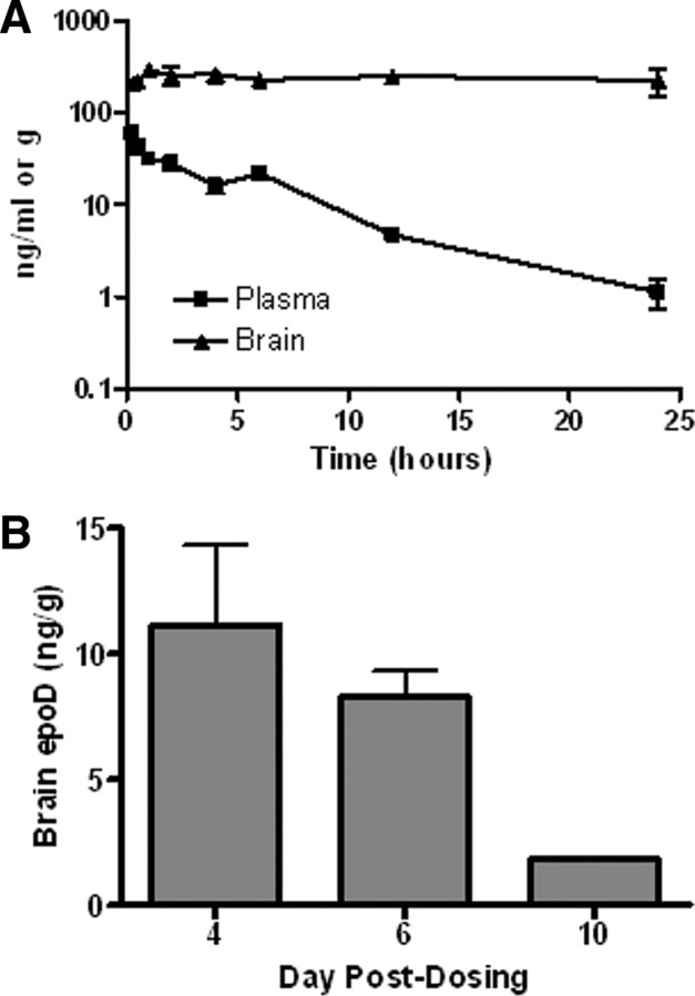
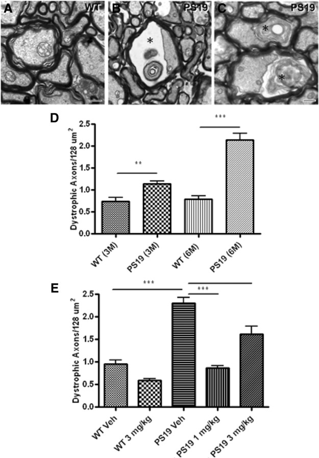
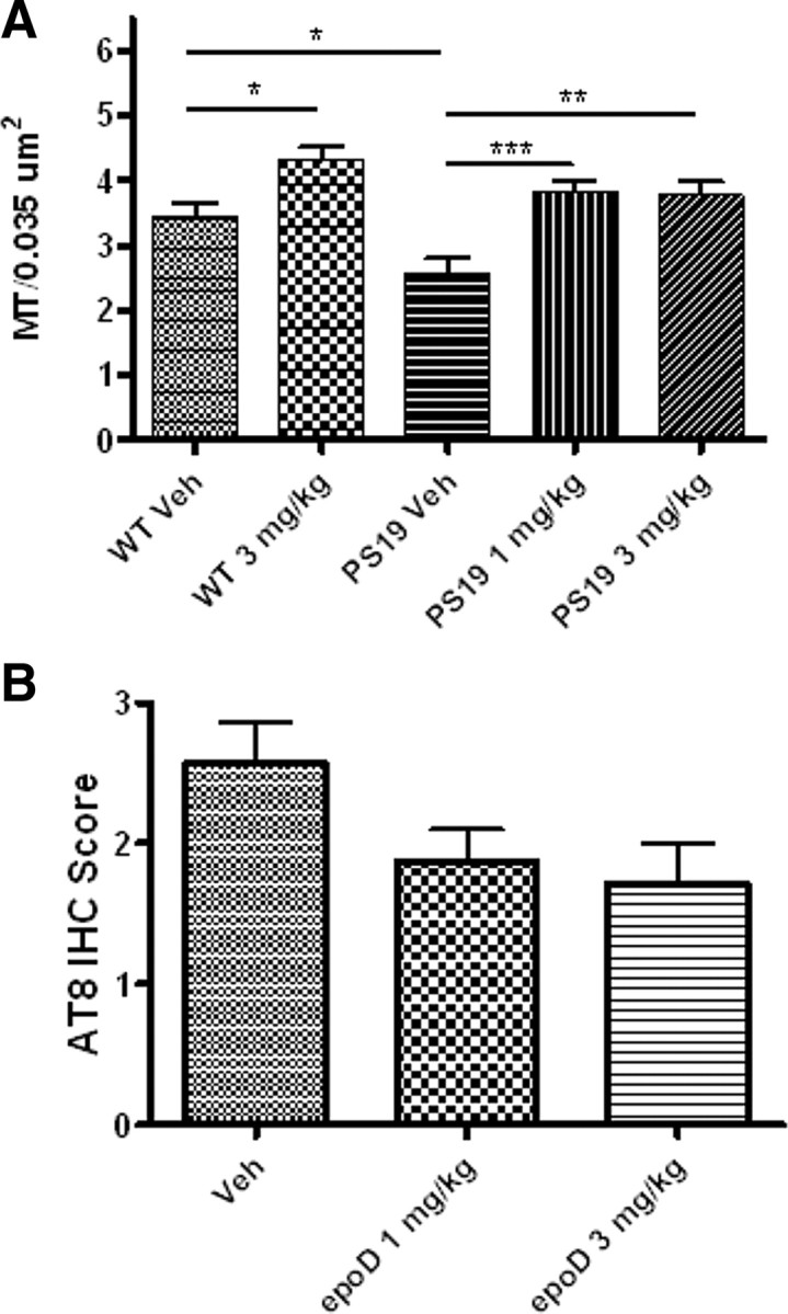
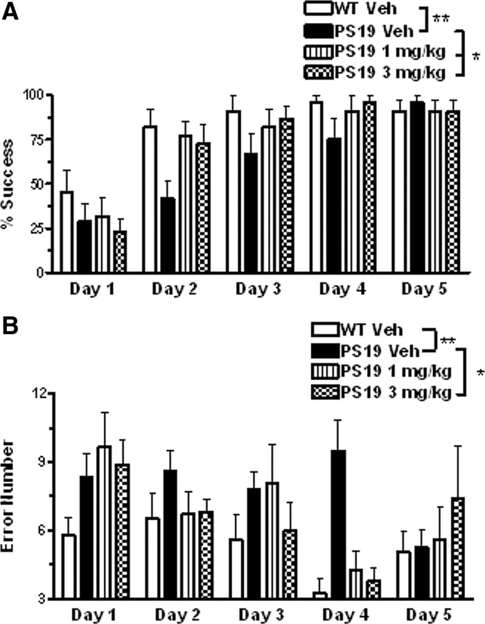
Neurons in the brains of those with Alzheimer's disease (AD) and many frontotemporal dementias (FTDs) contain neurofibrillary tangles comprised of hyperphosphorylated tau protein. Tau normally stabilizes microtubules (MTs), and tau misfolding could lead to a loss of this function with consequent MT destabilization and neuronal dysfunction. Accordingly, a possible therapeutic strategy for AD and related "tauopathies" is treatment with a MT-stabilizing anti-cancer drug such as paclitaxel. However, paclitaxel and related taxanes have poor blood-brain barrier permeability and thus are unsuitable for diseases of the brain. We demonstrate here that the MT-stabilizing agent, epothilone D (EpoD), is brain-penetrant and we subsequently evaluated whether EpoD can compensate for tau loss-of-function in PS19 tau transgenic mice that develop forebrain tau inclusions, axonal degeneration and MT deficits. Treatment of 3-month-old male PS19 mice with low doses of EpoD once weekly for a 3 month period significantly improved CNS MT density and axonal integrity without inducing notable side-effects. Moreover, EpoD treatment reduced cognitive deficits that were observed in the PS19 mice. These results suggest that certain brain-penetrant MT-stabilizing agents might provide a viable therapeutic strategy for the treatment of AD and FTDs.
Figures
Similar articles
-
The microtubule-stabilizing agent, epothilone D, reduces axonal dysfunction, neurotoxicity, cognitive deficits, and Alzheimer-like pathology in an interventional study with aged tau transgenic mice.J Neurosci. 2012 Mar 14;32(11):3601-11. doi: 10.1523/JNEUROSCI.4922-11.2012. J Neurosci. 2012. PMID: 22423084 Free PMC article.
-
Brain-penetrant microtubule-stabilizing compounds as potential therapeutic agents for tauopathies.Biochem Soc Trans. 2012 Aug;40(4):661-6. doi: 10.1042/BST20120010. Biochem Soc Trans. 2012. PMID: 22817712 Free PMC article. Review.
-
A brain-penetrant triazolopyrimidine enhances microtubule-stability, reduces axonal dysfunction and decreases tau pathology in a mouse tauopathy model.Mol Neurodegener. 2018 Nov 7;13(1):59. doi: 10.1186/s13024-018-0291-3. Mol Neurodegener. 2018. PMID: 30404654 Free PMC article.
-
Hyperdynamic microtubules, cognitive deficits, and pathology are improved in tau transgenic mice with low doses of the microtubule-stabilizing agent BMS-241027.J Neurosci. 2012 May 23;32(21):7137-45. doi: 10.1523/JNEUROSCI.0188-12.2012. J Neurosci. 2012. PMID: 22623658 Free PMC article.
-
Microtubule stabilizing agents as potential treatment for Alzheimer's disease and related neurodegenerative tauopathies.J Med Chem. 2012 Nov 8;55(21):8979-96. doi: 10.1021/jm301079z. Epub 2012 Sep 28. J Med Chem. 2012. PMID: 23020671 Free PMC article. Review.
Cited by
-
How do neurons age? A focused review on the aging of the microtubular cytoskeleton.Neural Regen Res. 2024 Sep 1;19(9):1899-1907. doi: 10.4103/1673-5374.390974. Epub 2023 Dec 15. Neural Regen Res. 2024. PMID: 38227514 Free PMC article.
-
Deleterious and protective effects of epothilone-D alone and in the context of amyloid β- and tau-induced alterations.Front Mol Neurosci. 2023 Oct 12;16:1198299. doi: 10.3389/fnmol.2023.1198299. eCollection 2023. Front Mol Neurosci. 2023. PMID: 37900942 Free PMC article.
-
Notch signalling in adult neurons: a potential target for microtubule stabilization.Ther Adv Neurol Disord. 2013 Nov;6(6):375-85. doi: 10.1177/1756285613490051. Ther Adv Neurol Disord. 2013. PMID: 24228073 Free PMC article.
-
Low dose tubulin-binding drugs rescue peroxisome trafficking deficit in patient-derived stem cells in Hereditary Spastic Paraplegia.Biol Open. 2014 May 23;3(6):494-502. doi: 10.1242/bio.20147641. Biol Open. 2014. PMID: 24857849 Free PMC article.
-
Mild and repetitive very mild axonal stretch injury triggers cystoskeletal mislocalization and growth cone collapse.PLoS One. 2017 May 4;12(5):e0176997. doi: 10.1371/journal.pone.0176997. eCollection 2017. PLoS One. 2017. PMID: 28472086 Free PMC article.
References
-
- Altmann KH. Recent developments in the chemical biology of epothilones. Curr Pharm Des. 2005;11:1595–1613. - PubMed
-
- Andrieux A, Salin P, Schweitzer A, Bégou M, Pachoud B, Brun P, Gory-Fauré S, Kujala P, Suaud-Chagny MF, Höfle G, Job D. Microtubule stabilizer ameliorates synaptic function and behavior in a mouse model for schizophrenia. Biol Psychiatry. 2006;60:1224–1230. - PubMed
-
- Arriagada PV, Growdon JH, Hedley-Whyte ET, Hyman BT. Neurofibrillary tangles but not senile plaques parallel duration and severity of Alzheimers disease. Neurology. 1992;42:631–639. - PubMed
Publication types
MeSH terms
Substances
Grants and funding
LinkOut - more resources
Full Text Sources
Other Literature Sources
Medical