. 2010 Dec;21(23):4275-86.
doi: 10.1091/mbc.E10-04-0332. Epub 2010 Oct 13.

Cell death or survival promoted by alternative isoforms of ErbB4

Affiliations

Cell death or survival promoted by alternative isoforms of ErbB4

Maria Sundvall et al. Mol Biol Cell. 2010 Dec.

Abstract

The significance of ErbB4 in tumor biology is poorly understood. The ERBB4 gene is alternatively spliced producing juxtamembrane (JM-a and JM-b) and cytoplasmic (CYT-1 and CYT-2) isoforms. Here, signaling via the two alternative ErbB4 JM isoforms (JM-a CYT-2 and JM-b CYT-2) was compared. Fibroblasts expressing ErbB4 JM-a demonstrated enhanced ErbB4 autophosphorylation, growth, and survival. In contrast, cells overexpressing ErbB4 JM-b underwent starvation-induced death. Both pro- and antisurvival responses to the two ErbB4 isoforms were sensitive to an ErbB kinase inhibitor. Platelet-derived growth factor receptor-alpha (PDGFRA) was identified as an ErbB4 target gene that was differentially regulated by the two ErbB4 isoforms. The soluble intracellular domain of ErbB4, released from the JM-a but not from the JM-b isoform, associated with the transcription factor AP-2 and promoted its potential to enhance PDGFRA transcription. Survival of cells expressing JM-a was suppressed by targeting either PDGFR-α or AP-2, whereas cells expressing JM-b were rescued from cell death by the PDGFR-α agonist, PDGF-BB. These findings indicate that two alternative ErbB4 isoforms may promote antagonistic cellular responses and suggest that pharmacological inhibition of ErbB4 kinase activity may lead to either suppression or promotion of cellular growth.


Figures
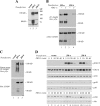
Figure 1.
Figure 1.
Expression and activity of ErbB4 isoforms in NR6 cells. (A) Expression of ErbB4 protein in NR6 transfectants was analyzed by Western blotting. (B) Cells were starved overnight without serum and stimulated with or without 50 ng/ml NRG-1 for 10 min. ErbB4 tyrosine phosphorylation was analyzed by immunoprecipitation with an anti-ErbB4 antibody followed by Western blotting with an anti-phosphotyrosine antibody. Membrane was reblotted with an anti-ErbB4 antibody. (C) For in vitro kinase assay, cell lysates were immunoprecipitated with an anti-ErbB4 antibody, incubated with ATP, and analyzed for ErbB4 tyrosine phosphorylation by immunoprecipitation with anti-ErbB4 antibody followed by Western blotting with an anti-phosphotyrosine antibody. (D) Cells were starved overnight and stimulated with or without NRG-1 for indicated time periods. Phosphorylation of Erk, Akt, and p38 was analyzed by Western blotting with phosphospecific antibodies. Subsequently, membranes were reblotted with anti-Erk, anti-Akt, anti-p38, or anti-actin antibodies.
Figure 2.
Figure 2.
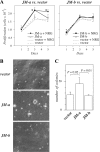
Growth of NR6 cells expressing ErbB4 isoforms. (A) NR6 transfectants were plated onto 24-well plates in the presence of 10% FCS. The next day the medium was replaced with medium containing 5% FCS with or without 50 ng/ml NRG-1. Adherent cells were counted at indicated time points with hemocytometer. *p < 0.05 for a difference between ErbB4 overexpression versus vector in the presence of NRG-1. (B and C) Cells were cultured in soft agar for 7 wk. Representative images (B) and quantitation of colonies (C) are shown.
Figure 3.
Figure 3.
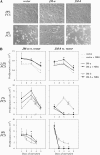
The effect of serum starvation on cells expressing ErbB4 isoforms. (A) NR6 transfectants were cultured for 2 d in medium containing 0 or 10% of FCS and photographed. (B) NR6 transfectants were plated onto 24-well plates in the presence of 10% FCS. The next day the medium was replaced with medium containing 0, 1, or 2.5% FCS with or without 50 ng/ml NRG-1. Adherent cells were counted at indicated time points with hemocytometer. *p < 0.05 for a difference between ErbB4 overexpression versus vector in the presence of NRG-1.
Figure 4.
Figure 4.
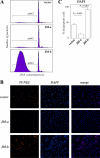
Starvation-induced death of NR6 cells expressing ErbB4 isoforms. (A) Cells were cultured for 72 h in the absence of FCS, and their DNA contents were analyzed by PI staining and FACS. The proportion of cells containing subG1 quantities of DNA per particle is indicated. (B) Cells were stained with TUNEL (red) and DAPI (blue) after starvation for 2 d and photographed under a fluorescence microscope. (C) The percentage of condensed nuclei stained with DAPI was calculated from five arbitrary fields per cell line under a microscope.
Figure 5.
Figure 5.
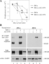
The effect of inhibition of ErbB tyrosine kinase activity on cells expressing ErbB4 isoforms. (A) NR6 cells expressing JM-a CYT-2 or JM-b CYT-2 were plated onto 24-well plates in the presence of 10% FCS. The next day the medium was replaced with serum-free medium containing 0 or 10 μM of the ErbB kinase inhibitor AG 1478. Adherent cells were counted at indicated time points with hemocytometer. *p < 0.05 for a difference between AG 1478-treated and control cells. (B) Cells were starved overnight without serum, treated for 6 h with 0 or 10 μM AG 1478, and stimulated for 15 min with 0 or 50 ng/ml NRG-1. ErbB4 tyrosine phosphorylation was analyzed by immunoprecipitation with an anti-ErbB4 antibody followed by Western blotting with an anti-phosphotyrosine antibody. Two different exposures of the phosphotyrosine blot are shown. Membrane was reblotted with an anti-ErbB4 antibody. Binding of the anti-phosphotyrosine antibody 4G10 masks the epitope for the anti-ErbB4 sc-283, resulting in an apparently reduced ErbB4 signal for the heavily phosphorylated ErbB4 species in the reblot (lanes 1 and 3).
Figure 6.
Figure 6.
Regulation of PDGFRA expression by ErbB4 isoforms. (A) NR6 transfectants were cultured for 8 h in the absence of serum, stimulated for 2 h with or without 50 ng/ml NRG-1, and analyzed for PDGFRA mRNA expression by RT-PCR. (B) NR6 transfectants were treated with or without 10 μM AG 1478 for 8 h in the absence of serum. PDGFRA mRNA expression was analyzed by RT-PCR. (C) Cells were cultured overnight in the presence or absence of 10% FCS. PDGFR-α protein expression was analyzed by Western blotting with anti-PDGFR-α antibody. n.s., nonspecific band also present in the vector control lanes. (D) Cells were starved overnight without serum and stimulated for 10 min with 0 or 50 ng/ml PDGF-BB. PDGFR-α tyrosine phosphorylation was analyzed by immunoprecipitation with an anti-PDGFR-α antibody followed by Western blotting with an anti-phosphotyrosine antibody. Membrane was reblotted with an anti-PDGFR-α antibody. (E) NR6 transfectants were plated onto 24-well plates. The next day the medium was replaced with serum-free medium containing or not 20 μM of PDGFR inhibitor AG 1296 or 50 ng/ml PDGF-BB. Adherent cells were counted at indicated time points with hemocytometer. *p < 0.05 for a difference between AG 1296– or PDGF-BB–treated and control cells. (F) NR6 cells expressing JM-a CYT-2 were cultured for 16 h in the presence or absence of 20 μM AG 1296 in serum-free medium. PDGFR-α tyrosine phosphorylation was analyzed by immunoprecipitation with an anti-PDGFR-α antibody followed by Western blotting with an anti-phosphotyrosine antibody. Membrane was reblotted with an anti-PDGFR-α antibody. ErbB4 tyrosine phosphorylation was analyzed by Western blotting with a phospho-specific anti-ErbB4 antibody followed by reblotting with an anti-ErbB4 antibody.
Figure 7.
Figure 7.
Regulation of PDGFRA transcription by ErbB4 activity. (A) SK-N-MC human neuroblastoma cells were treated for 48 h with or without control or ErbB4-specific siRNAs. Relative PDGFRA and ERBB4 mRNA expression was analyzed by real-time RT-PCR and normalized to the expression of the reference gene β-actin. (B) MCF-7 breast cancer cells were transfected with the PDGFRA promoter luciferase reporter constructs pSLA4PDGFRαluc–441/+118 (441), pSLA4PDGFRαluc–944/+118 (944), or pSLA4PDGFRαluc–1253/+118 (1253) together with a plasmid encoding EGFP. Cells starved for 16 h were treated for 2 h with or without 80 ng/ml NRG-1 to activate ErbB4 or for 18 h with 10 μM retinoic acid (RA) as a positive control known to stimulate PDGFRA promoter activity. Columns represent relative luciferase activity of promoter constructs normalized by the EGFP fluorescence signal (no treatment = 100% for each experiment). (C) MCF-7 cells expressing pSLA4PDGFRαluc–1253/+118 were treated for 48 h with or without control or ErbB4-specific siRNAs, starved for 16 h, and treated for 2 h with 0 or 80 ng/ml NRG-1. Columns represent relative luciferase activity of promoter constructs normalized by the EGFP fluorescence signal. (D) MCF-7 cells expressing pSLA4PDGFRαluc–1253/+118 were treated for 48 h with control siRNA or siRNAs targeting the indicated transcription factors (TF), starved for 16 h and treated for 2 h with 0 or 80 ng/ml NRG-1. PDGFRA promoter activity was determined as in C. The values represent the effect of NRG-1 treatment as percentages of the control siRNA sample. The transcription factor mRNA expression levels, as determined by real-time RT-PCR, are shown relative to control siRNA treatment (TF mRNA %).
Figure 8.
Figure 8.
Selective and functional association of soluble ErbB4 ICD with AP-2. (A) COS-7 cells were transfected with pcDNA3.1ICD2-HA or pcDNA3.1ErbB4JM-bCYT-2-HA, stained with anti-HA (green) and anti-AP-2α (red) antibodies. The nuclei were stained with DAPI (blue). The cells were visualized by confocal microscopy. Bar, 10 μm. (B) COS-7 cells were transiently transfected with constructs encoding HA-tagged ErbB4 JM-b CYT-2 or ErbB4 ICD2 and Myc-tagged AP-2α or AP-2-γ. Lysates were immunoprecipitated with anti-Myc antibody and anti-HA antibody was used in Western blotting. Expression levels were controlled by Western analysis with anti-HA and anti-Myc antibodies. (C) GST fusion proteins including the whole intracellular domain of the CYT-2-type (ICD), N-terminally truncated ICD (ICD-ΔN), or C-terminally truncated ICD (ICD-ΔC) were incubated with lysates of COS-7 cells transiently expressing Myc-tagged AP-2γ or Wwox. Material precipitating with glutathione Sepharose beads was analyzed by Western blotting with anti-Myc antibody. Membranes were reblotted with anti-GST antibody. (D) HEK-293T cells were cotransfected with the PDGFRA promoter-luciferase construct pSLA4PDGFRαluc–1253/+118 and a construct encoding Renilla luciferase together with an empty vector, constructs encoding the ErbB4 ICD of CYT-2-type (ICD), AP-2α, or AP-2γ, or a combination of constructs encoding ICD and AP-2. Columns represent relative luciferase activity of the PDGFRA promoter construct normalized by fluorescence signal from the Renilla luciferase. (E) NR6 cells stably expressing JM-a CYT-2 or JM-b CYT-2 were transfected with siRNA targeting AP-2α or with a nonsilencing control siRNA. After siRNA transfection the cells were starved for 3 d, and the number of viable cells was determined with MTT assay. *p < 0.05.

Similar articles

Cited by

References

    1. Afink G. B., Nister M., Stassen B. H., Joosten P. H., Rademakers P. J., Bongcam-Rudloff E., Van Zoelen E. J., Mosselman S. Molecular cloning and functional characterization of the human platelet-derived growth factor alpha receptor gene promoter. Oncogene. 1995;8:1667–1672. - PubMed
    1. Alaoui-Jamali M. A., Song D. J., Benlimame N., Yen L., Deng X., Hernandez-Perez M., Wang T. Regulation of multiple tumor microenvironment markers by overexpression of single or paired combinations of ErbB receptors. Cancer Res. 2003;13:3764–3774. - PubMed
    1. Aqeilan R. I., Donati V., Palamarchuk A., Trapasso F., Kaou M., Pekarsky Y., Sudol M., Croce C. M. WW domain-containing proteins, WWOX and YAP, compete for interaction with ErbB-4 and modulate its transcriptional function. Cancer Res. 2005;15:6764–6772. - PubMed
    1. Aqeilan R. I., Palamarchuk A., Weigel R. J., Herrero J. J., Pekarsky Y., Croce C. M. Physical and functional interactions between the Wwox tumor suppressor protein and the AP-2gamma transcription factor. Cancer Res. 2004a;22:8256–8261. - PubMed
    1. Aqeilan R. I., et al. Functional association between Wwox tumor suppressor protein and p73, a p53 homolog. Proc. Natl. Acad. Sci. USA. 2004b;13:4401–4406. - PMC - PubMed

Publication types

MeSH terms