. 2010 Dec;21(23):4173-83.
doi: 10.1091/mbc.E09-09-0782. Epub 2010 Oct 13.

Microautophagy of the nucleus coincides with a vacuolar diffusion barrier at nuclear-vacuolar junctions

Affiliations

Microautophagy of the nucleus coincides with a vacuolar diffusion barrier at nuclear-vacuolar junctions

Rosie Dawaliby et al. Mol Biol Cell. 2010 Dec.

Abstract

Nuclei bind yeast vacuoles via nucleus-vacuole (NV) junctions. Under nutrient restriction, NV junctions invaginate and release vesicles filled with nuclear material into vacuoles, resulting in piecemeal microautophagy of the nucleus (PMN). We show that the electrochemical gradient across the vacuolar membrane promotes invagination of NV junctions. Existing invaginations persist independently of the gradient, but final release of PMN vesicles requires again V-ATPase activity. We find that NV junctions form a diffusion barrier on the vacuolar membrane that excludes V-ATPase but is enriched in the VTC complex and accessible to other membrane-integral proteins. V-ATPase exclusion depends on the NV junction proteins Nvj1p,Vac8p, and the electrochemical gradient. It also depends on factors of lipid metabolism, such as the oxysterol binding protein Osh1p and the enoyl-CoA reductase Tsc13p, which are enriched in NV junctions, and on Lag1p and Fen1p. Our observations suggest that NV junctions form in two separable steps: Nvj1p and Vac8p suffice to establish contact between the two membranes. The electrochemical potential and lipid-modifying enzymes are needed to establish the vacuolar diffusion barrier, invaginate NV junctions, and form PMN vesicles.


Figures
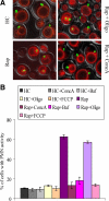
Figure 1.
Figure 1.
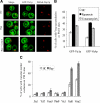
The V-ATPase inhibitor concanamycin A reduces PMN. (A) Wild-type cells expressing GFP-Nop1 under its endogenous promotor were stained with FM4-64 in HC media to visualize vacuoles. Cells were then incubated for a total of 3 h with 0.2 μΜ rapamycin and/or 1 μM concanamycin A, 5 μM oligomycin, or 5 μM FCCP. Cells were analyzed by fluorescence microscopy. (B) The percentage of cells carrying PMN vesicles or PMN structures was determined for each condition. For each experiment, 200 cells per condition were analyzed by confocal microscopy. Three independent experiments were averaged and standard deviations calculated.
Figure 2.
Figure 2.
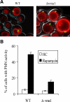
V-ATPase cells are defective in PMN. (A) Wild-type or Δvma1 cells expressing GFP-Nop1p under its endogenous promotor were stained with FM4-64 in HC media to visualize vacuoles. Cells were then treated with 0.2 μΜ rapamycin, incubated for a total of 3 h, and analyzed by fluorescence microscopy. (B) Cells carrying PMN vesicles or PMN structures were counted as in Figure 1.
Figure 3.
Figure 3.
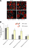
Effect of concanamycin A on PMN structures and vesicles, depending on the time of addition. (A) Cells expressing GFP-Nop1 under its endogenous promotor were stained with FM4-64 in HC and were then treated for a total of 3 h as follows: With 0.2 μΜ rapamycin for 3 h; with rapamycin and concanamycin A for 3 h; with rapamycin for 2 h before adding 1μΜ concanamycin A and continuing the incubation for another 1h; with rapamycin for 2h. (B) For PMN activity the number of cells with PMN vesicles or PMN structures inside the vacuoles was counted. The scheme illustrates the different PMN phenotypes monitored.
Figure 4.
Figure 4.
Vph1p exclusion is abolished by concanamycin A. (A) Wild-type yeast cells carrying Vph1p-GFP, DsRed-Nop1p, and Nvj1p-ECFP were treated in HC medium with 0.2 μΜ rapamycin and/or 1 μΜ concanamycin A for 3 h, or left untreated. Samples were analyzed by confocal microscopy. Arrowheads show Vph1p exclusion sites. (B) The percentage of wild-type cells in A that presented Vph1p exclusion from Nvj1p-CFP–positive sites was quantified. In a parallel experiment, we performed the same quantifications for a strain carrying Vma2-GFP instead of Vph1-GFP.
Figure 5.
Figure 5.
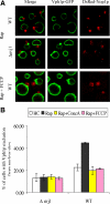
Vph1p exclusion depends on Nvj1. (A) Wild-type and Δnvj1 mutants carrying Vph1p-GFP and DsRed-Nop1p were treated with 0.2 μΜ rapamycin for 3 h. Samples were analyzed by confocal microscopy. (B) Wild-type cells carrying Vph1p-GFP and DsRed-Nop1p were treated together with 0.2 μΜ rapamycin and 5 μM FCCP for 3 h. Samples were analyzed by confocal microscopy. (C) The percentage of wild-type and Δnvj1 cells presenting Vph1p exclusion at DsRed-Nop1p-positive sites was determined.
Figure 6.
Figure 6.
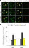
Presence of vacuolar proteins at PMN sites. (A) Wild-type cells expressing DsRed-Nop1 and GFP-Vtc1p or GFP-Vtc4p were incubated in HC medium with or without 0.2 μΜ rapamycin for 3 h before being analyzed by confocal microscopy. Only GFP-Vtc1p pictures are shown as examples. (B) Quantification of DsRed-Nop1p–positive sites showing an enrichment of GFP-Vtc1p or GFP-Vtc4p when compared with the rest of the vacuolar boundary membrane. (C) Cells expressing DsRed-Nop1 and GFP fusions of the indicated proteins were incubated and analyzed as in A. The frequency of cells showing exclusion of the respective GFP fusion from DsRed-Nop1–positive sites was determined.
Figure 7.
Figure 7.
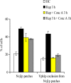
Concanamycin A inhibits Vph1p exclusion but not formation of Nvj1p patches. Wild-type cells carrying Vph1p-GFP and Nvj1p-CFP had been stained with FM4-64 in HC and were then treated for a total of 3 h as follows: With 0.2 μΜ rapamycin for 3 h; with rapamycin for 2 h before adding 1 μΜ concanamycin A and continuing the incubation for another 1 h; with rapamycin and concanamycin A for 3 h. Samples were analyzed by confocal microscopy. The percentage of cells showing Nvj1p patches adjacent to vacuoles and the percentage of cells showing Vph1p exclusion next to Nvj1p patches was determined.
Figure 8.
Figure 8.
Random exclusion of Vph1p upon overexpression of Nvj1p. (A) Cells overexpressing HA-Nvj1 from the GPD promoter and carrying Vph1p-GFP and DsRed-Nop1p were treated in HC medium for 3 h with 0.2 μΜ rapamycin and 1 μM concanamycin A as indicated, or left without additions. Cells were analyzed by confocal microscopy. Arrows indicate random exclusion sites. (B) Quantification of random and restricted exclusion of Vph1p in the cells from A. (C) Cells with GFP-Nvj1 overexpressed from the GPD promoter and carrying Vph1p-CFP were treated and analyzed as in A. Arrows indicate random exclusion sites. BF: bright field. (D) Quantification of random and normal exclusion of Vph1p in the cells from C.

References

    1. Ashman W. H., Barbuch R. J., Ulbright C. E., Jarrett H. W., Bard M. Cloning and disruption of the yeast C-8 sterol isomerase gene. Lipids. 1991;26:628–632. - PubMed
    1. Baars T. L., Petri S., Peters C., Mayer A. Role of the V-ATPase in regulation of the vacuolar fission-fusion equilibrium. Mol. Biol. Cell. 2007;18:3873–3882. - PMC - PubMed
    1. Bayer M. J., Reese C., Buhler S., Peters C., Mayer A. Vacuole membrane fusion: V0 functions after trans-SNARE pairing and is coupled to the Ca2+-releasing channel. J. Cell Biol. 2003;162:211–222. - PMC - PubMed
    1. Beh C. T., Cool L., Phillips J., Rine J. Overlapping functions of the yeast oxysterol-binding protein homologues. Genetics. 2001;157:1117–1140. - PMC - PubMed
    1. Beh C. T., Rine J. A role for yeast oxysterol-binding protein homologs in endocytosis and in the maintenance of intracellular sterol-lipid distribution. J. Cell Sci. 2004;117:2983–2996. - PubMed

MeSH terms

Substances

LinkOut - more resources