. 2010 Dec;84(24):12599-608.
doi: 10.1128/JVI.01437-10. Epub 2010 Oct 13.

Identification of specific determinants of human APOBEC3F, APOBEC3C, and APOBEC3DE and African green monkey APOBEC3F that interact with HIV-1 Vif

Affiliations

Identification of specific determinants of human APOBEC3F, APOBEC3C, and APOBEC3DE and African green monkey APOBEC3F that interact with HIV-1 Vif

Jessica L Smith et al. J Virol. 2010 Dec.

Abstract

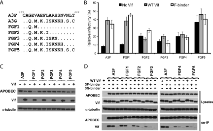
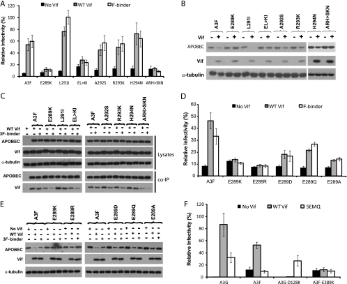
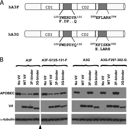
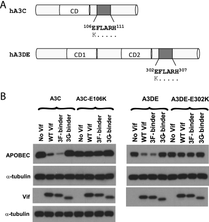
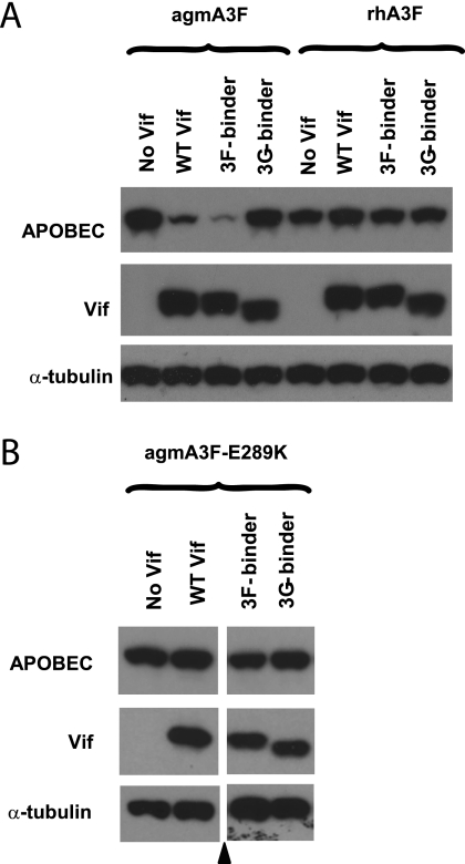
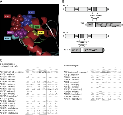
Human APOBEC3F (hA3F) and human APOBEC3G (hA3G) are potent anti-human immunodeficiency virus (anti-HIV) host factors that suppress viral replication by hypermutating the viral genome, inhibiting reverse transcription, and hindering integration. To overcome hA3F and hA3G, HIV-1 encodes Vif, which binds and targets these host proteins for proteasomal degradation. Previously, we reported that the hA3F-Vif interactions that lead to hA3F degradation are located in the region comprising amino acids 283 to 300. We have now performed mutational analysis of this region and found that the (289)EFLARH(294) amino acids contribute to hA3F-Vif binding and are critical for A3F's sensitivity to Vif. Mutants in which E289 is mutated significantly increase hA3F's ability to inhibit viral infectivity in the presence of Vif, and coimmunoprecipitation assays show that binding of Vif to the E289K mutant is decreased. We examined the role of the EFLARH sequence in other A3 proteins, including human A3C (hA3C), human A3DE (hA3DE), African green monkey A3F (agmA3F), and rhesus macaque A3F (rhA3F). hA3C, hA3DE, and agmA3F were all susceptible to degradation induced by HIV-1 Vif, while rhA3F was not. Mutagenesis of the glutamate in the EFLARH sites of hA3C, hA3DE, and agmA3F decreases the susceptibilities of these proteins to Vif-induced degradation. Together, these results indicate that the EFLARH region in hA3F, hA3C, hA3DE, and agmA3F interacts with HIV-1 Vif and that this interaction plays a role in the Vif-mediated proteasomal degradation of these A3 proteins. These studies identify a conserved region in 3 of 7 human A3 proteins that is critical for degradation mediated by HIV-1 Vif and provide structural insights into the hA3F-Vif interactions that could facilitate the development of a novel class of anti-HIV agents.


Figures

FIG. 1.
FIG. 1.
Localization of the Vif interaction domain in hA3F. (A) Schematic representation of hA3F mutants used to determine the residues in hA3F which are important for interactions with HIV-1 Vif. The top line depicts amino acids 283 to 300 of WT hA3F, and the second line shows the sequence within the same region in hA3G. The third line through the seventh line show the amino acid substitutions in hA3F for five FGF chimeras. Dots indicate identical amino acids. (B) Effect of hA3F and the FGF chimeras on HIV-1 infectivity in the presence and absence of HIV-1 Vif. Single-cycle viruses were produced from 293T cells that were transfected with pHDV-eGFP, pHCMV-G, an hA3F expression plasmid, and either no Vif, WT Vif, or the F binder. The p24 CA levels were determined and used to normalize the levels for virus samples prior to infection of TZM-bl indicator cells. The infectivity of the viruses was determined by quantitation of luciferase activity. Data are plotted as relative infectivity levels, with the level for the hA3F-free virus (not shown) set to 100%. Error bars indicate standard errors of the means (SEM) of results from six infectivity experiments performed with three independent virus stocks. (C) Effects of WT HIV-1 Vif on the degradation of WT hA3F or the FGF chimeras. Cotransfections of 293T cells were carried out with an hA3F expression plasmid and either no Vif or WT Vif expression plasmid. Cell lysates were analyzed by Western blotting using anti-FLAG, anti-Vif, or anti-α-tubulin antibodies. The results are representative of two independent experiments. (D) Co-IP assays to determine binding of Vif to FGF chimeras. 293T cells were cotransfected with expression plasmids for FLAG-A3F or FLAG-FGF chimeras along with expression plasmids for WT Vif, the F binder, or the G binder. Cell lysates and immunoprecipitated proteins were analyzed by Western blotting using anti-FLAG and anti-Vif antibodies. The cell lysates were also analyzed for α-tubulin as a loading control. The results are representative of three independent experiments. WT, wild type; +, with; −, without.
FIG. 2.
FIG. 2.
Identification of specific amino acids in hA3F that are critical for Vif interactions. (A) Effects of single amino acid substitutions within the hA3F region comprising amino acids 289 to 294 on HIV-1 infectivity. Viruses were produced from 293T cells that were transfected with pHDV-eGFP and expression plasmids for VSV-G, hA3F, or its mutants and either no Vif, WT Vif, or the F binder. The infectivity of the viruses was determined by infection of TZM-bl cells and quantitation of luciferase activity. Data are plotted as relative infectivity levels, with the level for the hA3F-free virus (not shown) set to 100%. Error bars indicate standard errors of the means (SEM) of results from six infectivity experiments performed with three independent virus stocks. (B) Effects of WT HIV-1 Vif on the degradation of WT hA3F or its mutants. Cotransfections of 293T cells were carried out with a hA3F expression plasmid and either no Vif or WT Vif expression plasmid. Cell lysates were analyzed by Western blotting using anti-FLAG, anti-Vif, or anti-α-tubulin antibodies. The results are representative of two independent experiments. (C) Co-IP assays to determine binding of Vif to hA3F mutants. 293T cells were cotransfected with expression plasmids for FLAG-A3F or FLAG-A3F mutants along with expression plasmids for WT HIV-1 Vif, the F binder, or the G binder. Cell lysates and immunoprecipitated proteins were analyzed by Western blotting using anti-FLAG and anti-Vif antibodies. The cell lysates were also analyzed for α-tubulin as a loading control. WT, wild type; +, with; −, without. The results are representative of three independent experiments. (D) Effects of substituting amino acids with different biochemical properties at position E289 on HIV-1 infectivity. Data are plotted as relative infectivity levels, with the levels for the hA3F-free virus (not shown) set to 100%. Error bars indicate standard errors of the means (SEM) of results from six infectivity experiments performed with three independent virus stocks. (E) Effects of WT HIV-1 Vif on the degradation of WT A3F or the E289 mutants. Lysates from the viral producer cells of one set of viruses used for panel D were analyzed by Western blotting using anti-FLAG, anti-Vif, or anti-α-tubulin antibodies. (F) Rescue of infectivity by HIV-1 Vif(SEMQ). Data are plotted as relative infectivity levels, with the level for the hA3F-free virus (not shown) set to 100%. Error bars indicate standard errors of the means (SEM) of results from six infectivity experiments performed with three independent virus stocks.
FIG. 3.
FIG. 3.
Sensitivity of hA3F-G125-131-F to G binder Vif and hA3G-F297-302-F to F binder Vif. (A) Schematic representation of hA3F and hA3G with the amino acid substitutions used to generate the hA3F-G125-131-F and hA3G-F297-302-F constructs. The relative locations of the catalytic domains (CD) are indicated, and dots indicate identical amino acids. (B) Effects of WT HIV-1 Vif, the 3F binder, and the 3G binder on the degradation of hA3F-G125-131-F and hA3G-F297-302-F. 293T cells were cotransfected with hA3F, hA3F-G125-131-F, A3G, or hA3G-F297-302-G expression plasmids and either no Vif, WT Vif, F binder, or G binder expression plasmids. Cell lysates were analyzed by Western blotting using anti-FLAG, anti-Vif, or anti-α-tubulin antibodies. The black triangle indicates removal of uninformative lanes from a single gel. The results are representative of two independent experiments.
FIG. 4.
FIG. 4.
Sensitivity of hA3C and hA3DE to HIV-1 Vif. (A) Schematic representation of hA3C and hA3DE with the relative locations of the catalytic domains (CD) and the EFLARH Vif interaction domains. The E106K and E302K mutants of hA3C and hA3DE, respectively, are indicated. Dots indicate identical amino acids. (B) Effects of WT HIV-1 Vif, the 3F binder, and the 3G binder on the degradation of hA3C, hA3C(E106K), hA3DE, and hA3DE(E302K). 293T cells were cotransfected with hA3C or hA3DE expression plasmids and either no Vif, WT Vif, F binder, or G binder expression plasmids. Cell lysates were analyzed by Western blotting using anti-FLAG, anti-Vif, or anti-α-tubulin antibodies. The results are representative of three independent experiments.
FIG. 5.
FIG. 5.
Sensitivity of agmA3F and rhA3F to HIV-1 Vif. (A) Effects of WT HIV-1 Vif, the 3F binder, and the 3G binder on the degradation of WT agmA3F, WT rhA3F, and the E289K mutant of agmA3F. Cotransfections of 293T cells were carried out with myc-A3F expression plasmids and either no Vif, WT Vif, F binder, or G binder expression plasmids. Cell lysates were analyzed by Western blotting using anti-myc, anti-Vif, or anti-α-tubulin antibodies. The results are representative of three independent experiments. (B) The E289K mutant of agmA3F is resistant to degradation by HIV-1 Vif. The black triangle indicates removal of uninformative lanes from a single gel. The results are representative of two independent experiments.
FIG. 6.
FIG. 6.
Model structure of the HIV-1 Vif binding domain of hA3F. (A) Model structure of the C-terminal domain of hA3G (Protein Data Bank accession number 2KEM) (17) is shown. hA3G amino acids 297 to 302 of α-helix 3, which are equivalent to hA3F amino acids 289 to 294, are shown in space-filling form, with color coding as indicated. The remaining hA3G protein is shown in ribbon form; the α-helices are shown in red, and the β-sheets are shown in cyan. The figure was generated using the Accelrys Discovery Studio Visualizer (version 2.5) software program. (B) Schematic representation of hA3F-Vif and hA3G-Vif interactions. The major determinants of hA3F-Vif interactions and hA3G-Vif are shown. The determinants that were used in this study are shown extending out from each protein. The residues shown are those that have been shown to decrease interactions with A3F (top) or A3G (bottom) upon mutagenesis but to retain binding to the other APOBEC3 protein or cullin 5. (C) Sequence alignments of seven human A3 proteins and the A3F, A3C, and A3DE proteins from several nonhuman primates. The sequences are compared to the hA3F C-terminal EFLARH sequence and surrounding amino acids (left) and the N-terminal EFLAEH sequence and surrounding amino acids (right). Protein alignments were done using the BioEdit Sequence Alignment Editor, version 7.0.5.3 (16). The GenBank accession numbers of the sequences used in these comparisons were NP_660341.2 (A3F isoform a; Homo sapiens), AAH38808.1 (A3F; H. sapiens*), AAH11739.1 (A3C; H. sapiens), NP_689639.2 (A3D; H. sapiens), NP_068594.1 (A3G; H. sapiens), EAW60281.1 (A3B; H. sapiens), NP_663745.1 (A3A; H. sapiens), ACK77772.1 (A3H; H. sapiens), NP_001035832.1 (A3F; M. mulatta), XP_525658.2) (A3F; P. troglodytes), AAT44387.1 (A3C; G. gorilla), ABY85203.1 (A3C; C. aethiops), ABY85204.1 (A3C; M. mulatta), XP_001094328.2 (A3D; M. mulatta), XP_525657.2 (A3D; P. troglodytes), XP_001094452.2 (A3G; M. mulatta), and NP_001009001.1 (P. troglodytes). The sequence for A3F (C. aethiops) was provided in reference .

Similar articles

Cited by

References

    1. Anderson, J. L., and T. J. Hope. 2008. APOBEC3G restricts early HIV-1 replication in the cytoplasm of target cells. Virology 375:1-12. - PMC - PubMed
    1. Bergeron, J. R., H. Huthoff, D. A. Veselkov, R. L. Beavil, P. J. Simpson, S. J. Matthews, M. H. Malim, and M. R. Sanderson. 2010. The SOCS-box of HIV-1 Vif interacts with ElonginBC by induced-folding to recruit its Cul5-containing ubiquitin ligase complex. PLoS Pathog. 6:e1000925. - PMC - PubMed
    1. Bishop, K. N., M. Verma, E. Y. Kim, S. M. Wolinsky, and M. H. Malim. 2008. APOBEC3G inhibits elongation of HIV-1 reverse transcripts. PLoS Pathog. 4:e1000231. - PMC - PubMed
    1. Bogerd, H. P., B. P. Doehle, H. L. Wiegand, and B. R. Cullen. 2004. A single amino acid difference in the host APOBEC3G protein controls the primate species specificity of HIV type 1 virion infectivity factor. Proc. Natl. Acad. Sci. U. S. A. 101:3770-3774. - PMC - PubMed
    1. Boussif, O., F. Lezoualc'h, M. A. Zanta, M. D. Mergny, D. Scherman, B. Demeneix, and J. P. Behr. 1995. A versatile vector for gene and oligonucleotide transfer into cells in culture and in vivo: polyethylenimine. Proc. Natl. Acad. Sci. U. S. A. 92:7297-7301. - PMC - PubMed

Publication types

MeSH terms

Substances

LinkOut - more resources